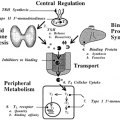
FUNCTIONS OF ADRENOCORTICOTROPIN
The primary action of ACTH is to promote steroidogenesis—that is, to enhance the synthesis and secretion of glucocorticoids, mineralocorticoids, and weak androgenic steroids of the adrenal cortex. However, the main physiologic controller of aldosterone release is the renin-angiotensin system (see Chap. 17). The N-terminal 18 amino acids are capable of cortisol release but are subject to rapid degradation; hence, the N-terminal 24 amino acids are used clinically to stimulate ACTH release (cosyntropin). ACTH acts on a specific 297-amino acid cell surface receptor that belongs to the Gs-protein–coupled 7-transmembrane superfamily of receptors. The ACTH receptor (also known as the melanocortin-2 receptor) gene is located at chromosome 18p11.1.12,13 The ACTH receptor acts via a cyclic adenosine monophosphate–dependent second messenger pathway to increase adrenal lipoprotein uptake from plasma and increase the transcription rates of genes for enzymes involved in steroidogenesis. Increased low-density lipoprotein uptake is facilitated by an increase in cell-surface low-density lipoprotein receptors. In contrast, ACTH has a smaller effect on zona fasciculata hydroxymethylglutaryl-coenzyme A reductase levels and hence cholesterol synthesis.14,15 Acutely, ACTH increases the activity of the rate-limiting enzyme of adrenal steroidogenesis, the P450SCC enzyme that catalyzes the conversion of cholesterol to Δ5-pregnenolone. Chronically, ACTH increases the activity of other adrenal enzymes involved in steroidogenesis. ACTH also stimulates protein synthesis, resulting in adrenal hypertrophy and hyperplasia. The activity of phenylethanolamine-N-methyltransferase, the enzyme that catalyzes epinephrine production (see Chap. 85), is dependent on cortisol, and hence ACTH levels. ACTH has a trophic effect on tyrosine hydroxylase activity, the enzyme responsible for catalyzing the rate-limiting step in catecholamine biosynthesis, and increases the rate of melanin synthesis in melanocytes, which leads to skin pigmentation.
REGULATION OF ADRENOCORTICOTROPIN SECRETION
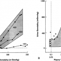
The anterior pituitary contains ˜600 μg of ACTH.16 ACTH, which is released on a pulsatile basis with peaks at ˜30-minute intervals,17 is subject to intravascular enzymatic degradation with a plasma disappearance half-life of 7 to 12 minutes.18 Cortisol pulses follow those of ACTH by ˜30 minutes, although not all cortisol peaks follow those of ACTH.19 The relationship between increasing levels of ACTH and consequent cortisol secretion is defined by a sigmoidal curve.20 Very high levels of ACTH do not further increase plasma cortisol concentrations, although the duration of a cortisol secretory burst continues to increase.21
Under some circumstances, the relation between ACTH and cortisol secretion is disturbed. In chronic stress, such as critical illness, a greater cortisol release occurs for an additional given ACTH stimulus because of adrenal hypertrophy and altered adrenal enzyme activities, which favor production of cortisol over that of adrenal androgens.22 Elevated ACTH levels after appropriate stimuli, with blunted or normal cortisol responses, are seen in myotonic dystrophy,23 a multisystem genetic disorder, and fibro-myalgia,24 an idiopathic pain syndrome. In these cases, the primary defect is thought to lie in ACTH regulation rather than an altered ACTH bioactivity or adrenal hyporesponsiveness.
Regulation of ACTH release is subject to three themes: stress, the circadian rhythm, and glucocorticoid negative feedback. Stress, defined as a threat to homeostasis, derives from such factors as sepsis, trauma, or emotion. In response to stress, ACTH release is greatly increased; consequently, the secretion of cortisol, the principal glucocorticoid in humans, can increase five-fold. The principal functions of cortisol during stress are to restrain the immune system, by reducing production of potentially damaging cytokines25; augment the effects of catecholamines on the vascular system; mobilize glucose and fatty acids for metabolic use; and sharpen cognition to allow appropriate behavioral responses. During starvation, cortisol acts to increase hepatic glucose production for use as energy.
ACTH secretion follows a light-entrained circadian rhythm with peak cortisol blood levels attained at ˜6:00 a.m. to 8:00 a.m. and the lowest concentrations at approximately midnight.26,27 and 27a In animals, the circadian rhythm is based on serotonergic pathways arising from the suprachiasmatic nucleus,28 although the mechanism—or indeed the function—of circadian ACTH release in humans is unknown. In humans, severe disturbances of circadian ACTH release following transmeridian travel require several days to be reentrained29 (see Chap. 6).
Related posts:


Stay updated, free articles. Join our Telegram channel

Full access? Get Clinical Tree


