Eyes
Age-Related Changes
Examination of the Fundus in Old Age
Age-related miosis can make it difficult to examine the fundi of older people. Short-acting eye drops are recommended, usually tropicamide 0.5% (rapid onset and effects last 4–6 h). Reversal with pilocarpine is usually unnecessary and can be painful. The risk of acute closed-angle glaucoma is minimal, but beware the small eyeball with a shallow anterior chamber and small-diameter cornea.
Loss of Vision
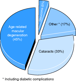
Approximately 70,000 persons over the age of 65 years in the UK are registered as partially sighted, i.e. about 1% of the elderly population. Figure 15.1 lists the causes of blindness in older people. Many more are visually disabled but remain unregistered. Registration of disability is essential for special benefits and visual aids.
Figure 15.1 Causes of blindness in old age. Source: ABPI (1991) The Challenges of Ageing. ABPI, London.

Criteria for being Registered Blind/Partially Sighted
- Severely sight impaired/blind:
 Visual acuity (VA) of less than 3/60 with a full visual field.
 VA between 3/60 and 6/60 with severe field reduction, such as tunnel vision.
 VA of 6/60 or above, but with a much reduced field of vision, especially in the lower part of the field.
- Sight impaired/partially sighted:
 VA of 3/60 to 6/60 with a full field of vision.
 VA of up to 6/24 with a moderate reduction of field of vision or central vision that is blurry.
 VA of up to 6/18 if a large part of the field, for example a whole half field or a lot of peripheral vision, is missing.
Advantages of being Registered Blind
- Fifty percent reduction in television licence fee; free for over 75s anyway!
- Blue badge scheme for the driver.
- Free eye tests; free for over 60 year olds anyway.
- Free loan of equipment from Low Visual Aid clinics.
The Painful Eye
 Conjunctivitis.
 Uveitis.
 Herpes zoster: when the ophthalmic branch of the facial nerve is affected, there may be involvement of the forehead, eyelids and conjunctiva. The patient must see an ophthalmologist urgently.
Primary Open Angle/Chronic Glaucoma
- A silent and progressive disease which may result in blindness if not treated.
- Accounts for 15% of blindness fulfilling criteria for registration in the UK.
- Risk factors include increasing age, family history, African or Caribbean descent, diabetes, myopia and hypertension.
- There may be cupping and atrophy of the optic disc.
- Early cases are best detected by regular eye tests, with ocular-pressure measurement.
- Affects peripheral vision first, only affecting central vision late in the disease.
- May affect one eye more than the other.
- Treatment is aimed at reducing the intraocular pressure:
 Once daily prostaglandin agonists give excellent results, e.g. latanaprost. Intraocular pressure is reduced by increased drainage of aqueous humour via the trabecular meshwork.
 Beta-blockers, e.g. timolol eye drops and alpha2 adrenergic agonists, e.g. brimonidine eye drops, reduce the production of aqueous humour by the ciliary body.
 Carbonic anhydrase inhibitors act on the carbonic anhydrase in the ciliary body, e.g. dorzolamide. They are less effective than other treatments so are often used as adjuncts.
 Educate the patient about the efficacy of treatment and the high risk of blindness without it, to engage them in persevering with eye drops for life.
- If treatment with eye drops fails, trabeculoplasty with an argon laser or surgical trabeculectomy will improve aqueous flow.
Acute Glaucoma
- Less common than chronic glaucoma.
- More common in females because the anterior chamber is more shallow.
- Most common in 6th and 7th decades.
- Easier to detect because it presents with sudden painful loss of vision in a red eye.
- Often associated with ipsilateral headache and vomiting.
- The patient may describe haloes around lights secondary to corneal oedema.
- The patient may report a precipitating event such as exposure to very dim light or mydriatic treatments such as anticholinergics and sympathomimetics.
- The pupil is mid-dilated and non-reactive.
- Visual acuity is reduced.
- Slit lamp examination may show corneal oedema and an irregular pupil.
- The intraocular pressure will be increased.
- Emergency treatment is IV and oral acetazolamide or IV mannitol plus topical beta-blocker eye drops. Steroid drops reduce inflammation.
- Analgesia and anti-emetics reduce the distress of the patient which also helps reduce the pressure.
- Laser iridotomy reduces the pressure acutely.
Table 15.1 Summary of differences between acute and chronic glaucoma
| Acute – closed-angle | Chronic – open-angle | |
| Symptoms | Sudden pain in eye, blurred vision, vomiting and prostration | Insidious loss of vision, leading to tunnel vision; family history common |
| Signs | Painful red eye with reduced vision. Eye tense, irregular fixed pupil, cornea and conjunctiva congested | Painless normal looking eye. |
| Raised pressure on tonometry, scotoma on field testing; cupped disc | ||
| Pathology | Sudden impairment of anterior-chamber drainage – may be precipitated by anticholinergics and mydriatics | Gradual increase in intraocular pressure – idiopathic |
| Treatment | Constrict pupil, analgesia, carbonic anhydrase inhibitor – urgent action needed | Prostaglandin, beta-blocker and/or pilocarpine drops, drainage operation |
Table 15.1 summarizes the differences between acute and chronic glaucoma.
Age-Related Macular Degeneration (AMD)
- Affects people aged 50 and over, and becomes more common with advancing age.
- The most common cause of visual impairment in the industrialized world.
- Affects 1.5 million people in the UK currently, and predicted to rise to 1.9 million by 2020 because of the ageing population.
- As the macula is the area damaged, central vision is affected more, with peripheral vision being preserved.
- Loss of central vision means loss of face recognition and difficulty reading.
- AMD is usually bilateral, but may affect one eye earlier or more than the other.
- Age-related maculopathy: mild or moderate non-exudative changes in the macula.
AMD is divided into two broad types:
- Develops slowly over months or years.
- No treatment, but encourage to stop smoking; some evidence that antioxidant supplements (vitamins A, C and E with zinc and copper) may be protective.
- Geographic atrophy (patchy loss of RPE cells) may signal development of wet AMD.
- Ten to twenty percent of patients progress to the wet type.
- More rapidly progressive and causes more visual impairment.
- New treatments have been developed to inhibit VEGF: pegaptanib sodium (a direct antagonist) and ranibizumab (a monoclonal antibody). Both are given as monthly intraocular injections and need to be continued for at least 2 years to preserve vision. ‘Rationing’ of these expensive treatments by imposing strict eligibility criteria (likely to increase in the current economic climate) creates controversy,
- NICE (2011) only recommended ranibizumab. Criteria for treatment include best corrected vision 6/12 and 6/96, no permanent structural damage of fovea (central macula), lesion less than 12 disc areas, evidence of disease progression and response to initial treatment.
- Side-effects: conjunctival haemorrhage, eye pain, vitreous floaters, vitreous haemorrhage, retinal detachment and increased intraocular pressure.
- Ranibizumab improves vision in 40% of cases and prevents further deterioration of sight in most patients.
- NICE supports photodynamic therapy for wet AMD with definite choroidal neovascularization. Verteporfin (a light-sensitive drug given intravenously) sticks to the new vessels and is activated using a cold laser to seal the vessels.
- Exciting developments in the genetics of AMD point to a single nucleotide polymorphism in complement factor H (CFH) gene on chromosomes 1 and 10. CFH is an inhibitor of the complement pathway and abnormal CFH leads to inflammation.
- If vision is severely impaired, refer to the Low Vision Service.
Table 15.2 summarizes the differences between dry and wet AMD.
Table 15.2 Differences between dry and wet AMD
| Dry AMD | Wet AMD | |
| Frequency | 80–85% | 10–15% |
| Symptoms | Asymptomatic until late stage | Rapid loss of central vision over days to weeks |
| May notice loss of central acuity | Metamorphopsia | |
| Peripheral vision preserved | Decreased colour and contrast sensitivity | |
| Sudden deterioration of vision suggests progression to wet AMD | Slow dark adaptation | |
| Appearance | Focal hyperpigmentation, drusen, geographic atrophy | Larger more confluent drusen, more pigmentation and new vessels, scarring |
| Management | Stop smoking | Intravitreal ranibizumab |
| Increase dietary antioxidants | Photodynamic therapy |
Driving
- Legally, the patient must be able to read a car registration plate at 20 m, in good light with glasses if appropriate.
- A patient with visual field defects, regardless of cause (e.g. glaucoma, stroke or pituitary disease), should not drive unless allowed by the ophthalmologist.
Cataracts
- So called because the world appears blurred as if seen behind a waterfall.
- Arise because of protein breakdown and dehydration of the lens.
- Are very common and easily detected and corrected, resulting in marked improvement of quality of life.
- In the USA 300,000–400,000 cases occur annually.
- Responsible for around 36% of cases of blindness in Africa.
- Short-sighted patients may report an improvement in near vision (‘second sight’) because progression temporarily increases the power of the lens. As the cataract progresses further, near vision is lost again.
- Other visual problems include reduced contrast sensitivity and increased glare from daylight or car headlights at night.
Types
Multifactorial pathogenesis.
Causes
- Ageing.
- Hereditary.
- Diabetes mellitus.
- Hypertension.
- Iatrogenic, e.g. steroids.
- Alcohol.
- Environmental – bright sunshine (increased incidence in the tropics).
- Nuclear cataracts seem to be related to smoking.
Examination
Thorough assessment of both eyes is essential to exclude problems, e.g. AMD which will not be resolved by cataract extraction, to ensure a good outcome from surgery. This includes acuity, fields, assessment of pupillary reflexes, indirect and direct ophthalmoscopy and slit lamp examination.
Treatment
Surgery
Timing depends on individual need, e.g. earlier in those who read a lot, but may be delayed in those whom the distortion, change in magnification and reduced visual fields caused by wearing glasses post-operatively would be a hindrance.
Contraindications
Surgical Procedure
- Usually done as a day case under local anaesthetic.
- The anterior chamber of the eye is incised. Phacoemulsification is the process of fragmenting the opacified lens by ultrasound. This debris is removed, leaving the lens capsule intact. An artificial lens is inserted into the capsule.
- The power of the new lens is selected to optimize vision so that the patient does not have to wear thick aphakic spectacles.
Complications of Treatment
Giant-Cell Arteritis (GCA)
See also Chapter 9.
Clinical Features
Stay updated, free articles. Join our Telegram channel

Full access? Get Clinical Tree


