(1)
Institute of Oncology “Prof. Ion Chiricuță”, Nuclear Medicine & Endocrine Tumors, Cluj-Napoca, Romania
Nature is the source of all true knowledge. She has her own logic, her own laws, she has no effect without cause nor invention without necessity.
Leonardo Da Vinci
5.1 Gamma Camera
The revolutionary metabolic image provided by nuclear medicine has in its background a tremendous effort for the improvement of instruments and equipments.
Especially during the last two decades, the spectacular development of positron emission tomography and of fusion/hybrid images was possible due to the new and astonishing equipment and software, able to give us the best functional and structural information of the human body.
With respect to pathology occurring in endocrinology, the most frequent instruments used are gamma cameras, also called gamma scanners; nevertheless, in the last years, it significantly increased the use of hybrid equipments, as PET/CT or SPECT/CT.
The principle of these procedures consists of radiopharmaceutical administration into patient’s body, mainly intravenously; after a period of time, the emitted gamma rays from patient’s organs are registered by gamma camera alone or fused with CT, and an image is obtained in the final software station.
The order of the procedures is presented in the next schema in Figs. 5.1, 5.2, and 5.3.
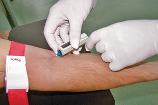
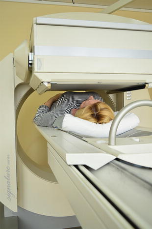
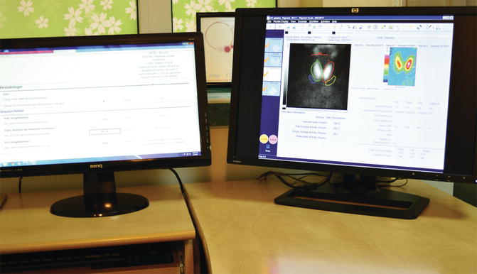

Fig. 5.1
Intravenous administration of radiopharmaceutical, protection holder of the syringe

Fig. 5.2
Patient examined by gamma camera (Model Siemens e.cam Signature)

Fig. 5.3
Image processing by gamma camera’s software station (Model Siemens e.cam Signature)
The main parts of gamma camera are the crystal, the collimator, the photomultiplier tubes, the lead shielding, and the electronic circuit and the image processing station.
5.1.1 Collimators
Gamma rays take random directions from the patient’s body. The collimator has the role to block the emitted rays that are not suitable for creating the image. It restricts the rays from the source so that each point in the image corresponds to a unique point in the source.
The collimator consists of a different number and types of holes distributed in a piece of lead (Fig. 5.4).


Fig. 5.4
The core of a collimator with hexagonal holes
The holes may be:
Hexagonal
Circular
Square
Triangular
Manufacturers have different preferences in order to maintain the best resolution and sensitivity at low costs and ease of manufacture. The difference in spacing creates differences in the thickness of the walls or of the septa. The collimator septa must be accurately aligned to avoid any distortion or artifacts.
Collimators are specially designed for:
Different energies of radionuclides
Different resolution
Different sensitivity
Different image, size, or shape
All these characteristics are reflected in different types of collimators: parallel, divergent, converging, and pinhole.
The parallel collimator creates an image of the object in its real dimensions. The design of the hole, the sensitivity, and the spatial resolution of the gamma camera are changing. The field of view (FOV) of this does not change with the distance between the object (the organ in the human body) and the collimator (Fig. 5.5).
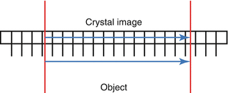

Fig. 5.5
Collimator with parallel holes
- (a)
Divergent collimator: The size of the object in the image from gamma camera’s crystal is smaller than the original object (Fig. 5.6).
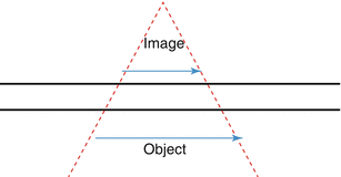
Fig. 5.6
Divergent collimator
- (b)
Convergent collimator: The size of the object in the image from gamma camera’s crystal is bigger than the original object (Fig. 5.7).

Fig. 5.7
Convergent collimator
- (c)
Pinhole collimator
This collimator acts just like a pinhole camera and produces a magnified and inverted image of the object. This is typically used to examine small objects such as the thyroid gland and the hip joint (Fig. 5.8).
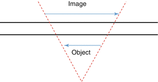
Fig. 5.8
Pinhole collimator
The level of energy of the radionuclide, which will be used for testing, dictates the thickness of lead from the collimator selected for examination. The septa and the hole’s size influence the resolution of the collimator (Table 5.1).
Table 5.1
The selection of different types of collimator according to the energy level of the radionuclide
Type of collimator | Energy (keV) |
|---|---|
Low-energy high sensitivity (LEHS) | 140 |
Low-energy general (all) purpose (LEGP/LEAP) | 140 |
Low-energy high resolution (LEHR) | 140 |
Medium-energy general purpose (MEGP) | 250 |
High-energy general purpose (HEGP) | 364 |
Low-energy pinhole (LEP) | 140 |
It is very important to underline that the resolution of the gamma camera degrades severely with the distance between the testing body and collimator.
Briefly, higher sensitivity means bigger holes of the collimators, while higher resolution means smaller holes or longer septa between the holes.
Regarding the performance of gamma camera, there are some characteristics determined by collimator proprieties and also by the intrinsic camera proprieties. All together, they express the following camera characteristics:
Resolution is the ability to determine two separate sources of radioactivity as different entities. Thus, it is a measure of the amount of blurring of these sources by the camera.
Sensitivity is the ability to register a certain fraction of gamma rays emitted by the source.
Uniformity is the ability of the instrument to faithfully reproduce an image of a uniformly distributed radioactive source.
Choosing the collimator is the first step in preparing the examination. A careful changing of the equipment leads to an accurate diagnostic test and better results in the patient’s management. In Fig. 5.9, the layout of three different collimators ready to be used is presented.
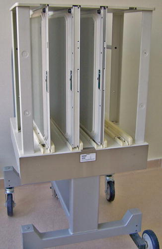

Fig. 5.9
Collimators in their deposit trolley
5.1.2 Crystals
The crystal is the “heart” of a gamma camera. This is a core of sodium iodide (NaI) doped with an activator (frequently thallium—Tl). The scintillations produced by the ionizing radiation from the patient’s body occur in this core. A gamma photon enters the crystal; the energy given to this crystal is released as light photons. Not all the photons are detected; in most cases, three light photons are detected for every 1 keV of energy absorbed.
Stay updated, free articles. Join our Telegram channel

Full access? Get Clinical Tree


