Epstein-Barr Virus and Gastric Carcinoma


Epstein-Barr Virus and Gastric Carcinoma


Masashi Fukayama

Aya Shinozaki

Rumi Hino


INTRODUCTION

Several human neoplasms, including gastric carcinoma (GC), develop in close association with infectious organisms. Helicobacter pylori and Epstein-Barr virus (EBV) are now regarded as causative microorganisms of GC.1,2 Like other human oncogenic microorganisms, these infectious agents cause malignant neoplasms to develop in a limited number of patients out of a large population of healthy or healthy-appearing carriers through unknown mechanisms.2 However, the roles of H. pylori and EBV in the development and progression of GC are quite different (Table 4-1).

EBV was the first human oncogenic virus to be identified when it was discovered in human neoplastic cells (Burkitt lymphoma cell lines) in 1964. Subsequent investigations have identified the virus in nasopharyngeal carcinoma (NPC), Hodgkin lymphoma, B-cell lymphomas with or without immunosuppression, and nasal natural killer/T-cell lymphoma. EBV was identified in GC tissues in 1990 (Fig. 4-1), when the in situ hybridization (ISH) method for detecting EBV-encoded small RNA (EBER) had just been introduced to the field of pathology (Fig. 4-1C and D).3 EBER is abundant in latently infected cells (up to 107 copies per infected cell) and is identified in nearly all neoplastic cells of tumor tissues when it is present in GC. Thus, detection of EBER by ISH is a gold standard for the identification of EBV-associated GC. Furthermore, ISH can be used to identify EBER in archived materials, such as formalin-fixed and paraffinembedded tissues, which can be used to investigate the epidemiology of this type of GC. EBVassociated GC occurs worldwide and its frequency is estimated to be 8.7% (95% confidence interval: 7.5%-10.0%) of all GC cases,4 which corresponds to an annual incidence of 70,000 to 80,000 patients worldwide.1


EBV-ASSOCIATED GC IS A DISTINCT CLINICOPATHOLOGICAL SUBGROUP OF GC

EBV-associated GC is defined as the presence of latent EBV infection in neoplastic cells. This type of GC (Table 4-2)4, 5 and 6 affects predominantly males and is usually located in the proximal stomach. Some reports suggest that this carcinoma occurs in patients of a relatively younger age compared to those with non-EBV GC, but meta-analyses did not confirm that observation.4,5 EBV is involved in 25% of remnant GCs, that is, GC, which occurs in the remnant stomach, at least >5 to 10 years after the initial gastrectomy for either benign or malignant stomach diseases, and several reports have suggested that multiple carcinomas are likely to occur in association with EBV6 EBV-associated GC has a lower rate of lymph node involvement, especially during its early stage
within the submucosa, and has a relatively favorable prognosis compared with EBV-negative GC, although both findings remain to be confirmed.5


Table 4-1 Comparison of H. pylori and the EBV as causative agents of GC


H. pylori


Epstein-Barr Virus


Carriers


More than 75% of people over age 40


More than 95% of people over age 20


Involvement in GC


Most GC (intestinal/diffuse types of histology)


10% of GC, including LE-like GC


Higher risk of GC in carriers compared to noncarriers


Monoclonal EBV present in all neoplastic cells


Carcinogenic mechanisms


Hummingbird phenomenon by transfection of the CagA gene into epithelial cells


Transformation of epithelial cells after transfection of the LMP2A gene.


Activation of ERK mediated by CagA/SHP2 or Grb2


Abnormal signal transduction mediated by LMP2A


Model animal/cell line


Mongolian gerbil, CagA transgenic mouse


GC strain (KT), transplantable to SCID mice/GC cell line (SNU-719)


Other related malignancies


MALT lymphoma


Lymphomas (Burkitt lymphoma, Hodgkin lymphoma, T/NK cell lymphoma)


Nasopharyngeal carcinoma


Neoplasms in immunocompromised hosts


GC, gastric carcinoma; LE, lymphoepithelioma-like.


EBV-associated GC often occurs as an ulcerated or saucer-like tumor accompanied by marked thickening of the gastric wall (Fig. 4-1A). These features are easily discernible on endoscopic ultrasonograms and CT scans of the stomach. There are two histological types of EBV-associated GCs, lymphoepithelioma (LE)-like (Fig. 4-1B and C) and ordinary (Fig. 4-1D), although there is a continuum between the types.6 The typical morphology of LE-like GC is described as poorly
differentiated carcinoma with dense infiltration of lymphocytes, resembling NPC.3 LE-like GC is nearly identical to the subgroup that Watanabe et al.7 reported as “GC with lymphoid stroma (GCLS),” but GCLS is relatively a broad category that includes LE-like GC. The relative ratio of the LE-like/GCLS type to the ordinary type reported in the literature varies considerably from 1:10 to 4:1.6 This variability is due to interpretation variability in diagnostic criteria of the LE-like/GCLS type, especially when there is heterogeneity within the tumor. More than 80% of the LElike/GCLS-type tumors were positive for EBV in most reports.3,6,8 The histopathological features of EBV-positive LE-like/GCLS tumors, which are not seen in EBV-negative LE-like/GCLS tumors, are mild cellular pleomorphism, rare mitotic figures, a marked degree of lymphoid stroma, and lymphoid infiltration within the cancer cell nests.8


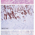
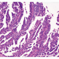
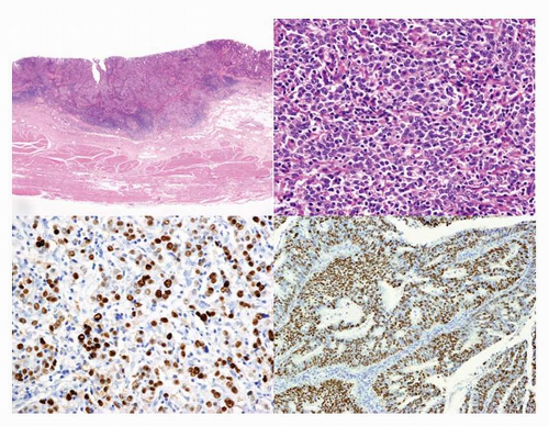
FIGURE 4-1 Histopathology of EBV-associated GC. A (left upper): Low-power view of EBV-associated GC. Lymphoepithelioma (LE)-like carcinoma shows expansive growth with prominent infiltration of lymphocytes. B (right upper): High-power view of LE-type of EBVassociated GC shown in (A). C (left lower): EBER in situ hybridization of LEtype of EBV-associated GC (A). D (right lower): EBER in situ hybridization of ordinary type of EBV-associated GC.


Table 4-2 Clinicopathological features of EBV-associated GC


Clinical


Nearly 10% of Total GC Male Predominance Remnant Stomach


Macroscopic


Location at gastric cardia/body


Ulcerated or saucer-like tumor


Marked thickening of the gastric wall


Multiple carcinomasa


Microscopic


Lymphoepithelioma-like histology


Moderate to poorly differentiated adenocarcinoma


Lymphocytic infiltration in various degrees


Lace pattern within the mucosa


Lymphoid follicle in the invasive sites


Behavioral


Lower rate of lymph node involvementa


Relatively favorable prognosisa


a Findings have been suggested, but more evidence is necessary for confirmation.


For EBV-associated GC, the incidence of intramucosal and submucosal carcinomas (early GC) at diagnosis is not statistically different from that of deeply invasive carcinomas (advanced GC). At the intramucosal stage, EBV-associated GC shows a “lace pattern,” which is formed by the connection and fusion of neoplastic glands within the mucosa proper. Further invasion of the carcinoma into the submucosa is frequently accompanied by the infiltration of massive lymphocytes (Fig. 4-1A) consisting of CD8-positive T lymphocytes, CD4-positive T lymphocytes, and CD68-positive macrophages, in a ratio of 2:1:1, respectively. EBV infection is observed in only a very limited number of these infiltrating lymphocytes. Infiltrating cells are sometimes variable, and their extreme effect is prominent lymphoplasmacytic infiltration with numerous Mott cells9 or a granulomatous reaction with many osteoclast-like giant cells (Ushiku et al., Pathol Int. 2010;60:551-558).


EBV INFECTION AND DEVELOPMENT OF CARCINOMA

The evidence regarding the lineage of EBV-infected cells, the clonality of EBV in infected cells, and the infection of epithelial cells by EBV, as described below, strongly suggests that stomach epithelial cells initiate clonal growth after they are infected with EBV from the reactivation of EBV-carrying lymphocytes at the mucosa (Fig. 4-2).


Lineage of EBV-infected Cells

Neoplastic cells from EBV-associated GC show characteristic profiles of differentiation markers, which might be associated with the lineage of EBV-infected cells. Claudin (CLDN) proteins
constitute the tight junction, and neoplastic cells of EBV-associated GC showed a high frequency of CLDN18 expression (84%) and a low frequency of CLDN3 expression (5%).10 This expression profile (CLDN18+, 3-) corresponds to that of the normal gastric epithelium in adults and fetuses, but not to that of intestinal metaplasia (CLDN18-, 3+). In accordance with the CLDN expression patterns, almost half of the EBV-associated GC cases in a study by Barua et al.11 had gastric-type mucin expression (MUC5AC, MUC6), and the other half lacked gastric- or intestinal-type mucin or CD10 expression. These results indicate that EBV-associated GC is very homogenous with regard to cellular differentiation and that it preserves the nature of the cells of origin. Thus, the cells that are targets of EBV infection and their subsequent transformation may be precursor cells with intrinsic differentiation potential toward the gastric cell type.


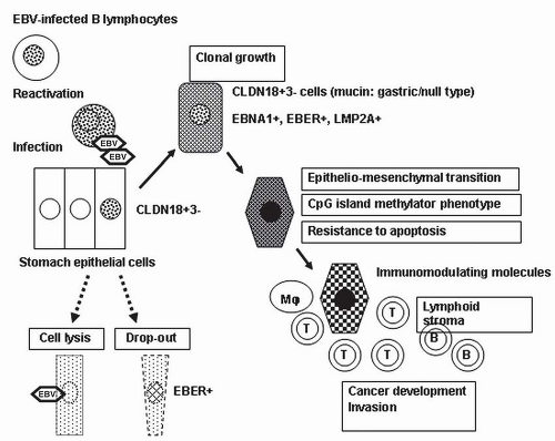
FIGURE 4-2 Schematic representation of the development and progression of EBV-associated GC. Stomach epithelial cells are infected with EBV from the reactivation of EBV-carrying lymphocytes at the mucosa. Some EBV-infected cells begin to grow clonally in the milieu of atrophic gastritis. The neoplastic cells retain the claudin 18-positive/claudin 3-negative (CLDN18+, 3-) gastric phenotype, which may correspond to the phenotype of the original EBV-infected cells. The CpG-island methylation phenotype is the primary abnormality in neoplastic cells of EBV-associated GC. Expression and secretion of immunomodulator molecules induce the characteristic stromal reaction.


Clonality of EBV in Infected Cells

EBV is a double-stranded DNA virus (184 kb in size), which is maintained in a linear form in the virus particles. After EBV enters infected cells, the viral DNA circularizes by fusing the terminal repeats (TRs, repetitive 500-bp structures) at both ends of the linear genome and maintains its circular form in the nuclei of latently infected cells.2 Southern blot analysis of EBV-TRs provides information about the clonality of EBV and the state of viral activation, that is, replicating (linear configuration) versus latent (episomal circular forms). In EBV-associated GC, TR analysis has demonstrated that monoclonal EBV is present in an episomal form without integration into the
host genome and infection is latent, with no viral replication. Monoclonal or biclonal EBV was observed in carcinoma tissues in the intramucosal stage; however, it was always monoclonal in the submucosal invasion stage (Fig. 4-3) and in more advanced carcinomas.12,13 Since all carcinoma cells show a positive signal in EBER-ISH in all cases of EBV-associated GC, EBV infection must occur at the initial or a very early stage of carcinoma development.


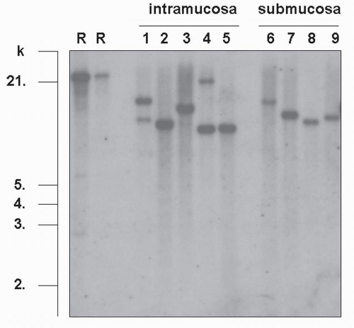
FIGURE 4-3 Clonal analysis of EBV derived from virus-associated GC. Southern blot hybridization analysis of TRs of EBV DNA, extracted from EBV-associated GC. DNA is cut with the BamHI restriction enzyme, electrophoresed, and hybridized with an EBV-TR-specific probe (R, R’: Raji cell used as a control, 1-5: samples of intramucosal carcinomas, 6-9: samples of submucosal carcinomas). Analysis of the blot shows either one or two bands larger than 6 kb in each sample. When the detected bands are short, it indicates that EBV takes the linear form in viral particles. Smeared signals indicate infection of the tumor with various EBV clones. (From Uozaki H, Fukayama M. Int J Clin Exp Pathol. 2008;1:198-216[2], with permission from the Journal.)

May 22, 2016 | Posted by in ONCOLOGY | Comments Off on Epstein-Barr Virus and Gastric Carcinoma

Full access? Get Clinical Tree

Get Clinical Tree app for offline access