Endothelial Cell-Blood Cell Interactions


Endothelial Cell-Blood Cell Interactions


Klaus Ley


Interactions between monocytes, platelets, and endothelial cells are intimately involved with and are of key importance for the regulation of hemostasis, thrombosis, inflammation, and atherosclerosis. Although other cells such as neutrophils and T lymphocytes participate, the monocyte-platelet-endothelial cell axis has emerged as a predominant factor in vascular disease and thrombosis. This chapter introduces the molecular basis of the adhesive interactions among the three cell types, explores the crosstalk between hemostasis, thrombosis, and inflammation, and discusses the pathophysiologic significance of these cell-cell interactions with an emphasis on vascular disease.


CELLULAR AND MOLECULAR BASIS


Monocytes

Monocytes are myeloid cells produced in the bone marrow. Although originally thought to be a homogeneous population, it has become clear that in human and mouse blood at least two subpopulations of monocytes exist.1,2 One type is characterized by a more inflammatory phenotype while the other type can migrate along the vascular endothelium.3 Dendritic cells are the most important antigen-presenting cells of the immune system. Both dendritic cells and monocytes are derived from macrophage-dendritic cell progenitors in the bone marrow.4,5 In mice, a committed blood precursor for dendritic cells (pre-DC) has been described, which is distinct from monocytes.6 Both “inflammatory” and “resident-type” monocytes express adhesion molecules, chemokine receptor, and cytokine receptor molecules that support interaction with platelets and endothelial cells (Table 46.1). It is unclear whether the human blood monocyte subsets correspond to the mouse subsets.7,8

Most adhesion molecules are integral transmembrane cell surface molecules that can bind to the same (homotypic), or other cell surface molecules (heterotypic), or molecules in the extracellular matrix. Most adhesion molecules provide mechanical strength to the interaction between cells by mechanically linking the extracellular environment to the intracellular cytoskeleton through reversible interactions. Many if not all cell adhesion molecules also transduce signals into the cells upon engagement by extracellular ligands.


Monocyte Adhesion Molecules


Integrins

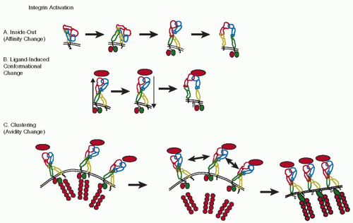
Integrins are transmembrane αβ heterodimers that bind many extracellular matrix proteins and certain immunoglobulin-like adhesion molecules to other cells.9,10 Most integrins require conformational activation to support binding. The mechanisms of integrin activation have been studied in detail for αVβ3,11 an integrin also expressed on monocytes, and lymphocyte function-associated antigen 1(LFA-1),12 an integrin highly expressed on monocytes and other leukocytes. Activation of αxβ213 and α4β1 integrins14 has also been studied. Both are expressed on blood monocytes at low and high levels, respectively. Integrin activation is probably initiated by the binding of the intracellular cytoskeletal adaptor molecules talin and kindlin-3 to the integrin β chains,15,16 which causes extension of the extracellular domain that exposes the ligand-binding site and greatly increases the on-rate of ligand binding (FIGURE 46.1A). This process is called inside-out signaling, because the change in the extracellular domain of the integrin is brought about by intracellular processes. The mechanism of activation-induced conformational change is thought to apply fairly generally to many, if not all integrins.10 However, the regulation of affinity by reducing the dissociation of the ligand from the integrin is much less clear. Although conformational changes in the ligandbinding domain have been reported,17 it is not clear whether these changes are directly linked to integrin extension or could be regulated separately.14

Integrins may first undergo some initial conformational change (probably extension) through inside-out signaling, followed by ligand binding, which then causes outside-in signaling that leads to full activation and strong binding10,18,19 (FIGURE 46.1B). However, not all experimental data are consistent with this model.20 In addition, integrins can also rearrange in the plasma membrane to cluster and form patches, which results in enhanced binding (FIGURE 46.1C). This rearrangement and clustering probably happens after ligand binding, results in increased binding of multivalent ligands, and is called avidity change. It does not result in increased affinity for the monovalent ligand.21 Under in vivo conditions, cell activation probably results in a combination of integrin affinity and avidity increase. Although this can be tested by looking at monovalent versus polyvalent ligand binding, the role of avidity and affinity regulation remains controversial.10,18,21

Of the 24 known integrins, mature blood monocytes express only a few. The most abundant monocyte integrin is α4β1 integrin or very late antigen-4 (VLA-4) (CD49d/CD29). It is composed of a 150 kDa α4 chain that can undergo proteolytic cleavage and a noncovalently associated 130 kDa β1 chain. α4β1 integrin is preferentially expressed on cell surface projections that are often called microvilli, but resemble ridges rather than true villous processes.22 This position is believed to facilitate the interaction of α4β1 integrin with its ligands under conditions of flow. The most important ligands for α4β1 integrin include
the vascular cell adhesion molecule 1 (VCAM-1) on endothelial and other cells (Table 46.2) and the heparin-binding region of alternatively spliced fibronectin expressed in the extracellular matrix and on the luminal surface of inflamed endothelial cells.23 Like other integrins, α4β1 integrin can probably undergo conformational activation.14,24,25 This process of affinity regulation can be triggered by monocyte activation, for example, through chemokines. Gene-targeted mice lacking either α426 or β127 are not viable. Blocking α4β1 with a monoclonal antibody or a peptide based on the fibronectin sequence ILDV reduces atherosclerosis in mice,28 suggesting that α4β1 is important in monocyte recruitment to atherosclerotic lesions.


Table 46.1 Cell surface molecules on monocytes relevant to cell-cell interaction


“Inflammatory” Monocytes


“Resident” Monocytes


Molecule


Mouse


Human


Mouse


Human


Main Function


Secondary Function


CD11a/CD18 (αLβ2, LFA-1)


+


+


+


+


Inducible adhesion to ICAM-1, 2


Reduces rolling velocity (neutrophils)


CD11b/CD18 (αMβ2, Mac-1)


+


+


+


+


Inducible adhesion to C3bi, ICAM-1, 2, fibrinogen


Phagocytosis


CD11c (αxβ2)


+


+


Inducible binding to VCAM-1


Complement binding


CD14


+


low


LPS coreceptor


CD16 (FcγRIII)


+


+


+


Fc receptor for IgG


CD64 (FcγRI)


+


CX3CR1


low


+


high


high


Binds CX3CL1, activates monocytes


May mediate adhesion


CCR1


n.d.


+


n.d.


Binds chemokines CCL3, 5, 7, 14, 15, 16, 23; causes activation


Chemokine binding causes arrest of rolling cells


CCR2


+


+


Binds chemokines CCL2, 7, 12, 13, causes activation, chemotaxis


Soluble CCL2 can cause monocyte arrest


CCR4


n.d.


+


n.d.


Binds chemokines CCL17, 22


CCR5


+


CCR7


n.d.


+


n.d.


Binds chemokines CCL19, 21


Promotes mature DC migration to lymphatic organs


CXCR1 (IL-8 receptor)


low


Binds chemokines, mainly CXCL8


CXCR2


n.d.


+


n.d.


Binds chemokines CXCL1, 2, 3, 5, 6, 7, 8


Chemokine binding causes arrest


CXCR3


n.d.


n.d.


Binds chemokines CXCL9, 10, 11


Promotes type 1 inflammation


CXCR4


n.d.


low


n.d.


+


Binds CXCL12, induces arrest


Release from bone marrow


Ly-6C (Gr-1)


+


No known function


CD49b (α2β1, VLA2)


+


n.d.


n.d.


Inducible binding to collagen


CD49d (α4β1, VLA4)


+


n.d.


+


n.d.


Inducible binding to VCAM- 1, fibronectin


Monocyte arrest on atherosclerotic lesions


CD62L (L-selectin)


+


n.d.


n.d.


Binds PSGL-1, secondary tethering


Binds PNAd, monocyte rolling in lymph nodes


CD162 (PSGL-1)


+


+


+


+


Binds P-selectin on platelets, endothelial cells, microparticles


Binds L-selectin for secondary tethering, binds E-selectin


Myeloperoxidase


high


Low


MHC-II


low


High


Tissue


factor Inducible, but unknown in which subsets


Initiator of coagulation


Signaling into monocyte


PECAM-1 (CD31)


Most or all


Transendothelial migration


Monocyte activation


FIGURE 46.1 Regulation of integrin ligand binding activity. A: Integrin affinity regulation by conformational changes in the α and β chains (red-yellow and blue-green, respectively), resulting in increased availability and accessibility of ligand binding sites (third diagram) and, through an additional activation step, increased affinity for monovalent ligands (last diagram). During conformational activation, talin and kindlin-3 (green and red ellipses) bind to the integrin β cytoplasmic tail, causing the integrin “legs” to move apart. B: Ligand-induced activation and outside-in signaling. After activation as in (A, inside-out signaling, arrow up), ligand (red ellipse) binding to integrin induces outside-in signaling (arrow down) and bond maturation. C: Integrin avidity regulation by lateral mobility/clustering. Transient release of integrins from cytoskeletal anchorage (actin filaments, represented as strings of circles) allows integrin rearrangement and clustering in the plane of the cell membrane, resulting in increased avidity for multivalent ligands. It is not known whether the release occurs at the level of talin-actin binding. Integrins bind actin through talin, and various other linker proteins (not shown).

The integrin αMβ2 (CD11b/CD18) is also known as Macrophage-1 or Mac-1. Mac-1 antibodies were some of the earliest monocyte-macrophage specific antibodies described. Although Mac-1 is also expressed on neutrophils, most of its function seems to be related to monocyte-macrophages. First, Mac-1 participates in monocyte adhesion to various substrates including endothelial cells. Second, Mac-1 is an important receptor for the complement and is also known as complement receptor 3 (CR3). Mac-1 binds complement C3bi and is critically involved in phagocytosis of complement-coated bacteria and particles. Mac-1 engagement promotes a proinflammatory response, including a respiratory burst with vigorous oxygen radical production, actin polymerization, induction of nitric oxide synthase, and shape change. Interestingly, under flow conditions such as those achieved in flow chambers in vitro, or isolated perfused vessels ex vivo, Mac-1 does not appear to contribute to monocyte adhesion to endothelial cells.29 Mac-1 has been shown to lower the rolling velocity of neutrophils,30 but the role of Mac-1 in monocyte rolling has not been studied. Mice lacking Mac-1, prepared by targeting the gene for αM, are viable, fertile, and healthy under vivarium conditions.31 There is no evidence that these mice are protected from atherosclerosis, but their response to vascular injury is blunted.32 Like all β2 integrins, Mac-1 has an I-domain with homology to the von Willebrand factor (vWF) A-domain inserted in its α subunit, which contains the ligand-binding site. Mac-1 binds many other ligands
including fibrinogen and coagulation factor × (Table 46.2). Mac-1 is thought to be involved in assembling prothrombinase on the monocyte surface and may be able to support platelet binding to monocyte through a fibrinogen bridge between αIIbβ3 on platelets and Mac-1 on monocytes.33 Although Mac-1 deficient mice have no obvious defect in hemostasis, Mac-1 could participate in monocyte activation and the delivery of tissue factor to sites of thrombosis.34 Human, but not mouse monocytes also express a closely related integrin, αxβ2, which is also a complement receptor, alternatively known as CR4. Abundant αx expression is found on dendritic cells. Like Mac-1, αxβ2 binds the intercellular adhesion molecule 1 (ICAM-1), C3bi, and denatured proteins,35 but in addition VCAM-1 also.36


Table 46.2 Ligands for monocyte and platelet integrins


Integrin


Ligands


Function


α4β1


VCAM-1
Fibronectin


Adhesion to endothelial cells
Adhesion to extracellular matrix


Mac-1
αMβ2


Complement C3bi
Coagulation factor Xa
Fibrinogen
Intercellular adhesion molecule-1 (ICAM-1)
Intercellular adhesion molecule-2 (ICAM-2)
Denatured collagen
Denatured albumin
Leishmania GP63
Bortedella FHA
Fibronectin


Phagocytosis of opsonized particles
Assembly of prothrombinase complex
Bridging between monocytes and platelets
Adhesion to endothelial and other cells
Adhesion to endothelial cells and platelets
Migration, phagocytosis?
Uptake of leishmania
Uptake of Bortedella
Adhesion to extracellular matrix


CD11c/CD18
αxβ2


Complement C3bi
VCAM-1, ICAM-1
Fibrinogen


Phagocytosis of opsonized particles
Monocyte adhesion
Bridging between monocytes and platelets


LFA-1
αLβ2


Intercellular adhesion molecule-1 (ICAM-1)
Intercellular adhesion molecule-2 (ICAM-2)


Adhesion to endothelial and other cells
Adhesion to endothelial cells and platelets


αVβ3


Vitronectin
Entactin
L1


Bone remodeling?
Unknown
Unknown


αIIbβ3


Fibrinogen
Fibronectin
vWF
Vitronectin
CD40L


Binds immobilized Fg without activation
Induced by GPIb binding to vWF
Promotes CD40L shedding


α2β1


Collagens


On platelets, requires activation by GPVI


The LFA-1, or αLβ2 integrin (CD11a/CD18) is expressed on all leukocytes including monocytes. While LFA-1 is responsible for lymphocyte arrest, the sudden stopping of rolling cells upon activation37 and participation in neutrophil arrest under flow, little is known about its function in monocytes. LFA-1 binds to cell surface immunoglobulins including InterCellular Adhesion Molecules ICAM-1 and-2, and has no known extracellular matrix ligands. Mice lacking LFA-1 were prepared by targeting the gene for αL38 and are viable, healthy, and fertile under vivarium conditions. There are no reports of these mice having altered thrombosis, hemostasis, or atherosclerosis. Like the other β2 integrins, LFA-1 has an I-domain and undergoes extensive conformational changes of the extracellular domain upon activation.12

The integrin αVβ3 is expressed on blood monocytes at a low copy number. Its expression increases upon differentiation to osteoclast-like cells. This integrin was initially called leukocyte response integrin39 because it participates in inducing the respiratory burst associated with NADPH oxidase activation and oxygen free radical production in neutrophils. Ligands for αVβ3 integrin include vitronectin, entactin, and possibly the immunoglobulin-like adhesion molecule L1. Gene-targeted mice lacking αV are not viable, whereas mice lacking β3 have a defect in both αVβ3 on monocytes, neutrophils, and proliferating endothelial cells, and αIIbβ3 on platelets, which share the common β3 subunit. The phenotype of these mice is dominated by the platelet defect (Glanzmann thrombasthenia-like), and these mice also have osteosclerosis, suggesting defective osteoclast function.40 αVβ5 integrin is also a vitronectin receptor. Monocytes express α2β1 integrin, a collagen receptor, at the mRNA and protein levels. α5β1, a fibronectin receptor, α10β1, a collagen receptor, and αEβ7, a receptor for E-cadherin, are found at the message level, but functional data have not been published.


Immunoglobulins

Blood monocytes express many immunoglobulin-like molecules. Of importance for this chapter is ICAM-1, because it supports homotypic aggregation of monocytes via LFA-1 and Mac-1 and because it can bind fibrinogen.41 Mice with hypomorphic mutations in the ICAM-1 gene or lacking ICAM-1 entirely have no overt defect in hemostasis or thrombosis, but are somewhat protected from atherosclerosis in the C57BL/6 and apolipoprotein E knockout models.42 However, these mice also lack ICAM-1 on endothelial cells, smooth muscle cells, lymphocytes, and many other cells. ICAM-2 and ICAM-3 are found in monocytes at the message level, but have no known function in monocytes.

Platelet endothelial cell adhesion molecule 1(PECAM-1) (CD31) is a homotypic adhesion molecule expressed on blood monocytes and has an important role in transendothelial migration and in monocyte activation.43,44 Monocyte PECAM-1 interacts with PECAM-1 on endothelial cells during transmigration. PECAM-1-deficient C57BL/6 mice have no apparent defect in leukocyte and monocyte transmigration, demonstrating that PECAM-1-independent pathways of transmigration exist.45

Other immunoglobulin-like molecules expressed on monocytes include major histocompatibility complex (MHC) class II, which is important in antigen presentation, but is not fully induced until monocytes differentiate to macrophages. CD83 is also expressed and has costimulatory functions in macrophages, but no known function in blood monocytes.


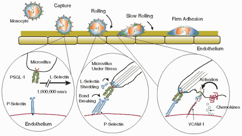
FIGURE 46.2 Sequence of monocyte capture, rolling, slow rolling, and adhesion on endothelial cells. Flow from left to right, endothelial surface layer shown in green. Primary capture or tethering is initiated by monocyte PSGL-1 binding to endothelial P-selectin (left insert). Note high velocity of monocyte (1 mm/s). The middle insert shows a P-selectin/PSGL-1 bond at the trailing edge of a rolling monocyte. Applied stress induces faster bond breakage and may also activate cleavage of L-selectin. The right insert shows VLA-4/VCAM-1-dependent bond required for slow rolling. Monocyte activation via surface-immobilized chemokine binding to chemokine receptor resulting in integrin affinity upregulation and firm binding to endothelial ligands such as VCAM-1 (shown here).


Selectins and their Ligands

L-selectin (CD62L) is expressed on “inflammatory” blood monocytes and most other leukocytes. Its most important function is in lymphocyte homing to secondary lymphatic organs,46 but it is also involved in inflammation.47 Like the other selectins, L-selectin can mediate leukocyte rolling, a passive motion of leukocytes down a vessel wall driven by the blood flow and the forces exerted on the loosely attached cell. During rolling, molecular bonds form at the leading edge and continually break at the trailing edge of the cell, allowing the leukocyte to stay in contact with the endothelium without actually stopping48 (FIGURE 46.2). Rolling is thought to serve to “scan for” inflammatory stimuli, and rolling cells can stop (arrest) in response to appropriate stimuli.18,49 On neutrophils, L-selectin is expressed on the tips of microvilli50 and can be rapidly shed upon cell activation by a protease-dependent mechanism involving tumor necrosis factor-α-converting enzyme (TACE) (ADAM-17).51 Although L-selectin has been shown to support monocyte rolling on L-selectin ligands in flow chambers, it is not known whether L-selectin-mediated monocyte rolling on endothelial cells serves a physiologic function. Endothelial ligands for L-selectin known as peripheral node addressins are expressed in endothelial cells of lymphoid organs, but L-selectin ligands on endothelial cells outside lymphoid organs have not been identified. It is tempting to speculate that monocyte L-selectin may enhance monocyte recruitment to atherosclerotic lesions by nucleating monocyte-monocyte interactions through binding
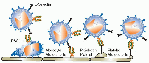
to P-selectin glycoprotein ligand-1 (PSGL-1, CD162, see below) in a process called secondary capture52 (FIGURE 46.3). Secondary capture is initiated when a vessel wall-adherent monocyte or monocyte-derived microparticle exposes PSGL-1 to other monocytes that pass by in the free stream. These cells can transiently bind to the adherent monocyte through L-selectin and then attach to the endothelium downstream from the nucleation site. While this process is certainly plausible and has been observed to occur in mouse aortas,53 no direct evidence has been provided to support its importance in atherosclerosis, thrombosis, or hemostasis.


FIGURE 46.3 Secondary capture or secondary tethering. Monocytes can be captured by binding to other, already adherent monocytes or other leukocytes (left) or to monocyte-derived microparticles (middle). Both require L-selectin on the circulating and PSGL-1 on the adherent monocyte or microparticle. Monocytes can also adhere to platelets or platelet-derived microparticles (right), which requires monocyte PSGL-1 and platelet P-selectin.

The most important selectin ligand on monocytes is PSGL-1, (CD162). PSGL-1 is somewhat of a misnomer, because PSGL-1 also binds L- and E-selectins with similar affinity as P-selectin.54 All monocytes, like most leukocytes, express PSGL-1 protein on the cell surface, but not all cell surfaceexpressed PSGL-1 can bind selectins. PSGL-1 binding to selectins is regulated by a series of glycosyltransferases and sulfotransferases required to make it a functional selectin ligand (Table 46.3). Blood monocytes express fucosyltransferase VII, core2 N-acetylglucosaminyltransferase-I, and at least one sialyltransferase. Therefore, PSGL-1 on monocytes is constitutively active and can bind all three selectins. PSGL-1 is a covalent homodimer and, like VLA-4 and L-selectin, is expressed on microvilli.55


Table 46.3 Glycosyl- and sulfotransferases relevant to selectin binding


Enzyme


Abbreviation


Involved in Ligands for:


Expression


Fucosyl transferase-VII


FucT-VII


L, E, P


myeloid, act. T cells, HEV


Fucosyl transferase-IV


FucT-IV


L, E, Pa


myeloid, some HEV


core2 β1,6 glucosaminyl transferase


C2GlcNAcT-I


L, P


myeloid, act. T cells B cells


sialyl 3-galactosyltransferase


ST3Gal


L, E, P


ubiquitous


β1,4 galactosyl transferase


β1,4GalT-I


Pβ


ubiquitous


tyrosine protein sulfotransferases


1, 2 TPST-1, 2


L, P


ubiquitous


HEC-glucosaminyl sulfotransferase


HEC-GlcNAc6ST


L


HEV, chronically inflamed ECs


a Only in myeloid cells, not T cells.b Not yet tested for E, L; probably involved for L.


From Ley K, Kansas GS. Selectins in T-cell recruitment to non-lymphoid tissues and sites of inflammation. Nat Rev Immunol 2004;4:325-335.


Recently, PSGL-1 has been shown to be of key importance for the delivery of tissue factor to sites of thrombosis.56,57 PSGL-1-expressing microparticles, presumably derived from monocytes, deliver tissue factor to sites of thrombosis in a PSGL-1 and P-selectin-dependent process. Apparently, the generation of these microparticles can be induced by soluble P-selectin58 in a process that requires PSGL-1 expression. Taken together, PSGL-1 is one of the most important molecules connecting inflammation with hemostasis and thrombosis. PSGL-1 deficient mice show reduced inflammatory responses in many models and have a remarkable defect in tissue factor recruitment to sites of vascular injury and thrombosis.56,57 The importance of PSGL-1 is underlined by epidemiologic evidence that humans expressing PSGL-1 alleles that encode shorter isoforms (fewer tandem repeats) are relatively protected from cardiovascular disease.59 The shorter versions of PSGL-1 support less monocyte-platelet adhesion.

Recently, P-selectin was also found to be expressed on peritoneal macrophages60 and in foam cells found in the neointima
formed after vascular injury in atherosclerotic mice.61 There is no evidence for P-selectin expression on blood monocytes.


Integrin-Associated Molecules

The urokinase plasminogen activator receptor (UPAR) is expressed on blood monocytes and sharply upregulated by activation. UPAR associates with αLβ2, αMβ2, and various other integrins. UPAR coexpression increases the affinity of integrins and can change ligand specificity.62 Tetraspanins have also been reported to associate with integrins and change their activation status.63 A third integrin-associated molecule is CD47, which is associated with leukocyte activation and transendothelial migration.64 These integrin-associated cell surface glycoproteins can change integrin affinity for ligand, ligand specificity, and serve signaling functions.65,66


Monocyte Chemokines

Chemokines are small peptides that bind to G-protein-coupled receptors on monocytes, lymphocytes, neutrophils, platelets, and many other cells.67 Most chemokines are classified according to the location of two conserved pairs of cysteins that either do (CXC), or do not (CC) have an intervening amino acid. Chemokines are named for their ability to induce a chemotactic response, that is, initiate migration of a receptor-bearing cell from areas of lower to areas of higher concentration of chemokines. However, chemokines have many other important functions. Some, but not all can induce rapid integrin activation by inducing conformational changes and/or clustering of integrins (see above). When chemokines act on rolling leukocytes, such integrin activation often results in arrest, converting the rolling motion to firm adhesion.49,68 Inflammatory chemokines such as interleukin-8 (CXCL8) also induce degranulation of secondary and tertiary granules in neutrophils and monocytes, and induce a respiratory burst resulting in production of oxygen-derived free radicals. Stromal cell-derived factor-1α (SDF-1α, CXCL12) has homeostatic functions in the bone marrow where it supports progenitor cells and hematopoiesis. Other chemokines participate in the organogenesis of lymphatic organs.

In the context of this chapter, we will discuss chemokines elaborated by monocytes, platelets, and endothelial cells. These chemokines can have autocrine, paracrine, and remote effects. Monocytes express low levels of CCL2, 3, 4, 20 and CXCL8, which are heavily and immediately upregulated upon activation. CCL7 and 8 as well as CXCL9 and 10 show only modest upregulation. By contrast, CCL5 is downregulated in activated monocytes. CCL22 (macrophage-derived chemokine [MDC]) is an inflammatory chemokine that shows low expression on monocytes, but is induced by macrophage differentiation. CCL2 (monocyte chemotactic protein-1[MCP-1]), CCL3 (macrophage inflammatory protein 1 alpha (MIP-1α)), CCL4 (MIP-1β), CCL7 (MCP-3), CCL8 (MCP-3) and CXCL8 (interleukin-8) are considered proinflammatory chemokines, because they can activate neutrophils and monocytes. Injection of these chemokines into experimental animals results in a rapid and significant accumulation of monocytes and/or neutrophils. CCL20 (liver activation regulated chemokine [LARC]), MIP-3α) is considered a constitutive chemokine that was first described to be expressed in the intestine and skin. Its function in monocytes is not known. CXCL9 (Mig) and CXCL10 (IP-10) are considered classical T-helper1 chemokines that are proinflammatory and can attract effector T lymphocytes by binding to their CXCR3 receptor. Although CCL5 is considered an inflammatory chemokine, it is constitutively expressed in many organs including the lung and in some blood cells, predominantly platelets.69 Overall, it is clear that monocytes produce proinflammatory chemokines that can sustain and increase inflammation, attract more monocytes, neutrophils, and inflammatory T cells. In the context of this chapter it is interesting to note that monocytes can synthesize several chemokines including CCL3, CLL5, and CCL22 that can help activate platelets and promote platelet aggregation in the presence of suboptimal concentrations of agonist. Platelets express the corresponding chemokine receptors CCR1, 3, and 4 (Table 46.4).


Monocyte Chemokine Receptors

Chemokine receptors are G-protein-coupled receptors that transduce signals via phospholipase C (PLC) and phosphatidylinositol-3-kinase (PI3K). CCR2 is the main receptor for CCL2 (MCP-1), CCL8 (MCP-2), CCL7 (MCP-3), and CCL13 (MCP-4). MCP-1 was the first monocyte-specific chemoattractant to be discovered. Atherosclerosis-prone mice lacking CCL270 or CCR271 are about 50% protected from atherosclerosis in all models tested. CCR2 is not only important in monocyte recruitment to sites of inflammation, but also crucial for their release from the bone marrow,72 and CCR2-deficient mice have reduced circulating monocyte numbers. Monocyte exit from the bone marrow compartment is mainly controlled by the CCR2 ligand CCL7 (MCP-3).73 Beyond CCR2, monocytes express many other chemokine receptors, including CCR1, 5, CXCR1, 2, and 4, and CX3CR1. CXCR1 and 2 are of special interest, because they elicit no chemotactic response in monocytes, but can very effectively activate monocyte integrins to promote arrest.29 In fact, CXCR2 on monocytes is the first example of a “pure” arrest chemokine receptor, because it only induces arrest of rolling monocytes but no other responses like chemotaxis. CCR1 is upregulated in activated monocytes and stays expressed in macrophages, which suggests possible autocrine effects of CCR1 chemokines such as MIP-1α and RANTES, which are also produced by monocytes. CCR1 is the receptor responsible for RANTESinduced monocyte arrest.74 CCR5 is also expressed on monocytes, another receptor for the chemokines RANTES, MIP-1α and MIP-1β. RANTES has been shown to induce plateletmonocyte and monocyte-endothelial interactions.69,75 CCR7 is not highly expressed in monocytes, but induced upon macrophage differentiation. CCR7 binds and responds to the secondary lymphoid tissue-expressed chemokines CCL19 and CCL21, attracting macrophages (and dendritic cells) to lymph nodes and Peyer patches. CXCR4 is very highly expressed on resting monocytes and downreguated upon activation. It stays low in macrophages. CXCR4 is believed to be involved in retaining monocytes in the bone marrow, but is also a very effective arrest chemokine76 (Table 46.1). The fractalkine receptor CX3CR1 is highly expressed on “resident-type” blood monocytes and is involved in controlling monocyte development and survival.77


Endothelial Cells

Endothelial cells line all blood and lymph vessels. Endothelial cells are specialized by organ and even by vessel size (large artery, arteriole), or position in the circulatory system
(arteriole, capillary, venule). The endothelial cell properties and molecules mentioned below apply to endothelial cells in a general sense. Most data are derived from experimental systems using cultured human umbilical vein endothelial cells.


Table 46.4 Cell surface molecules on platelets relevant to cell-cell interaction


Molecule


Main Function


Secondary Function


GPIbα


Binds vWF, mediates adhesion under high shear


Binds P-selectin


GPIIb/IIIa


Promotes platelet aggregation by binding fibrinogen


Binds monocyte Mac-1


GPVI


Main platelet receptor for collagen


CD16


Obligatory co-receptor for GPVI


Binds Fc


P-selectin


Binds PSGL-1 on monocytes, microparticles, neutrophils


Anchors vWF


vWF


Binds GPIbα on other platelets, binds P-selectin


ICAM-2


Binds LFA-1 on monocytes, neutrophils, lymphocytes


CCR1


Receptor for MIP1α, RANTES


Augment responses to low-dose ADP


CCR3


Receptor for RANTES, eotaxin


CCR4


Receptor for MDC


CXCR4


Receptor for SDF-1α


CD40L


Binds CD40 on monocytes and endothelial cells


GPIIb/IIIa on platelets


FcγRIIa


Binds Fc portion of IgG


Activates platelets

Only gold members can continue reading. Log In or Register to continue

Stay updated, free articles. Join our Telegram channel

Jun 21, 2016 | Posted by in HEMATOLOGY | Comments Off on Endothelial Cell-Blood Cell Interactions

Full access? Get Clinical Tree

Get Clinical Tree app for offline access