Endometrial Carcinoma


Endometrial Carcinoma


Anais Malpica MD


In this chapter, a brief overview of the current classifications of endometrial carcinoma is presented, with a more detailed review of the histopathology of endometrial carcinoma and an emphasis on features that are clinically relevant.


PATHOGENETIC CLASSIFICATION OF ENDOMETRIAL CARCINOMA

From a pathogenetic standpoint, sporadic endometrial carcinoma has been classified into two categories: type I and type II.1 These two categories differ in their pathologic, molecular, and clinical findings, as summarized in Table 4-1.1,2,3 and 4 Some cases of endometrial carcinoma may have overlapping features of these two categories, underscoring the existence of exceptions to this dualistic model of endometrial tumorigenesis (author’s unpublished observations).2


HISTOLOGIC CLASSIFICATION OF ENDOMETRIAL CARCINOMA

The current World Health Organization (WHO) histologic classification of endometrial carcinoma is shown in Table 4-2.5 In this chapter, we review the histologic subtypes and variants included in this classification, as well as other variants not included in it.


Endometrioid Adenocarcinoma

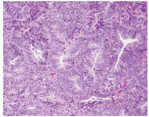
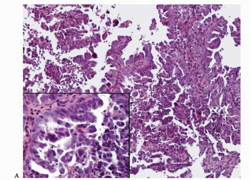
Endometrioid adenocarcinoma is the most common type of endometrial carcinoma, accounting for 80% of cases.
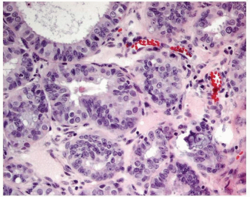
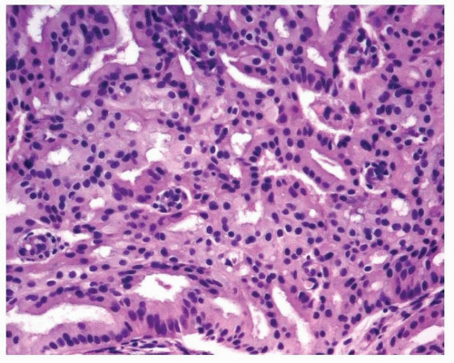
3 This tumor is composed of glands that resemble those seen in the normal endometrium. The variably sized glands seen in this type of tumor are usually oval or round, although they can be angulated or branched. They are lined by columnar cells with stratified or pseudostratified nuclei. The degree of nuclear atypia is usually mild to moderate. The cytoplasm is either basophilic, amphophilic, or slightly eosinophilic (Fig. 4-1). Intraluminal mucin can be seen, and in some cases, this becomes a prominent finding. Intracellular mucin, usually focal and less frequently diffuse, may also be noted (Fig. 4-2).
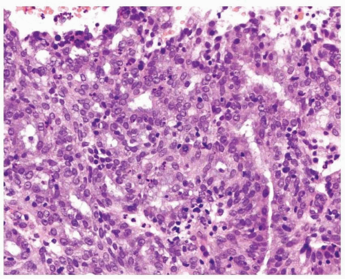
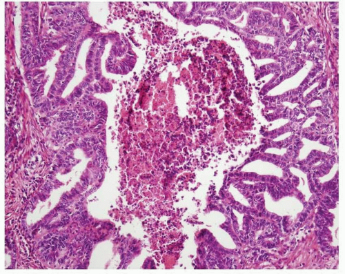
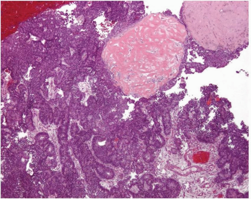
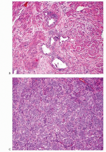
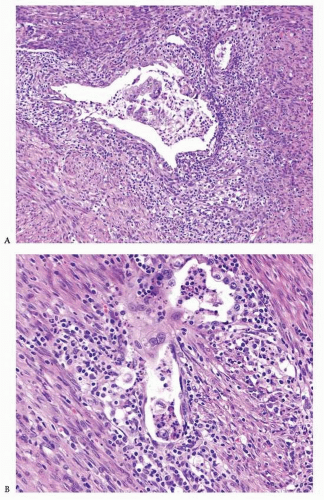
6,7 Ciliated cells, necrosis of the glands or within luminal spaces (Fig. 4-3), foamy stromal cells, signet ring cells (
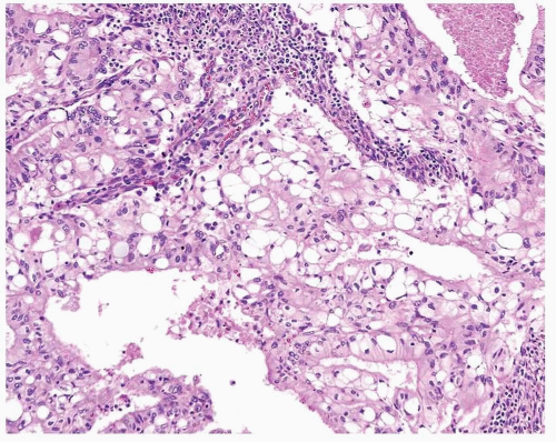
Fig. 4-4), and infrequently, intestinal differentiation, including goblet cells and argentaffin cells, can be seen.
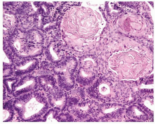
7,8,9,10 and 11 In addition, psammoma bodies and focal, bland-looking heterologous elements (fat, cartilage, osteoid) are occasionally found (Fig. 4-5).12,13


Table 4-1 Pathogenetic Classification of Sporadic Endometrial Carcinoma


Type I


Type II


Age


Premenopausal and perimenopausal


Postmenopausal


Unopposed estrogen stimulation


Present


Absent


Background endometrium


Hyperplasia


Atrophy


Histologic type


Endometrioid


Serous


Mucinous


Clear cell


Estrogen/progesterone receptors expression


Present


Absent or weakly positive


Behavior


Favorable


Aggressive


Molecular alterations


PTEN inactivation


Microsatellite instability


Mutations of K-ras and β-catenin


p53 mutation


Loss of heterozygosity at different loci


Inactivation of p16 and E-cadherin


Amplification of HER2/neu


Table 4-2 World Health Organization (WHO) Classification of Endometrial Carcinoma


Endometrioid adenocarcinoma


Variants


With squamous differentiation


Villoglandular


Secretory


Ciliated cell


Mucinous adenocarcinoma


Serous carcinoma


Clear cell carcinoma


Mixed carcinoma


Squamous carcinoma


Transitional cell carcinoma


Small-cell carcinoma


Undifferentiated carcinoma


Others


FIGURE 4-1: Endometrioid adenocarcinoma; glands are lined by columnar cells with stratified nuclei. The luminal contour of the glands is smooth.


FIGURE 4-2: Endometrioid adenocarcinoma with intracellular (intracytoplasmic) mucin.


FIGURE 4-3: Endometrioid adenocarcinoma with dirty necrosis within glandular luminal spaces.


FIGURE 4-4: Endometrioid adenocarcinoma with signet ring-like cells.


FIGURE 4-5: Endometrioid adenocarcinoma with osseous metaplasia.


Variants of Endometrioid Adenocarcinoma


Variant with Squamous Differentiation

Squamous differentiation in endometrioid adenocarcinoma can have a variable appearance: (1) morular (rounded intraluminal masses of bland-looking eosinophilic or amphophilic cells), (2) nests or sheets of tumor cells resembling keratinized squamous carcinoma, (3) nests or sheets of squamous cells rich in glycogen, (4) sheets of spindled cells with minimal evidence of squamous differentiation, and (5) papillae with associated mucinous metaplasia and neutrophils (Fig. 4-6). The previously used terms “adenoacanthoma” and “adenosquamous carcinoma” are considered obsolete and should not be used.5


Villoglandular Variant

This variant is characterized by the presence of slender, finger-like papillae lined by columnar cells with low-grade cytologic atypia. The nuclei of the neoplastic cells preserve their polarity in relation to the basement membrane of the epithelium, and the contour of the papillae is smooth (Fig. 4-7). The presence of this variant within the myoinvasive component of an endometrial adenocarcinoma has been found to be associated with higher incidences of vascular/lymphatic invasion and lymph node metastasis.14


Secretory Variant

This rare variant of endometrioid adenocarcinoma is usually seen in postmenopausal patients, although it can also occur in premenopausal patients, with or without a history of endogenous or exogenous progestational effect. Its histologic hallmark is the presence of intracytoplasmic vacuoles in most of the neoplastic cells. These vacuoles can be supranuclear, subnuclear, or both (Fig. 4-8).15,16


Ciliated Variant

This uncommon variant of endometrial adenocarcinoma shows neoplastic glands lined predominantly by ciliated cells (Fig. 4-9).8,17


FIGURE 4-6: Endometrioid adenocarcinoma with squamous differentiation.


FIGURE 4-7: Endometrioid adenocarcinoma with a villoglandular pattern. Finger-like papillae with fibroconnective tissue cores, lined by cells with nuclei that have preserved their polarity. Note the smooth outer contour of the papillae (inset).


FIGURE 4-8: Endometrioid adenocarcinoma, secretory variant.


FIGURE 4-9: Endometrioid adenocarcinoma, ciliated cell variant.


Other Changes


SARCOMATOID (SPINDLE CELL) FEATURES

Sarcomatoid (spindle cell) features also can be seen in endometrioid adenocarcinoma. This is usually a focal finding, and the recognition of the area where the spindle cells merge with the glandular component of the tumor is crucial to avoid the misinterpretation of the spindle
cells as a true sarcoma (Fig. 4-10A-B). The use of a keratin cocktail and epithelial membrane antigen (EMA) may assist in making the correct diagnosis; however, these immunostains can be only focally positive.


FIGURE 4-10: A. Endometrioid adenocarcinoma with spindle cell features. B. Note how the cells lining the neoplastic endometrial glands merge with the spindle cells (arrows).


CLEAR CELL CHANGES

The cytoplasm of the neoplastic cells in endometrioid adenocarcinoma can be focally clear. The specific cause of this finding is usually unknown, although it may be related to the presence of intracytoplasmic lipid.18


MICROGLANDULAR HYPERPLASIA-LIKE PATTERN

Endometrioid adenocarcinoma with a prominent microglandular hyperplasia-like pattern is uncommon and usually seen in postmenopausal women with a history of exogenous hormone use (i.e., medroxyprogesterone acetate, estradiol, or a combination of both).19 This type of neoplasm shows a proliferation of small glands lined by one or more layers of cuboidal, columnar, or flattened cells, with a variable amount of amphophilic or mucin-rich cytoplasm. In certain areas, the glands can be large or cystic or the neoplastic cells can be arranged in solid nests. The degree of cytologic atypia is variable, from none to moderate, and the mitotic index is also variable, ranging from one mitosis per 10 high-power fields (HPFs) to frequent mitoses per 10 HPFs (Fig. 4-11). Abnormal mitotic figures can be seen. The luminal spaces contain mucin, and there are numerous neutrophils. In addition, lymphocytes and plasma can be found. Immunohistochemically, this type of neoplasm stains variably with carcinoembryonic antigen (CEA), vimentin, and Ki-67.20,21,22 and 23 Recently, some investigators have found p16 immunostaining to be positive in endometrial adenocarcinoma with a microglandular hyperplasia-like pattern and negative in microglandular hyperplasia of the uterine cervix.24 Because experience with p16 immunostaining is limited, the definitive diagnosis of this type of tumor requires the recognition of continuity between the tumor and endometrium.


FIGURE 4-11: Endometrioid adenocarcinoma, microglandular hyperplasia-like.


SMALL NONVILLOUS PAPILLAE

This pattern consists of small, simple or complex papillae protruding with the glandular component of an otherwise typical International Federation of Gynecology and Obstetrics (FIGO) grade 1 or 2 endometrioid adenocarcinoma or from the finger-like papillae of a villoglandular adenocarcinoma. The neoplastic cells have eosinophilic cytoplasm and low-grade nuclear features (Fig. 4-12).25


SERTOLIFORM PATTERN

The presence of a sertoliform pattern in endometrioid adenocarcinoma arising in the endometrium is rare. This pattern is characterized by tubules, cords, and nests of columnar cells with eosinophilic or clear apical cytoplasm mimicking a Sertoli cell tumor. This pattern can be focal or diffuse. Immunohistochemically, the tumor cells are positive for EMA and vimentin and positive or negative for keratin. A rare case has also expressed α-inhibin, calretinin, WT-1, and Melan-A, indicating true sex cord differentiation.26 Smooth muscle markers such as desmin, smooth muscle actin (SMA), and HHF35 are negative.26,27 and 28


SEX CORD-LIKE PATTERN AND HYALINIZATION

This rare pattern of endometrioid adenocarcinoma shows cords or clusters of epithelioid and/or spindle cells that are intermixed and usually merge with typical endometrioid adenocarcinoma. Usually, the neoplastic cells are embedded in a hyalinized matrix. The degree of hyalinization can be so pronounced that it can resemble osteoid material. Squamous differentiation is common (Fig. 4-13). Immunohistochemically, the tumor cells have a variable expression of keratin and EMA, ranging from no staining to diffuse staining; however, the expression is typically seen in less than 50% of the cells. Although this pattern could be mistaken for a malignant mixed müllerian tumor, attention to the cytologic features and mitotic activity (this pattern displays less cytologic atypia and less mitotic activity than a malignant mixed müllerian tumor) is necessary to make the correct diagnosis.29


FIGURE 4-12: Endometrioid adenocarcinoma with small nonvillous papillae.


FIGURE 4-13: Endometrioid adenocarcinoma with sex cord-like pattern (inset).


OXYPHILIC (ONCOCYTIC) CHANGES

This rare pattern of endometrioid adenocarcinoma is seen mostly in postmenopausal patients. Tumors with this pattern display, either predominantly or entirely, large oxyphilic cells (Fig. 4-14). Some cases have been found to be rich in mitochondria.30,31 and 32


FIGURE 4-14: Endometrioid adenocarcinoma, oxyphilic variant.


Differential Diagnosis


Endometrioid Adenocarcinoma versus Complex Endometrial Hyperplasia with or without Atypia

One of the three following criteria is required for the diagnosis of endometrioid adenocarcinoma:


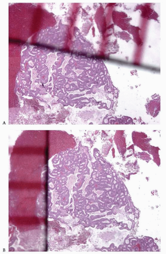
  • Back-to-back endometrial glandular proliferation occupying an area of at least 2 × 2 mm (Fig. 4-15)


  • An extensive papillary pattern (Fig. 4-16)


  • A desmoplastic or fibroblastic stroma infiltrated by irregular glands (Fig. 4-17)

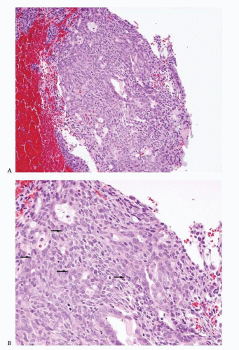
The last criterion mentioned earlier must be used with caution because the stromal reaction secondary to a recent endometrial curettage and the fibroblastic stroma of an endometrial polyp with torsion may mimic this feature.


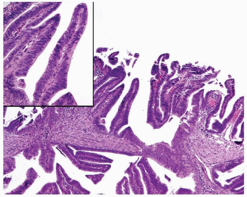
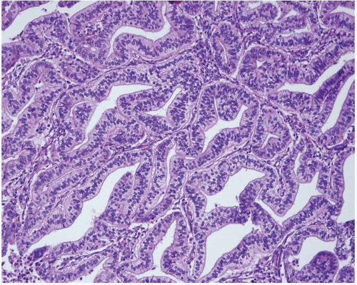
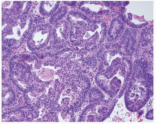
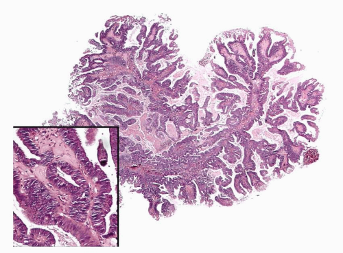
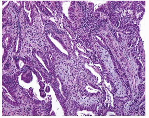
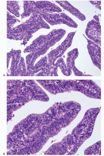
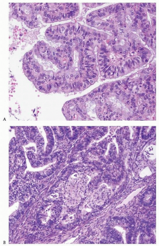
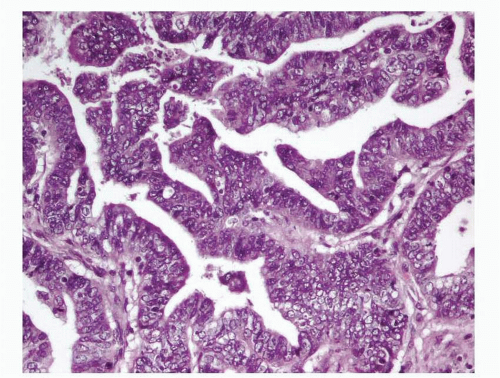
Endometrioid Adenocarcinoma with a Villoglandular Pattern versus Papillary Endometrial Adenocarcinoma of Intermediate Grade versus Serous Carcinoma

The papillae seen in the villoglandular pattern are slender and finger-like and lined by neoplastic cells with low-grade cytologic features and nuclei that preserve their polarity. In addition, the outer surface of the papillae is smooth. In contrast, the papillae seen in papillary endometrial adenocarcinoma of intermediate grade are long or short and lined by cells with moderate cytologic atypia and nuclei that have partially lost their polarity. The outer surface of the papillae is somehow irregular (Figs. 4-18A-B and 4-19A-B). Serous carcinoma can have either long or blunt papillae, with or without fibroconnective cores, lined by markedly atypical cells usually showing conspicuous mitotic activity. Immunohistochemically, villoglandular adenocarcinoma and intermediate-grade papillary endometrial adenocarcinoma are usually either negative or focally positive for p53 (author’s unpublished observations), whereas serous carcinoma is usually positive for this immunomarker.33,34


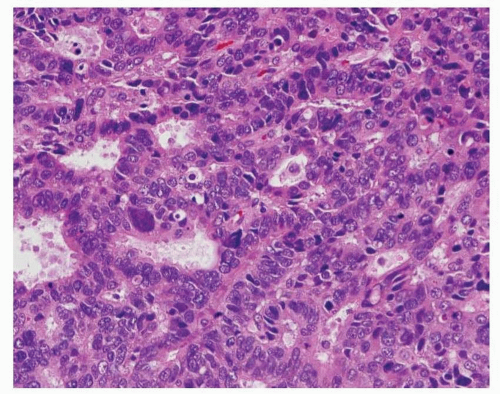
Endometrioid Adenocarcinoma versus Glandular Variant of Serous Carcinoma

The presence of marked cytologic atypia in an endometrioid-looking tumor composed mostly of glands should raise the diagnostic possibility of serous carcinoma, glandular variant (Fig. 4-20). The use of an immunohistochemical panel that includes p53, progesterone receptor (PR), and PTEN will assist in making the correct diagnosis. The combination of lack of p53 expression, positive PR expression, and loss of PTEN is in keeping with the diagnosis of endometrioid adenocarcinoma.34


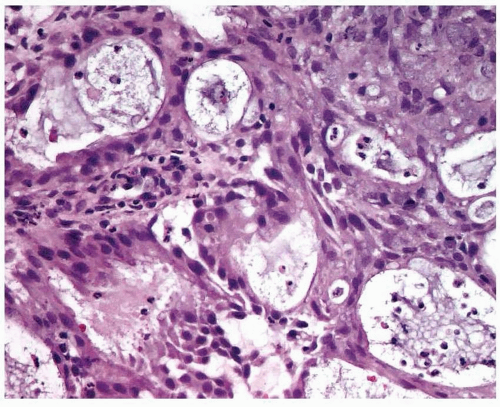
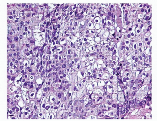
Endometrioid Adenocarcinoma with Glycogenated Squamous Differentiation versus Clear Cell Carcinoma

Attention to histologic features is of utmost importance in making the correct diagnosis. In cases of endometrioid adenocarcinoma with glycogenated squamous differentiation (Fig. 4-21), there are areas with overt squamous differentiation in the vicinity. In contrast, clear cell carcinoma usually demonstrates a combination of architectural patterns: solid, tubuloglandular, and papillary.


Endometrioid Adenocarcinoma with Sarcomatoid (Spindle Cell) Features or with Sex Cord-Like Pattern/Hyalinization versus Malignant Mixed Müllerian Tumor

Careful attention to histologic features will allow the correct diagnosis of these neoplasms. As mentioned earlier, endometrioid adenocarcinoma with sarcomatoid (spindle cell) features demonstrates areas where the spindle cell component merges with the glandular component, whereas endometrioid adenocarcinoma with sex cord-like pattern and hyalinization shows a constellation of typical features such as the formation of cords and clusters of spindle and epithelioid cells in a background of hyalinized material usually associated with squamous differentiation. The value of immunohistochemical studies, such as EMA and keratin, to enhance the epithelial nature of the spindle cells is limited because they can be negative or only focally positive in these neoplasms.29 In addition, the sarcoma component of a malignant mixed müllerian tumor can be focally or diffusely positive for keratin and EMA.35


FIGURE 4-15: Endometrioid adenocarcinoma; the diagnosis is based on a back-to-back glandular proliferation occupying an area measuring 2 × 2 mm (A and B).


FIGURE 4-16: Endometrioid adenocarcinoma; the diagnosis is based on the presence of an extensive papillary pattern. Note the absence of marked cytologic atypia in the cells lining the papillae (inset).


FIGURE 4-17: Endometrioid adenocarcinoma; the diagnosis is based on the presence of irregular glands embedded in a desmoplastic stroma.


FIGURE 4-18: Papillary endometrial adenocarcinoma of intermediate-grade, papillary structures (A), lined by cells that have lost the polarity of their nuclei (B).


FIGURE 4-19: Papillary endometrial adenocarcinoma of intermediate grade. Frequently seen features include mucinous metaplasia (A) and squamous differentiation (B).


FIGURE 4-20: Serous carcinoma, not to be mistaken for endometrioid adenocarcinoma. Note the presence of marked cytologic atypia, including the loss of polarity of the nuclei of the cells lining the neoplastic glands.


FIGURE 4-21: Endometrioid adenocarcinoma with glycogenated squamous differentiation.


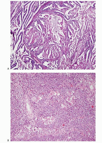
Endometrioid Adenocarcinoma Associated with Primitive Neuroectodermal Tumor versus Malignant Mixed Müllerian Tumor

Rare cases of endometrioid carcinoma associated with primitive neuroectodermal tumor (PNET) have been described (
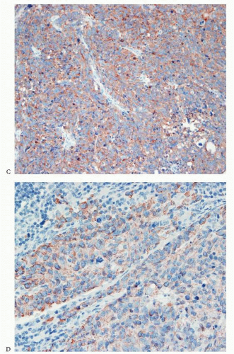
Fig. 4-22A-D). The neuroectodermal component is characterized by the presence of uniform oval, round, or slightly elongated cells with a scanty or moderate amount of cytoplasm arranged in sheets, nests, or trabeculae. The presence of perivascular rosettes, Homer Wright rosettes, ependymal-type rosettes, astrocyte-like cells in a fibrillary background, or ganglion-like cells is variable. Immunohistochemical studies show that cells with neuroectodermal differentiation are positive for neurofilament, synaptophysin, and CD99 (although some cases show nonspecific staining for the latter). These cells also are either negative or focally positive for keratin.36,37 When molecular studies detect the rearrangement of the EWSR1 gene, the diagnosis of peripheral PNET can be made. In contrast, cases lacking this finding should be designated as tumors with neuroectodermal differentiation or, alternatively, as central-type PNET rather than “PNET, not otherwise specified” to avoid confusion with the former. Of interest, some cases of uterine PNET are part of a malignant mixed müllerian tumor.37 Combined endometrioid adenocarcinoma and PNET have been described in patients ranging from 47 to 71 years of age and have been associated with advanced-stage disease and a poor prognosis.36,37


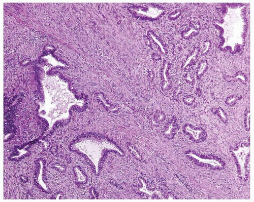
Endometrioid Adenocarcinoma, FIGO Grade 1, with Myometrial Invasion versus Atypical Polypoid Adenomyoma

The diagnosis of atypical polypoid adenomyoma (APA) should be considered whenever a proliferation of endometrial glands is seen within a smooth muscle or fibromuscular background arranged in intersecting fascicles (Fig. 4-23). In the typical case of APA, the glandular proliferation is similar to that seen in hyperplasia and tends to have squamous morules. The glandular size and shape are variable, as is the presence of cytologic atypia.38 The following features distinguish APA from myoinvasive endometrioid adenocarcinoma, FIGO grade 1:


  • Young age (average age, 39 years)


  • The presence of intersecting fascicles of smooth muscle or fibro muscular tissue


  • A more cellular smooth muscle than the normal myometrium seen in cases of myoinvasive endometrioid adenocarcinoma


  • The presence of endometrioid proliferation exclusively in fragments containing smooth muscle or fibromuscular tissue


Endometrioid Adenocarcinoma Arising in the Endometrium versus Endometrioid Adenocarcinoma Arising in the Uterine Cervix

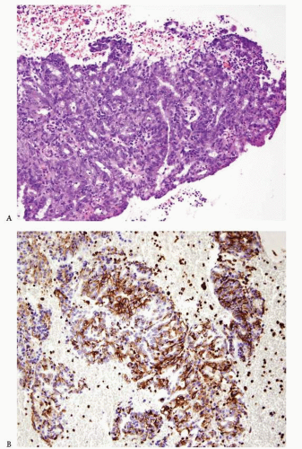
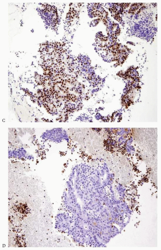
Carcinomas arising in the endometrium or endocervix may have overlapping histologic features. In cases where, upon review of the hematoxylin and eosin-stained slides, the origin of the tumor (i.e., endometrium versus endocervix) cannot be determined, the use of immunohistochemical studies is indicated. Tumors positive for vimentin and estrogen receptor (ER) and negative for CEA are favored to have an endometrial origin (Fig. 4-24A-D), whereas tumors that are positive for CEA and negative for vimentin and ER are favored to be endocervical in origin.39 p16 is another immunomarker recommended to assist in the distinction between these two types of tumor. It is usually diffusely expressed by adenocarcinomas of endocervical origin and has patchy expression in endometrioid endometrial adenocarcinomas40; however, there are two caveats to bear in mind: (1) p16 is also diffusely expressed in high-grade endometrial adenocarcinomas (serous, clear cell, and grade 3 endometrioid), and (2) in a small biopsy, a tumor with patchy expression of p16 can falsely appear to have diffuse expression of this marker. Human papillomavirus (HPV) in situ hybridization is another tool used to confirm the cervical origin of a given tumor; however, up to 30% of cases of cervical carcinoma can be negative for this test.41 In a small subset of cases, the results of these ancillary tests are discordant, and imaging studies and/or fractioned curettage are required to determine with certainty the site of origin of the tumor.


FIGURE 4-22: Endometrioid adenocarcinoma, FIGO grade 1 (A), associated with primitive neuroectodermal tumor. Note the ganglion cells within nests of small blue cells (B), synaptophysin-positive staining (C), and neurofilament-positive staining (D). (continued)


FIGURE 4-22: (continued)


FIGURE 4-23: Atypical polypoid adenomyoma displaying a proliferation of endometrial glands embedded in a background of intersecting myometrial fascicles, not to be mistaken for endometrial adenocarcinoma with myometrial invasion.


Serous Carcinoma


Overview

Serous carcinoma accounts for 10% of all endometrial carcinomas.42 This tumor is usually found in postmenopausal patients, although it can be seen in patients younger than age 56 years.43 An association with breast carcinoma is noted in approximately 16% of patients; this association increases to 23% in patients younger than age 56 years.44 In some cases, there is an association between serous carcinoma and pelvic irradiation, tamoxifen use, BRCA1 mutation, cervical or axillary lymph node metastasis at presentation, paraneoplastic hypercalcemia, or high levels of serum CEA.45,46,47,48,49,50,51 and 52


FIGURE 4-24: Endometrioid adenocarcinoma with mucinous metaplasia. Tumor difficult to differentiate from an endocervical adenocarcinoma on hematoxylin and eosin-stained slide (A). Immunoperoxidase studies show positive staining for vimentin (B) and estrogen receptor (C) and negative staining for carcinoembryonic antigen (CEA) (D). (continued)


FIGURE 4-24: (continued)


Serous carcinoma is characterized by the presence of marked cytologic atypia. The neoplastic cells are arranged in papillary, glandular, or solid patterns (Fig. 4-25A-C). Usually, there is tufting with detachment of the neoplastic cells. The mitotic activity is high. Hobnail cells, tumor giant cells, abnormal mitotic figures, and psammoma bodies can also be seen.53


Differential Diagnosis


Serous Carcinoma versus Endometrioid Adenocarcinoma, Villoglandular Variant

In the villoglandular variant of endometrioid adenocarcinoma, the papillae are finger-like, with smooth contours, and are lined by columnar cells with preserved polarity of the nuclei and bland cytology. In serous carcinoma, there is marked cytologic atypia and no preservation of the nuclear polarity.


Serous Carcinoma versus Papillary Endometrial Adenocarcinoma of Intermediate Grade

Papillary endometrial adenocarcinoma of intermediate grade is not included in the WHO classification; however, it is recognized by some investigators.54,55 This tumor has features that are between those of endometrioid adenocarcinoma with villoglandular features and serous carcinoma. Papillary endometrial adenocarcinoma of intermediate grade appears as papillae that are lined by cells with nuclei that are mildly or moderately disorganized, without preservation of their polarity but with less atypia than is seen in serous carcinoma (Fig. 4-26). Foci of squamous and mucinous differentiation are usually seen (Fig. 4-27A-B). In addition, the pattern of invasion described as microcystic, elongated, and fragmented (MELF pattern) is commonly seen. This type of tumor is associated with a higher incidence of vascular/lymphatic invasion, advanced stage at presentation, and recurrences than a typical FIGO grade 2 endometrioid adenocarcinoma.55


FIGURE 4-25: Serous carcinoma. Tumor characterized by the marked cytologic atypia of its neoplastic cells. Papillary pattern (A) (inset), glandular pattern (B), and solid pattern (C). (continued)


FIGURE 4-25: (continued)


Serous Carcinoma versus Endometrial Adenocarcinoma, FIGO Grade 1, and Complex Endometrial Hyperplasia

These two processes are characterized by a proliferation of well-formed glands lacking marked cytologic atypia. In contrast, the glandular variant of serous carcinoma shows open glands lined by cells with marked cytologic atypia (i.e., increased nuclear-to-cytoplasmic ratio, nuclear pleomorphism, and loss of nuclear polarity).


Serous Carcinoma versus Clear Cell Carcinoma

The papillae seen in clear cell carcinoma are usually lined by a single row of neoplastic cells with clear or eosinophilic cytoplasm and tend to show hyalinization of the stromal cores. In contrast, the papillae of serous carcinoma are usually more complex than the ones seen in clear cell carcinoma, and the neoplastic cells tend to have a basophilic or amphophilic cytoplasm. However, occasionally the neoplastic cells are eosinophilic or show focal clearing of the cytoplasm.


Serous Carcinoma versus Papillary Syncytial Metaplasia

This process lacks the marked cytologic atypia and the high mitotic index seen in serous carcinoma.


FIGURE 4-26: Papillary endometrial adenocarcinoma of intermediate grade.


FIGURE 4-27: Papillary endometrial adenocarcinoma of intermediate grade, with typical pattern of invasion seen in this type of neoplasm. Note the incomplete glands with attenuated epithelium and prominent inflammatory infiltrate (A and B).


Clear Cell Carcinoma


Overview

Clear cell carcinoma represents approximately 5% of all endometrial carcinomas.56 This tumor is composed of clear, eosinophilic (oncocytic), or hobnail cells arranged in tubulocystic, papillary, or solid patterns (Figs. 4-28A-C and 4-29). The cells can be polygonal, cuboidal, or flattened. The degree of atypia is variable, usually marked and less frequently mild or absent. The mitotic index is also variable, ranging from 0 mitoses per 10 HPFs to greater than 10 mitoses per 10 HPFs. The stroma can be hyalinized. Epithelial hyaline bodies, eosinophilic granular or homogeneous intraluminal material, and psammoma bodies also can be seen.57,58 Immunohistochemically, clear cell carcinoma is negative for PR and has a high Ki-67 proliferation index and variable expression of p53 and ER.59,

Only gold members can continue reading. Log In or Register to continue

Stay updated, free articles. Join our Telegram channel

May 22, 2016 | Posted by in ONCOLOGY | Comments Off on Endometrial Carcinoma

Full access? Get Clinical Tree

Get Clinical Tree app for offline access