Endocrine Tumors


Endocrine Tumors


Steven G. Waguespack

Winston W. Huh

Andrew J. Bauer


INTRODUCTION

Endocrine tumors comprise a variety of benign and malignant neoplasms that arise from the endocrine glands or neuroendocrine tissues. Functioning tumors are associated with typical clinical syndromes related to the specific hormone(s) being secreted, whereas nonfunctioning tumors present incidentally or secondary to symptoms related to mass effect. While most childhood endocrine tumors are sporadic without an identifiable mutation in germline DNA, others are familial, such as those that occur as part of the multiple endocrine neoplasia (MEN) syndromes. Endocrine tumors represent a minority of all neoplasms observed in the pediatric population and are generally clinically benign or low-grade cancers, although a small percentage of these tumors are high-grade malignancies requiring multimodality therapies. As with any rare childhood disease, treatment is best provided at tertiary care centers with multidisciplinary expertise in the management of such tumors.


PITUITARY TUMORS


Pituitary Adenomas


Staging and Prognosis

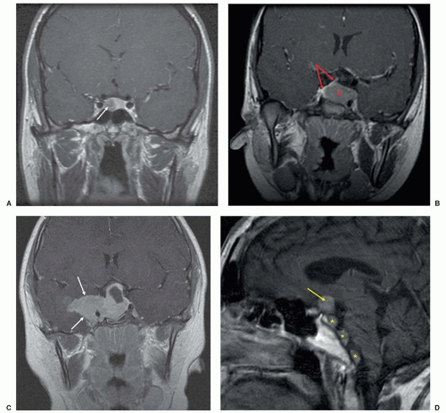
PAs are classified by the World Health Organization (WHO) into typical PAs, atypical PAs, and pituitary carcinomas. Atypical PAs are nonmetastatic tumors with morphologic features suggestive of aggressive behavior, including extensive nuclear staining for p53, an elevated mitotic index, and Ki-67 labeling index >3%.9 PAs may be locally invasive (Fig. 36.1), but an invasive growth pattern does not necessarily portend a more dire clinical course. In pediatric patients with non-adrenocorticotropin hormone (ACTH)-secreting PAs, disease-free survival is lower for macroadenomas, and these children typically require more aggressive multimodality therapy and have higher rates of hypopituitarism.2 While the mortality for children with PAs is low, there may be significant morbidity due to surgical complications and/or hypopituitarism.


Treatment and Follow-Up

In general, transsphenoidal resection (TSR)7 is the treatment of choice for hormonally active tumors, with the notable exception of prolactinomas (see below). Surgery is also considered in macroadenomas causing mass effect or contributing to hypopituitarism, pituitary apoplexy, ineffectiveness or intolerance of medical therapy, and for smaller sellar masses associated with chronic severe headaches. In the right hands, TSR is a safe and effective procedure, and it is imperative to identify a high-volume pituitary neurosurgeon.10


TABLE 36.1 Genetic Syndromes Associated with Endocrine Neoplasia


Gene (Chromosome)


Type(s) of Endocrine Neoplasia


Other


APC-associated polyposis (familial adenomatous polyposis [FAP], attenuated FAP, Gardner syndrome, and Turcot syndrome)


APC (5q21-q22)


  • ACT


  • PTC (cribriform-morular variant)


  • Colorectal polyps/colorectal carcinoma


  • Osteomas


  • Desmoid tumors


  • Pancreas adenocarcinomas


  • Medulloblastoma


  • Hepatoblastoma


Beckwith-Wiedemann syndrome


Several genes on chromosome 11p15.5 (BWS critical region)


  • ACT/Adrenocortical cytomegaly


  • ?PHEO (case reports)


  • ?Thyroid carcinoma (case report)


  • Macrosomia/organomegaly/macroglossia


  • Hemihypertrophy


  • Embryonal tumors (Wilms tumor, hepatoblastoma, neuroblastoma, pancreatoblastoma, rhabdomyosarcoma)


  • Omphalocele


  • Neonatal hypoglycemia


  • Anterior linear ear lobe creases/posterior helical ear pits


  • Renal abnormalities


CNC


PRKAR1A (17q24.2) and unknown gene at chromosome locus 2p16


  • Mammosomatotroph hyperplasia/GH-secreting PA


  • PPNAD/ACT


  • Thyroid neoplasia


    ♦ Follicular adenomas


    ♦ Thyroid carcinoma (PTC and FTC)


  • Large cell calcifying Sertoli cell tumors


  • GH excess and hyperprolactinemia


  • Spotty skin pigmentation


  • Blue nevus


  • Cardiac, cutaneous, or breast myxomas


  • Psammomatous melanotic schwannoma


CDC73-related HPT (HPT-jaw tumor syndrome)


CDC73 (1q25)


  • Parathyroid adenoma


    ♦ Often cystic with atypical histology


  • PTCa


  • ?Thyroid neoplasia (case reports)


  • Ossifying fibroma(s) of the maxilla and/or mandible


  • Renal cystic disease/renal hamartomas


  • Wilms tumor


  • Benign and malignant uterine tumors


FIPAs


AIP (11q13.3)


  • PA


  • Primarily GH-secreting PA


Familial PGL syndromes


PGL1


PGL2


PGL3


PGL4


PGL5


SDHD (11q23)


SDHAF2 (11q12.2)


SDHC (1q21)


SDHB (1p36.1-p35)


SDHA (5p15)


  • PHEO/PGL


  • PA


  • GIST


  • RCC


Hereditary nonpolyposis colorectal cancer (Lynch syndrome)


MLH1 (3p22.3)


MSH2 (2p21)


MSH6 (2p16)


PMS2 (7p22.1)


EPCAM (2p21)


  • ACT


  • Colorectal cancer


  • Endometrial and ovarian cancer


  • Gastric cancer


  • Small bowel cancer


  • Urinary tract cancers


  • Pancreatic cancer


  • Brain tumors


  • Sebaceous skin neoplasms


LFS


TP53 (17p13.1)


  • ACT


  • ?Thyroid carcinoma (case reports)


  • Sarcoma


  • Breast cancer


  • Leukemia


  • Melanoma


  • Gastrointestinal (GI) malignancies


  • CNS tumors/choroid plexus carcinomas


  • Gonadal germ cell tumors


MAS


GNAS (20q13)


  • Mammosomatotroph hyperplasia/GH-secreting PA


  • Functioning thyroid nodules


  • Ovarian cysts


  • Macronodular ACT


  • Peripheral precocious puberty


  • Polyostotic fibrous dysplasia


  • Café-au-lait macules with irregular margins


  • CS due to ACTs


  • GH excess and hyperprolactinemia


MEN1


MEN1 (11q13)


  • Parathyroid adenomas/hyperplasia


  • PA


  • GEP-NET (pancreas, duodenum, bronchopulmonary, thymus)


  • ACT


  • PHEO (rare)


  • Lipomas/angiofibromas/collagenomas


  • Meningiomas


  • Ependymomas


  • Leiomyomas


MEN2A


RET (10q11.2)


  • MTC


  • PHEO/PGL


  • Parathyroid adenoma/hyperplasia


  • Codon-specific risk of MTC


  • Variants with cutaneous lichen amyloidosis and Hirschsprung disease


  • Adrenal ganglioneuroma in rare cases


MEN2B


RET (10q11.2)


  • MTC


  • PHEO


  • Very high risk for early onset and metastasis of MTC


  • Mucosal neuromas of the lips, tongue, and eyelids


  • Medullated corneal nerve fibers


  • Distinctive facies with enlarged lips


  • Megacolon/ganglioneuromatosis of the GI tract


  • “Marfanoid” body habitus


  • Absent tears in infancy


  • Feeding difficulties and constipation in infancy


  • Adrenal ganglioneuroma in rare cases


MEN4


CDKN1B (12p13.1-p12)


  • PA


  • Parathyroid adenoma/hyperplasia


  • GEP-NET


?Adrenal and thyroid neoplasia


NF1


NF1 (17q11.2)


  • PHEO/PGL


    ♦ Composite PHEO (rare)


  • GEP-NET


    ♦ Primarily periampullary duodenal somatostatinomas but also insulinomas, gastrinomas, and nonfunctioning tumors


  • ?Parathyroid tumors (case reports)


  • ?ACT (case reports)


  • Café-au-lait macules with smooth borders


  • Axillary and inguinal freckling


  • Dermal and plexiform neurofibromas


  • Lisch nodules of the iris


  • Learning disabilities


  • Scoliosis, vertebral dysplasia, pseudarthrosis, and bony overgrowth


  • Optic and other central nervous system gliomas


  • Malignant peripheral nerve sheath tumors


  • Vasculopathy


  • Macrocephaly


Peutz-Jeghers syndrome


STK11 (19p13.3)


  • Sertoli cell tumors of the testis in males


  • Ovarian sex cord tumors with annular tubules (SCTAT) in females


  • ?DTC (case reports)


  • Hamartomatous polyps in the GI tract


  • Mucocutaneous hyperpigmentation


  • Various intestinal and extraintestinal malignancies


PTEN hamartoma tumor syndrome (Cowden syndrome, Bannayan-Riley-Ruvalcaba syndrome, PTEN-related Proteus syndrome, and Proteus-like syndrome)


PTEN (10q23)


  • Thyroid adenomas and multinodular goiter


  • Thyroid carcinoma (PTC and FTC)


  • Mucocutaneous lesions


    ♦ Papillomatous papules


    ♦ Trichilemmomas


    ♦ Acral keratoses


    ♦ Pigmented macules of the glans penis


  • Breast cancer


  • Macrocephaly


  • Endometrial carcinoma/uterine fibroids


  • Genitourinary tumors


TSC


TSC1 (9q34)


TSC2 (16p13.3)


  • GEP-NETs (insulinomas predominant)


  • ?Parathyroid adenoma/hyperplasia (case reports)


  • ?PHEO (case report)


  • ?PA (case reports)


  • ?Thyroid carcinoma (case report)


  • Cutaneous findings


    ♦ Hypomelanotic macules (“ash-leaf” spots)


    ♦ Facial angiofibromata (adenoma sebaceum)


    ♦ Ungual or periungual fibromas


    ♦ Shagreen patch


  • Neurologic disorders


    ♦ Epilepsy


    ♦ Mental retardation


    ♦ Autism


  • CNS lesions


    ♦ Cortical tuber


    ♦ Subependymal nodule


    ♦ Subependymal giant cell astrocytoma


  • Multiple retinal nodular hamartomas


  • Cardiac rhabdomyoma


  • Lymphangiomyomatosis


  • Renal angiomyolipoma


VHL


VHL (3p25.3)


  • PHEO/PGL


  • Nonfunctioning PETs


  • ?Lipid cell ovarian tumor (case reports)


  • Hemangioblastomas of the CNS and retina


  • Renal cysts and clear cell RCC


  • Pancreatic cysts and cystadenomas


  • Endolymphatic sac tumors


  • Papillary cystadenomas of the epididymis (males) and round ligament (females)


Werner syndrome


WRN (8p12)


  • PTC


  • PTC


  • ?ACT (case reports)


  • Premature aging


  • Cancer predisposition


    ♦ Sarcomas


    ♦ Acral lentiginous melanomas


FTC, follicular thyroid carcinoma; GIST, gastrointestinal stromal tumor; MEN, multiple endocrine neoplasia; NF, neurofibromatosis; PET, pancreatic endocrine tumor; RCC, renal cell carcinoma.


Figure 36.1 Various presentations of PAs on T1-weighted coronal MRI status post-gadolinium contrast. (A) Pituitary microadenoma (white arrow); (B) Pituitary macroadenoma (red asterisk); note the normal pituitary gland and pituitary stalk (red arrows) deviated to the right; (C) Invasive pituitary macroadenoma with extension through the right cavernous sinus into the right middle cranial fossa (white arrows); (D) Pituitary carcinoma with residual primary tumor (yellow arrow) and drop metastases (yellow asterisks).

Microadenomas have a high surgical cure rate, whereas complete resection of pituitary macroadenomas is often not possible, particularly with tumors >2 cm and when cavernous sinus invasion and/or significant suprasellar extension is present. Radiation therapy has a limited role in the management of children with PAs, but is considered in uncontrolled functional tumors or unresectable nonfunctioning tumors that are progressive.7,11 Long-term follow-up should include periodic MRI and hormonal assessment based on the functional status of the PA and previous treatments required.


Pituitary Adenoma Subtypes

PRL-secreting Adenomas (Lactotroph Adenomas). Prolactinomas are the most common PA in childhood, representing at least 50% of cases, and they are diagnosed most commonly in postpubertal girls. Prepubertal children present with symptoms of mass effect and/or delayed puberty, whereas pubertal children can also present with oligo- or amenorrhea (girls), galactorrhea (both genders), and rarely gynecomastia (boys).12,13 Males are more likely to have macroadenomas and therefore tend to present with symptoms related to tumor size.2,12


The degree of hyperprolactinemia typically correlates with tumor size, and the diagnosis of a prolactinoma is highly likely when the PRL level is >250 ng/mL.14 In cases where the PRL level and clinical findings are discordant, the differential diagnosis includes hyperprolactinemia due to stalk compression, the presence of macroprolactinemia, and assay artifact secondary to the high-dose hook effect that can occur with macroprolactinomas and extremely elevated PRL levels.14 Patients with elevated PRL levels and no tumor on MRI should have an evaluation for secondary hyperprolactinemia. Primary hypothyroidism should always be ruled out as a cause of hyperprolactinemia and sellar mass. Because of the possibility of GH cosecretion, especially with tumor-predisposing genetic syndromes (Table 36.1), an insulin-like growth factor 1 (IGF-1) level should also be included in the initial evaluation.

Prolactinomas can effectively be treated with one of the available dopamine agonists (DAs), either bromocriptine or cabergoline, even in the presence of visual changes. Cabergoline is preferred due to its less-frequent administration, fewer side effects, and greater efficacy.14 Oral contraceptive pills are a reasonable alternative in adolescent girls with microadenomas and nonbothersome galactorrhea.15,16 Sex steroids can exacerbate PRL hypersecretion and promote tumor growth, so caution should be undertaken when prescribing testosterone or estrogen replacement to adolescents with macroprolactinomas. The addition of an aromatase inhibitor may facilitate the use of testosterone replacement in males.16

Clinical improvement is usually seen before radiologic tumor reduction. In most patients, treatment is continued until the PRL level normalizes (or at least stabilizes) and the tumor demonstrates significant shrinkage. Some patients will never achieve normal PRL levels, but many patients may ultimately be able to stop DA therapy.15,17

ACTH-secreting Adenomas (Corticotroph Adenoma; CD). ACTH-secreting adenomas causing CD are the second most common PA in children, occurring in approximately one-third of children operated on for a pituitary tumor.2,3 Corticotroph tumors are the most likely PA to be diagnosed in prepubertal children, and there is a female-to-male predominance, except in prepubertal children.18,19

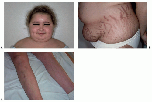
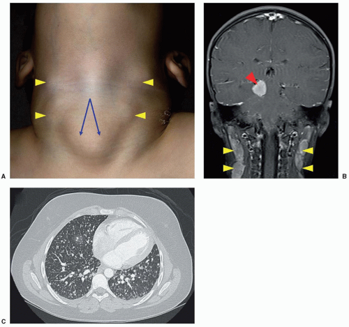
The onset of symptoms is usually insidious, and most children have a typical Cushingoid phenotype (Fig. 36.2) with generalized weight gain, obesity, impairment of growth (in prepubertal patients), violaceous striae (chiefly in older children), hirsutism/premature pubarche, acne, and hypertension.18,20,21,22 Clinical presentation can also include rounding of the face (moon facies), plethora, abnormal fat distribution with increased fat deposition in the supraclavicular fossae and upper back (Buffalo hump), acanthosis nigricans, easy bruising, hyperpigmentation (with significant ACTH elevation), muscle weakness, edema, impaired glucose tolerance/diabetes mellitus, pubertal delay, emotional lability, fatigue, headache, menstrual disturbance, and low bone mass for age and fractures. Not every case of Cushing syndrome (CS) is classic, and the diagnosis can be hard to differentiate from exogenous obesity based on phenotype alone, as not all children with CS are short or have delayed bone ages as would be expected.18,20 One of the most sensitive indicators of possible CS in a growing child is weight gain associated with a low growth velocity.23

These tumors are usually microadenomas and are often not visualized on routine MRI.18,21 Because of that, the diagnosis of CD is not always straightforward and care must be taken to differentiate an ACTH-secreting PA from ectopic ACTH secretion, such as from a thymic or bronchial neuroendocrine tumor (NET).24 Fortunately the latter situation is rare in children.

The diagnostic studies for CS have not been extensively validated in children. Screening studies to make the diagnosis include measurement of 24-hour urine-free cortisol, the overnight 1 mg (low dose) dexamethasone (DEX) suppression test, the 2-day low-dose DEX suppression test, and assessment of impaired circadian rhythm through the measurement of late evening salivary or serum cortisol levels.18,23,25 The DEX/CRH test is utilized to distinguish CS from pseudo-CS states, although this test may not be as reliable in very obese children.26 Once the diagnosis of ACTH-dependent CS (i.e., ACTH levels not suppressed and generally >15 pg/mL) has been made, the source of ACTH can generally be attributed to the pituitary if a clear adenoma is identified. If MRI findings are negative or equivocal, it is necessary to undertake additional testing to differentiate a pituitary from a peripheral source of ACTH production. These tests can include an overnight 8-mg (high-dose) DEX suppression test, 2-day high-dose DEX suppression test, CRH test, CRH/desmopressin test, and CRH-stimulated inferior petrosal sinus sampling (IPSS).18,23 IPSS obtained at the time of hypercortisolism appears to be the most definitive diagnostic test in the workup of ACTH-dependent CS and is safe and useful in children at experienced centers. Although IPSS can confirm a central source of ACTH production, its use solely for tumor lateralization is questionable.

The medical management of CD includes the use of inhibitors of cortisol synthesis (etomidate, ketoconazole, metyrapone, mitotane [o, p′-DDD; rarely used in nonmalignant conditions]) or action (mifepristone) as well as agents that can directly inhibit tumoral production of ACTH (cabergoline, pasireotide).27,28 In the patient with CD who is not cured with surgery or radiotherapy and who continues to suffer from CS despite optimization of medical therapy, bilateral adrenalectomy is favored. Although patients are committed to life-long glucocorticoid and mineralocorticoid replacement, removal of all adrenocortical tissue is the best way to cure CS in the treatment-refractory patient. The development of Nelson syndrome (the association of an expanding pituitary tumor and a high ACTH concentration after adrenalectomy in patients with CD) is a theoretical concern in such children.29

GH-Secreting Adenomas (Somatotroph Adenomas; Gigantism/Acromegaly). Somatotroph adenomas are the third most common PA in childhood, have a higher prevalence in males, are usually macroadenomas, and represent about 8% of all cases and 0.63% of surgically treated pituitary tumors in children.3,30 Gigantism is the term used to describe the overgrowth that occurs because of GH excess in children with open epiphyses, whereas acromegaly describes GH excess occurring after epiphyseal closure. The clinical presentation is usually one of accelerating linear growth. Other common features include coarse facial features, frontal bossing, prognathism, and disproportionately large hands and feet with thick fingers and toes. Symptoms related to tumor mass effect, pubertal delay, and menstrual irregularities can also be present at diagnosis.30 Other signs and symptoms of GH excess that are more common in the patient with acromegaly are reviewed elsewhere.31

During early childhood, gigantism is typically caused by mammosomatotroph tumors, secreting both GH and PRL, underscoring the genetic basis of these tumors9 (Table 36.1). Sporadic adenomas can harbor somatic GNAS mutations in up to 40% of adult patients, but this is not generally seen in sporadic tumors identified in childhood.32

The best initial diagnostic test is measurement of an IGF-1 level, comparing it to age-matched normative ranges.33 Confirmatory testing is accomplished through a 1.75 g/kg (max 75 g) oral glucose tolerance test, during which GH levels should fall to <1 ng/mL (<0.4 ng/mL in sensitive assays) during the 2-hour testing period.33 The clinician must also consider the gender and pubertal status of the adolescent patient undergoing testing, as the nadir of GH after oral glucose ingestion may be higher than adults, especially in mid-pubertal girls.34

When surgery is not curative, pharmacologic inhibition of GH secretion and suppression of tumor growth are accomplished through the use of the somatostatin (SST) analogs (octreotide
and lanreotide) and/or the DA cabergoline.31,35 Pegvisomant, a competitive GH receptor antagonist that lowers IGF-1 levels but has no direct impact on tumor size, is another treatment alternative. Primary medical therapy can be considered in patients who have surgically incurable tumors that are not compromising vision and in patients who are not good candidates for or refuse surgery.


Figure 36.2 Signs of CS in three different patients. A: Typical appearance of CS with moon facies, acne, and abnormal fat distribution in the supraclavicular fossae. B: Central adiposity with classic violaceous wide striae. C: Thin extremities with easy bruising and thinning of the skin (Photo courtesy of Dr. Rachel Edelen).

Clinically Nonfunctioning Adenomas (Null Cell Adenomas; Gonadotropin-secreting Adenomas). Clinically nonfunctioning tumors include those neoplasms that silently produce hormones (e.g., ACTH) without clinical signs or symptoms, in addition to those PAs that produce no pituitary hormones (null cell adenomas), bioinactive hormones, or hormones such as the gonadotropins (luteinizing and/or follicle-stimulating hormone) and α-subunit of the glycoprotein hormones that do not always cause clinical manifestations.9 In contradistinction to adults, these tumors represent <5% of pediatric PAs.2,3 Surgery is the mainstay of treatment, particularly in macroadenomas, which have a predisposition for enlargement and/or apoplexy over time.36 Medical management with a DA or SST analog can also be considered in the appropriate clinical setting but is typically ineffective.

Thyroid-stimulating Hormone-secreting Adenomas (Thyrotroph Adenomas). PAs secreting thyroid-stimulating hormone (TSH) are diagnosed because of elevated thyroxine and triiodothyronine levels and a nonsuppressed (and often elevated) TSH in the presence of a pituitary tumor on MRI. These tumors usually secrete excessive quantities of α-subunit, and the ratio of α-subunit to TSH is high, allowing for this diagnosis to be distinguished from pituitary resistance to thyroid hormone.37 Medical treatment includes SST analogs and DA, in addition to interventions directed at controlling the hyperthyroidism.


Pituitary Carcinoma


Staging and Prognosis

The prognosis of pituitary carcinoma is poor.38,39,40 Mean survival is 2 years, although cases of prolonged survival have been documented.41 There is no formal staging system for this rare cancer.


PARATHYROID TUMORS


Parathyroid Adenomas/Hyperplasia


Treatment and Follow-Up

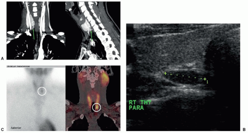
Treatment of HPT consists of surgical removal of the affected parathyroid gland(s). For all pediatric patients with primary HPT, but especially in situations where multigland disease is suspected, imaging is negative, or a second surgical exploration is needed, locating an experienced surgeon is of paramount importance.52,53 If preoperative localization studies identify a solitary adenoma, minimally invasive parathyroidectomy with the use of a rapid intraoperative PTH assay is generally the procedure of choice.54 In patients with genetic syndromes predisposing to multigland involvement, such as MEN1, possible surgical approaches include subtotal parathyroidectomy with viable remnant left in situ with a clip (the preferred approach in most centers) or total parathyroidectomy with forearm autograft.55,56 The appropriate timing
and indications for surgery in children with hereditary HPT have not yet been clearly defined. The medical management of hypercalcemia focuses on maintaining hydration, increasing urine calcium excretion, and diminishing bone resorption. Antiresorptive agents (bisphosphonates, subcutaneous calcitonin [CTN]) or the calcimimetic, cinacalcet, can also be used in the management of hypercalcemia in recalcitrant HPT or as a bridge to definitive surgical therapy.

Vitamin D (25-hydroxy vitamin D) levels should be checked preoperatively and supplemented to reduce the risk of postoperative hypocalcemia. More severe postoperative hypocalcemia can occur in patients with preexisting bone disease or vitamin D deficiency, a condition known as the “hungry bone syndrome” and manifested by hypocalcemia, hypophosphatemia, and hypomagnesemia.57

PTH levels drop immediately after successful surgical resection, and calcium levels follow in the next 24 to 48 hours.58 Therefore, cure can be determined shortly after surgery. Despite normal calcium levels, PTH levels may remain elevated in a secondary fashion and should not be misinterpreted as recurrent or residual disease.59 In pediatric patients with primary HPT, there is up to a 6% rate of recurrence.46 This is more likely in patients with genetic syndromes of HPT, but recurrent adenomas can occur in sporadic cases. Thus, annual screening with calcium and PTH levels should be considered.


Parathyroid Carcinoma


Staging and Prognosis

Two systems for the prognostic risk classification of parathyroid cancer that are based on histologic findings have been proposed.61 In general, PTCa is a slow-growing yet progressive tumor that tends to recur locally or spread to contiguous structures in the neck. Metastases occur later in the course of the disease via lymphatic or hematogenous spread. Cervical lymph nodes, lung, and liver are the most common metastatic sites. Recurrence rates are high, and the average time between surgery and first recurrence is approximately 3 years, noting that prolonged disease-free intervals have been reported. Disease-specific mortality usually results from uncontrolled hypercalcemia, and a median overall survival of 14.3 years has been observed.64


THYROID TUMORS


Benign Thyroid Neoplasms


Treatment and Follow-up

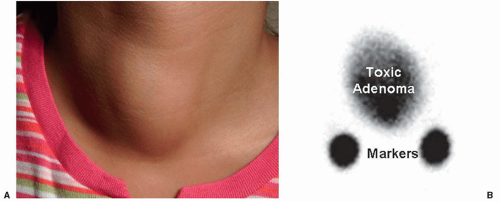
In most children with functional thyroid nodules, surgery is the preferred treatment, although 131I therapy can also be considered. For nonfunctional thyroid neoplasms, treatment and follow-up are determined by the cytopathologic diagnosis.67,73,74,75 For patients with malignant/suspicious cytology or a diagnosis of follicular neoplasm, surgery is recommended. In patients with nondiagnostic or indeterminate results, such as follicular lesion of undetermined significance, repeat FNAB or surgery is the most appropriate next step. Given the lack of established molecular or biochemical markers to accurately distinguish benign from malignant disease in children with an indeterminate biopsy, the decision to proceed to surgery is based on clinical criteria such as age of the patient, family history, nodule size, US criteria, history of radiation exposure, and others.66,76 In patients with benign
cytology, annual physical exam and US are appropriate, with repeat FNAB if the nodule grows significantly.75 TSH suppression with exogenous levothyroxine is no longer recommended in the euthyroid patient.66,67


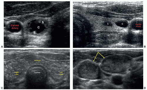
Figure 36.5 US findings of benign and malignant thyroid neoplasms. A: Benign colloid nodule with mixed solid [S] and cystic [C] components. B: Multiple benign thyroid cysts [C] in a different patient. C: PTC diffusely involving the thyroid gland with characteristic scattered microcalcifications. D: Metastatic lymphadenopathy (yellow arrows) identified in the same patient as (C).


Malignant Thyroid Neoplasms

Thyroid carcinoma is the most common endocrine malignancy, with an estimated 62,980 new cases diagnosed in the United States during 2014, of which only 1.8% occur in individuals <20 years of age.77,78 The incidence is ≤1 case/million/y in children under 10 years of age to 18 cases/million/y in adolescents ages 15 to 19, the most commonly affected pediatric age group.79,80

Thyroid malignancies arise from the thyroid follicular epithelium (differentiated thyroid carcinoma [DTC]: papillary thyroid carcinoma [PTC], follicular thyroid carcinoma [FTC], and their variants) or the neural crest-derived parafollicular C cell (medullary thyroid carcinoma, MTC).9 Anaplastic (undifferentiated) thyroid carcinomas and poorly DTCs are exceedingly rare in childhood, as are primary thyroid lymphomas and metastases to the thyroid gland. Several multidisciplinary guidelines have been created to assist with clinical decision-making in the care of adult patients,67,74,81 and specific guidelines for the evaluation and management of DTC in children are in development.


Differentiated Thyroid Carcinoma


Staging and Prognosis

Although several prognostic staging systems have been described for thyroid cancer, specifically PTC, a thorough discussion of these is beyond the scope of the current chapter. The pathologic tumor-node-metastasis (TNM) classification was adopted by the American Joint Committee on Cancer and the International Union against Cancer Committee (UICC) as the international reference staging system for thyroid cancer.103 By definition, however, the highest TNM stage that anyone less than age 45 can achieve is stage II, distinguished from stage I only by the presence of distant metastases. Another frequently used staging system, the MACIS score, may also be useful in children and adolescents but requires further validation.104

Most children with DTC having an excellent prognosis and survival over decades is generally the norm for these patients, even in the presence of distant metastases at diagnosis. Remission rates are high, and 10-year survival is almost universally 100% in this age group.80,83,105,106 For patients with stage II disease, those with micronodular lung metastases and iodine-avid disease (i.e., disease that retains good expression of the sodium-iodide symporter) have the best prognosis.107 Some children with DTC will ultimately succumb to their disease or die from treatment-related complications.105,106,108 Children diagnosed prior to age 10 appear to have a higher risk of recurrence and ultimately death from their disease,84,109,110 although this has not been observed universally.111 It remains unknown why children with a similar extent of disease presentation have a much better prognosis compared with adults, but it is suspected that underlying mutational differences play a role, in addition to the knowledge that pediatric thyroid cancer tends to be more iodine-avid and responsive to TSH suppressive therapy.


Treatment and Follow-up

Surgery. Surgery is the cornerstone of therapy for DTC and total thyroidectomy is the initial procedure of choice, since it has been associated with lower recurrence rates and better survival compared with lobectomy alone, particularly for tumors greater than 1 cm in size.105,112,113 In addition, total thyroidectomy facilitates 131I therapy, if indicated. All lymph node dissections should be comprehensive and compartment focused because the rates of recurrence are higher when “berry picking” alone is undertaken.114 Although total thyroidectomy and central compartment lymph
node dissections are associated with higher risks, such as permanent hypoparathyroidism and recurrent laryngeal nerve injury, these risks should be minimized when the surgery is performed by a high-volume surgeon.52,53

The diagnosis of FTC is established only after the pathologic identification of capsular and/or vascular invasion of a resected “follicular lesion” or “follicular neoplasm.” For patients with a solitary nodule and a minimally invasive FTC, lobectomy alone may suffice.67,115 Total thyroidectomy should be considered if there are bilateral nodules and/or there is significant vascular invasion. In addition, total thyroidectomy facilitates postoperative staging to determine if there are distant metastases that would benefit from 131I treatment. The lymph nodes should be managed similar to PTC in poorly differentiated tumors and more aggressive variants such as Hürthle cell carcinoma.

Radioactive Iodine/External-Beam Radiation Therapy. In previous decades, the majority of patients with DTC were administered 131I to ablate normal remnant thyroid tissue, thus improving the sensitivity to identify residual or recurrent disease via nuclear scintigraphy and serial TG measurements. Data from prior studies reported that 131I lowered recurrence rates and cancer-related mortality in patients whose tumors concentrate iodine.116,117 However, recent studies in adults have demonstrated that low-risk patients may not clearly benefit from adjuvant 131I.67,118 This issue has not been prospectively studied in pediatric DTC, and the possible benefits of 131I must be weighed against the potential risks of therapy.

Following thyroidectomy, the child with DTC is usually rendered hypothyroid via thyroid hormone withdrawal with plans to administer 131I when the TSH is >30 µU/mL.76 Recombinant human TSH (rhTSH) can also be utilized in low-risk patients, and its use may result in a lower absorbed dose to the blood compared with 131I treatment after withdrawal.119 However, prospective studies in children using rhTSH are lacking. A low-iodine diet is generally advised for 2 weeks to facilitate uptake by any remaining thyroid tissue. Most centers routinely obtain a pretherapy (or diagnostic) whole-body scan using 131I or 123I (preferred) to screen for persistent cervical disease, to assess for distant metastases, and to determine whether a patient may benefit from 131I.76,120,121 In addition to the results from the diagnostic scan, the decision on whether to administer 131I is based on histopathologic features/TNM staging and, in the absence of anti-TG antibodies, the stimulated TG. There are no standardized recommendations for the use of therapeutic 131I in children, and the dose is generally based on a weight (or Body Surface Area (BSA)) adjustment of the typical adult dose used for a similar extent of disease. Dosimetric studies to limit whole-body retention at 48 hours to less than 80 mCi and blood/bone marrow exposure to less than 200 cGy should be considered in those children anticipated to have significant diffuse lung uptake with 131I therapy and those children with more widespread distant metastases.122 A posttreatment thyroid scan, sometimes coupled with SPECT imaging123(Fig. 36.7), should be obtained 5 to 8 days after the administration of 131I to identify other sites of persistent disease that were not apparent on the diagnostic study.101,122

Only gold members can continue reading. Log In or Register to continue

Stay updated, free articles. Join our Telegram channel

Aug 25, 2016 | Posted by in ONCOLOGY | Comments Off on Endocrine Tumors

Full access? Get Clinical Tree

Get Clinical Tree app for offline access