(1)
State University of New York at Buffalo Kaleida Health, Buffalo, NY, USA
Carcinomas of the distal pancreas are found more often than those of the head to be locally and hematogenously advanced and therefore unresectable. Carcinomas of the head of the pancreas commonly present with itching and jaundice owing to obstruction of the terminal choledochus at or near the ampulla of Vater, at a time when about 25 % of the patients coming to exploration have no detectable evidence of distant disease. Carcinomas of the distal pancreas, on the other hand, occur in a relatively “silent” area, so that when symptoms develop, the disease is usually beyond the scope of extirpative surgery.
For carcinomas of the distal pancreas without evidence from CT scans, MRI, or PET-CT of hematogenous spread to the liver or other organs or of distant lymphogenous spread, an exploratory laparotomy is advisable, in order to perform a resection if no distant spread can be identified. Some surgeons prefer laparoscopic exploration first, and then proceed with an open laparotomy if that is negative.
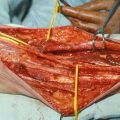
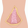
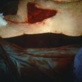
A midline incision from the xiphoid to several centimeters below the umbilicus can be used, or a bilateral subcostal incision with the patient in supine position. If the bulk of the tumor is concentrated at the tail of the pancreas, the patient may be placed in a right lateral position with the operating table tilted to the left, and a midline incision may be combined with an oblique incision from the left costal margin to the midline incision a few centimeters above the umbilicus. One starts with the midline incision first, and after exploration showing an absence of distant disease, one should be able to best judge the optimal placement of the oblique incision in providing the best exposure. For large tumors invading the diaphragm, the oblique incision may be extended in the corresponding intercostal space by dividing the costal margin. The semilateral position using a midline incision combined with an oblique incision to the left costal margin provides excellent exposure and facilitates the surgical maneuvers as the force of gravity works to the benefit of the surgeon when the left upper quadrant viscera are mobilized.
The gastrohepatic ligament (lesser omentum) is divided at its membranous, avascular portion near the liver to allow entry into the lesser sac. The superior border of the body of the pancreas is visualized. One can locate the hepatic artery coming off the celiac axis and coursing in a rightward direction above the body of the pancreas. Giving off the gastroduodenal artery, it enters the left side of the hepatoduodenal ligament as the proper hepatic artery, which soon bifurcates into the right and left hepatic arteries. The gastroduodenal artery bifurcates into the superior posterior pancreaticoduodenal artery and an anterior branch bifurcating into the superior anterior pancreaticoduodenal artery and the right gastroepiploic artery. The right gastric artery, usually arising from the left hepatic artery, may be divided, as well as the vein, as they come close to the lesser curvature of the stomach proximal to the pylorus. It is best, however, to avoid dividing the right gastric vessels or the gastroduodenal artery, as would be done in pancreaticoduodenectomy in order to expose the portal vein above the neck of the pancreas. In distal pancreatectomy, it is best to mobilize the tail of the pancreas and spleen first and then dissect around the body of the pancreas as needed in order to come well to the right of any palpable evidence of tumor.
Stay updated, free articles. Join our Telegram channel

Full access? Get Clinical Tree


