Disorders of Connective Tissue and Extracellular Matrix


Disorders of Connective Tissue and Extracellular Matrix


Fransiska Malfait

Anne De Paepe


THE CONNECTIVE TISSUE AND THE EXTRACELLULAR MATRIX

The connective tissue is the most abundant tissue throughout the body and serves both as a structural support and as a metabolic reservoir for tissue interactions. During recent years, the historical view on connective tissue as solely a supportive structure has been abandoned. It has become evident that the connective tissue actively participates in several important physiologic processes, including cell expression patterns and development, proliferation, differentiation, cell migration, and apoptosis, mainly acting through various cell signaling pathways. Three main connective tissues can be recognized: the loose or soft connective tissues (that embed organs and organ parts), the hard connective tissues (cartilage and bone), and blood. They consist of an extracellular matrix (ECM) and metabolically active cells that synthesize and maintain the ECM. Embryologically, connective tissue develops mainly from the mesoderm, the middle layer of the three-layered embryo. Mesodermal cells develop a loosely organized embryonic connective tissue, called mesenchyme, and differentiate further in specific cell types, including fibroblasts/fibrocytes, chondroblasts/chondrocytes, and osteoblasts/osteocytes. These cells synthesize and maintain the ECM in, respectively, the loose connective tissues, cartilage, and bone, according to the tissue’s specific needs. The ECM consists of four main components: elastic fibers and microfibrils, collagen fibers, proteoglycans, and glycoproteins.

Elastic fibers provide elasticity and resilience to all deformable tissues. They are abundantly present in the aorta, arterial vessels, lung, and, to a lesser degree, skin. They are composed of two ultrastructurally distinguishable components: an amorphous component, “elastin”, which contributes to the elasticity of the fiber, and a surrounding sheet of microfibrils. The microfibrillar component of the elastic fibers consists of fibrillin-rich microfibrils. Fibrillins are highly homologous glycoproteins, of which five isoforms have been described with the same overall arrangement of repetitive domains. Fibrillin-rich microfibrils interact with many other nonfibrillar components, which can be integral parts of the microfibrils or may associate with them. These microfibrils have a structural role and are functionally important in different processes such as microfibril assembly, elastogenesis, interactions with other ECM proteins, anchoring microfibrils to basement membranes and cell surfaces, and growth factor sequestration. For many of these molecules, however, the exact spatiotemporal relation and function still need to be addressed. Examples of nonfibrillar components include microfibril-associated glycoproteins, fibulins (a seven-member family of extracellular glycoproteins), and the latent transforming growth factor-beta-binding proteins (LTBPs), which are important players in the bioavailability of transforming growth factor-beta (TGF-β) in the ECM.

Collagens are the most abundant proteins in the body. They are trimeric proteins that are characterized by the presence of triple-helical domains. To date, 43 collagen genes have been described, the products of which combine to form at least 28 different collagen molecules. Most collagens form supramolecular assemblies, such as fibrils and networks, and the collagen superfamily has been divided in several subfamilies on the basis of their genomic structures, their assembly, and other structural features. The fibril-forming or fibrillar collagens, which include the collagen types I, II, III, V and XI, represent the most widespread and abundant class of collagens, and defects in these proteins are found in different heritable disorders of connective tissue (HCTD), such as osteogenesis imperfecta (OI), (type I collagen), Stickler syndrome (type II and XI collagen), and the Ehlers-Danlos syndromes (EDS) (type I, III, and V collagen). Fibrillar collagens are observed in tissues as long, highly ordered fibrils with a characteristic banding pattern. Type I collagen, a heterotrimer of two α1-chains and one α2-chain, is the major collagen type in the body and has a widespread tissue distribution. Type II and type XI collagens are predominantly found in cartilage. Type III collagen, a homotrimer consisting of three identical α1-chains, is an essential component of stretchable tissues such as the blood vessel walls, the gastrointestinal tract, the uterus, and the skin. Type V collagen is a minor fibrillar collagen that coassembles with type I collagen, and acts as a regulator of collagen fibril diameter through the retention of a noncollagenous amino-terminal domain of the proα1(V) collagen chain.1 Each fibrillar collagen has a central uninterrupted triple-helical domain with short nonhelical domains at the carboxy- and amino-terminal ends. The presence of glycine, the smallest amino acid, in every third position of each chain is a prerequisite for the formation of a stable collagen helix. The biosynthesis of fibrillar collagens in the fibroblast is a complex process and starts with the synthesis of soluble precursor molecules, procollagens. These contain globular amino- and carboxy-terminal propeptide extensions, called the N– and the C-propeptide. Intracellular association of three proα chains occurs through interaction and disulphide bonding at the C-propeptide. In this way, correct alignment of the growing polypeptide chain is obtained as required for formation and propagation of the triple helix from the C– to the N-terminal end of the molecule. During helix propagation, the proα chains undergo extensive enzymatic modifications (e.g., hydroxylation of prolyl and lysyl residues), which cease when the helix is formed. Mature triple-helical procollagen molecules are secreted into the extracellular
environment where they are converted to collagen by enzymatic removal of the N– and the C-propeptides. Individual collagen molecules spontaneously assemble in a nonenzymatic process to form fibrils and fibers, which are stabilized by covalent cross-linking.

Proteoglycans consist of a core protein to which short oligosaccharides or longer chains of glycosaminoglycans (GAGs) are covalently attached. There is a large variety of proteoglycans based on the types and lengths of GAGs as well as the sequence and length of the protein core. Chondroitin sulfate, keratan sulfate, heparan sulfate, and dermatan sulfate are four different forms of GAGs that consist of repeating disaccharide units. The sulfates that are present on the GAGs create a highly negatively charged environment that is hydrophilic. In addition to water, proteoglycans also bind cationic proteins, such as growth factors, resulting in a mechanism by which tissues can store growth factors in the ECM.

A large group of noncollagenous matrix glycoproteins make up a substantial part of the ECM. Important members of this group include thrombospondins (TSP), fibronectin, tenascins, laminin, and osteopontin. As “ground substance” of the ECM, they display a variety of functions in tissue morphogenesis and remodeling, including cell adhesion, cell cycle regulation, cell-matrix interactions, and interactions with growth factors and various other matrix molecules.


HERITABLE DISORDERS OF CONNECTIVE TISSUE


The Ehlers-Danlos Syndrome


Epidemiology and Criteria for Diagnosis

EDS comprise a clinically and genetically heterogeneous group of HCTD, of which the principal clinical features are due to varying degrees of tissue fragility of the soft connective issues, including the skin, ligaments, blood vessels, and internal organs. The prevalence of EDS is estimated to be approximately 1 in 5,000 births, with no racial predisposition.2 However, the incidence rises with increased physician awareness. The first attempts to classify EDS resulted in the Berlin nosology in 1986, in which 10 subtypes were recognized.3 Elucidation of the molecular basis of several types of EDS resulted in revision of this classification, established in 1997 as the Villefranche nosology4 that recognizes six subtypes, based on clinical characteristics, mode of inheritance, and the nature of the underlying biochemical and molecular defect(s). For each subtype, major and minor clinical diagnostic criteria were defined. Several EDS subtypes are caused by mutations in the genes for collagen type I (arthrochalasis type), type III (vascular type), or type V (classic type), or in the genes involved in the processing of type I collagen (kyphoscoliosis and dermatosparaxis type) (Table 69.1). In terms of management and counseling, it is very important to establish the correct subtype of EDS. Over the past few years, however, it has become clear that the Villefranche classification remains insufficient and that many patients present with overlapping forms of EDS, which cannot be classified unambiguously into one of the six recognized subtypes. Recent insights into the molecular and biochemical basis of some EDS variants call for a refinement of the Villefranche classification, which will likely improve genetic counseling of affected families.5


Clinical Characteristics


Cutaneous Manifestations

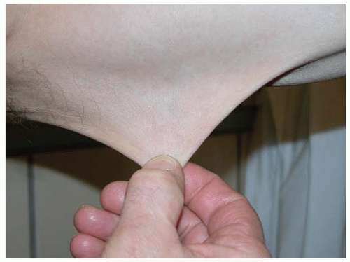
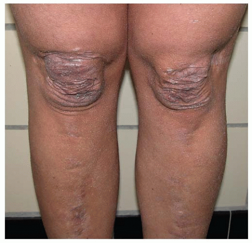
Cutaneous hyperextensibility (FIGURE 69.1) is one of the cardinal features of EDS in general, except for the vascular subtype. Skin extends easily and snaps back after release (unlike the lax and redundant skin observed in the cutis laxa (CL) syndromes). The skin is typically very smooth and velvety to the touch. It is also very fragile, as manifested by splitting of the dermis following relatively minor trauma. Wounds take a longer time to heal, and stretching of scars after apparently successful primary wound healing is characteristic. Scars become wide, with a “cigarette paper”-like or papyraceous appearance (FIGURE 69.2). Other dermatologic features in EDS include molluscoid pseudotumors, subcutaneous spheroids, and piezogenic papules. Less frequent cutaneous manifestations include acrocyanosis, chilblains, and, rarely, elastosis perforans serpiginosa.


Musculoskeletal Manifestations

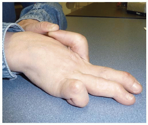
Joint hypermobility (FIGURE 69.3) should be assessed using the Beighton scale (Table 69.2), the most widely accepted grading system for the objective semiquantification of joint hypermobility.6 It may lead to major articular complications, such as habitual subluxation and dislocation of the joints most often affecting shoulder, patella, digits, hip, radius, and clavicles. When they occur, these dislocations usually resolve spontaneously or are easily managed by the patient. Many individuals with EDS suffer from chronic joint and limb pain, despite normal skeletal radiographs. Other problems related to the joint hypermobility are joint instability, foot deformities such as congenital clubfoot or pes planus, temporomandibular joint dysfunction, joint effusions, and osteoarthritis. At birth, uni- or bilateral dislocation of the hip may be present.


Bruising and Bleeding

The bleeding tendency primarily manifests itself as easy bruisability, bleeding of gums, prolonged bleeding after dental or surgical procedures, and prolonged menstrual episodes. Easy bruising is, to a variable degree, a common complaint in all subtypes of EDS. This bleeding diathesis is explained by an abnormal capillary structure with deficiency of normal perivascular collagen, resulting in poor support of cutaneous blood vessels that rupture when subjected to shearing forces. Laboratory investigation of platelet aggregation, clotting factors, and bleeding time in patients with EDS is usually normal. The Rumpel-Leede (or Hess) test may be positive, indicating capillary fragility.7,8 To perform this test, pressure is applied to the forearm with a blood pressure cuff inflated to between systolic and diastolic blood pressure for 10 minutes. After removing the cuff, the number of petechiae in a 5-cm diameter circle of the area under pressure is counted. Normally, <15 petechiae are seen. Fifteen or more petechiae indicate capillary fragility. In children with EDS, excessive bruising is often the presenting complaint to the pediatrician. If pronounced, it can cause suspicion of a hematologic disorder, a malignancy, or child abuse. Careful evaluation of the medical and family history, and rigorous clinical examination with special attention to skin features that are characteristic for EDS, are mandatory to distinguish between a HCTD and other causes of bruising.

Spontaneous arterial rupture leading to life-threatening complications is a hallmark of the vascular subtype of EDS. It may also occur in the rare individual with a severe form of the classic or the kyphoscoliotic type of EDS. Rupture of middle-sized
arteries has also been observed in individuals with a classic/vascular EDS-overlap phenotype, which results from mutations causing specific arginine-to-cysteine substitutions in COL1A1, the gene encoding the proα1(I) chain of type I collagen.9


Table 69.1 The Villefranche nosology for EDS


Clinical Manifestations


Type


Major Criteria


Minor Criteria


IP


Protein


Gene


Classic (gravis type I, mitis type II)


• Skin hyperextensibility


• Widened atrophic scarring


• Joint hypermobility


• Easy bruising


• Smooth and velvety skin


• Molluscoid pseudotumors


• Subcutaneous spheroids


• Muscular hypotonia


• Complications of joint hypermobility


• Surgical complications


• Positive family history


AD


Type V procollagen


COL5A1


COL5A2


Hypermobility (type III)


• Generalized joint hypermobility


• Mild skin involvement


• Recurring joint dislocations


• Chronic joint pain


• Positive family history


AD


mostly unknown


Tenascin-X (5%-10%)


mostly unknown


TNX-B


Vascular (type IV)


• Excessive bruising


• Thin, translucent skin


• Arterial/intestinal/


• Uterine fragility or rupture


• Characteristic facial appearance


• Acrogeria


• Early-onset varicose veins


• Hypermobility of small joints


• Tendon and muscle rupture


• Arteriovenous or carotid-cavernous sinus fistula


• Pneumo(hemo)thorax


• Positive family history, sudden death in close relative(s)


AD


Type III procollagen


COL3A1


Kyphoscoliotic (type VI)


• Severe muscular hypotonia at birth


• Generalized joint laxity


• Kyphoscoliosis at birth


• Scleral fragility and rupture of the globe


• Tissue fragility, including atrophic scars


• Easy bruising


• Arterial rupture


• Marfanoid habitus


• Microcornea


• Osteopenia


AR


Lysyl hydroxylase-1


PLOD1


Arthrochalasis (type VIIA&B)


• Severe generalized joint hypermobility with recurrent subluxations


• Congenital bilateral hip dislocation


• Skin hyperextensibility


• Tissue fragility, including atrophic scars


• Easy bruising


• Muscular hypotonia


• Kyphoscoliois


• Mild osteopenia


AD


Type I procollagen


COL1A1


COL1A2


Dermatosparaxis (type VIIC)


• Severe skin fragility


• Sagging, redundant skin


• Excessive bruising


• Soft, doughy skin texture


• Premature rupture of membranes


• Large herniae


AR


Procollagen-N-proteinase


ADAMTS-2


IP, inheritance pattern; AD, autosomal dominant; AR, autosomal recessive.


Modified from Beighton P, De Paepe A, Steinmann B, et al. Ehlers-Danlos syndromes: revised nosology, Villefranche, 1997. Ehlers- Danlos National Foundation (USA) and Ehlers-Danlos Support Group (UK). Am J Med Genet 1998;77(1):31-37.


FIGURE 69.1 Skin hyperextensibility in a patient with classic EDS.


Neuromuscular Involvement

Mild to moderate neuromuscular involvement is common in various types of EDS. Primary muscular hypotonia may cause delayed motor development and problems with ambulation. Muscle weakness, muscle cramps, musculoskeletal pain, and fatigue are frequent complaints. Muscle rupture can occur, especially in the vascular subtype of EDS.10 Cerebrospinal fluid leak has rarely been reported to cause postural hypotension and headache in individuals with classic EDS.11


FIGURE 69.2 Atrophic scarring on the knees and shins in a patient with classic EDS.


FIGURE 69.3 Severe joint hyperlaxity in a patient with EDS arthrochalasis type.


Cardiovascular Manifestations

Structural cardiac malformations are uncommon in EDS. Mitral valve prolapse (MVP) and, less frequently, tricuspid valve prolapse may occur. Stringent criteria should be used for the diagnosis of MVP. Severe cardiac valvular anomalies necessitating surgery at young adult age are observed in a rare autosomal recessive form of EDS, caused by mutations that lead to a total absence of the α2(I)-procollagen chain.12,13


Manifestations of Generalized Tissue Fragility

Manifestations of tissue extensibility and fragility may be observed in multiple organs. Patients with EDS may suffer from repetitive hernia, such as inguinal, umbilical, hiatal, or incisional hernia. In early childhood, recurrent rectal prolapse may be observed. Pregnancy-related manifestations include increased risk for extension of episiotomy incisions, tearing of the perineal skin, and prolapse of the uterus, and/or the bladder may occur after delivery. Prematurity, mostly due to premature rupture of the membranes, occurs more often than normal, especially in more severe forms of EDS and if the fetus is affected. Because of hypotonia, breech presentation is more frequent if the baby is affected and may lead to dislocation of the hips or shoulder of the newborn.


Vascular Ehlers-Danlos Syndrome

The vascular type of EDS is an autosomal dominant disorder that is caused by defects in the proα1(III) chain of type III collagen, encoded by the COL3A1 gene. Of all EDS subtypes, it has the worst prognosis because of the risk of potentially fatal vascular and intestinal complications.14,15 Unlike other types of EDS, the skin is not hyperextensible, but rather thin and translucent, showing a visible venous pattern over the chest, abdomen, and extremities.2,16 Excessive wrinkling and thinness of the skin over hands and feet may produce an old-looking appearance, referred to as “acrogeria.”

Excessive bruising is the most common sign and is often the presenting complaint. Other early manifestations include premature rupture of the membranes, congenital clubfoot or congenital
hip dislocation, inguinal hernia, pneumothorax, and recurrent joint dislocation or subluxation.17 Patients with vascular EDS often display a characteristic facial appearance, with prominent eyes (due to lack of subcutaneous adipose tissue around the eyes), a thin, pinched nose and small lips, hollow cheeks, and lobeless ears. Hypermobility is usually limited to the small joints of the hands.


Table 69.2 Beighton’s criteria for joint hypermobility


Joint/Finding


Negative


Unilateral


Bilateral


Passive dorsiflexion of the fifth finger >90 degrees


0


1


2


Passive flexion of thumbs to the forearm


0


1


2


Hyperextension of the elbows beyond 10 degrees


0


1


2


Hyperextension of the knees beyond 10 degrees


0


1


2


Forward flexion of the trunk with knees fully extended and palms resting on the floor


0


Present = 1


A total score of at least 5/9 defines hypermobility


Modified from Beighton P, Solomon L, Soskolne CL. Articular mobility in an African population. Ann Rheum Dis 1973;32(5):413-418.


The generalized vascular fragility largely dominates the clinical picture. Apart from excessive bruising and bleeding, it may cause precocious and severe varicosities and arterial rupture, potentially resulting in sudden death, usually in the third or the fourth decade of life. The vascular fragility affects large as well as small blood vessels, and bleeding may occur at every possible site in the body. The most common location of arterial bleeding is the abdominal cavity due to rupture of medium-sized arteries, such as the renal or splenic arteries, rather than of the aorta itself. Acute myocardial infarction due to coronary dissection or rupture is a rare complication. Some affected individuals may harbor predisposing lesions such as aneurysms or arteriovenous fistulae, but in other patients ruptures occur at locations that appear completely normal by angiography.2 Besides the vascular ruptures, dangerous internal complications such as spontaneous rupture of the bowel (usually the colon, sometimes the intestine), the gravid uterus, and hemorrhagic pneumothorax may occur.14,18,19,20,21

Although uncommon, vascular EDS is a cause of stroke in young adults. The mean age of intracranial aneurismal rupture, spontaneous carotid-cavernous sinus fistula, and cervical artery aneurysm is 28 years.22 Obstetrical complications are frequent and include vascular, intestinal, or uterine rupture, vaginal lacerations, prolapse of uterus and bladder, and premature delivery because of cervical insufficiency or fragility of the membranes. Patients with vascular EDS who are pregnant should be enrolled in a high-risk obstetrical program.

The clinical appearance of patients with vascular EDS may deviate from the typical picture, and especially the facial and cutaneous features, such as the acrogeria, may be very subtle or even absent. In the absence of a positive family history or a major vascular or intestinal complication, clinical diagnosis is difficult, especially in children.


From Genotype to Phenotype

EDS is extremely heterogeneous not only at the clinical but also at the molecular level. Abnormalities in the expression or structure of the fibrillar collagen types I, III, and V, as well as enzymatic abnormalities in the posttranslational modification and processing of these collagens, and more recently also in molecules involved in intracellular trafficking and extracellular organization of these molecules, have been identified in a number of EDS subtypes. Studies suggest that approximately 50% of individuals with classic EDS have an identifiable mutation in the COL5A1 or COL5A2 gene, the genes encoding the α1– and the α2-chain of type V collagen, respectively.23 However, since no prospective molecular studies of COL5A1 and COL5A2 have been performed in a clinically well-defined patient group, this number may underestimate the real proportion of classic EDS patients harboring a mutation in one of these genes.24

The vascular type of EDS results from mutations in the COL3A1 gene, encoding the proα1 chain of type III collagen. A wide spectrum of COL3A1 mutations has been identified, the majority of which are point mutations leading to substitutions for the obligatory glycine in the triple-helical region of the collagen molecule. Other types of mutations include splice-site mutations, partial gene deletions, and, rarely, mutations resulting in COL3A1 haplo-insufficiency.2,25 Parental mosaicism for COL3A1 mutations has been documented in vascular EDS26,27,28,29 and may explain unexpected recurrences in families in which a “new” dominant mutation has been identified.

Whereas defects in the genes encoding type I collagen generally result in different forms of OI, a subset of mutations in the COL1A1 or the COL1A2 gene result in specific subtypes of EDS. Mutations that cause loss of the N-proteinase cleavage site of either the proα1(I) or the proα2(I) collagen chain give rise to the arthrochalasia type of EDS. Mutations residing in the most amino-terminal part of the collagen type I triple helix result in a distinct EDS/OI overlap phenotype with variable joint hypermobility and bone fragility.30 In addition, some of these patients display increased vascular fragility with bleeding diathesis (personal observation). These mutations have been shown to interfere with removal of the N-terminal propeptide, even though the N-proteinase cleavage site remains intact.31 Specific arginine-to-cysteine (R-to-C) substitutions at different positions of the α1(I) helical domain have been associated with a range of EDS phenotypes, including a classic EDS-like phenotype in childhood, and a vascular EDS-like phenotype with increased risk for spontaneous arterial rupture in young adulthood.9,32,33 Finally, mutations leading to
total absence of the proα2(I) chain, due to a homozygous or compound heterozygous COL1A2-mutation, can be associated with severe cardiac valvular anomalies necessitating surgery at young adult age.12,13

Homozygous or compound heterozygous mutations in genes encoding enzymes involved in collagen biosynthesis have been documented in several autosomal recessive forms of EDS. Homozygous mutations in PLOD1 (procollagen-lysine, 2-oxoglutarate 5-dioxygenase or lysyl hydroxylase-1), the gene encoding lysyl hydroxylase-1, have been identified in patients with the kyphoscoliotic type of EDS.34 Lysyl hydroxylase-1 is required for the hydroxylation of specific lysine residues to hydroxylysines, which act as precursors for the cross-linking process that is essential for the tensile strength of collagen. A deficient activity of procollagen-N-proteinase due to homozygous and compound heterozygous mutations in the ADAMTS-2 gene is responsible for the dermatosparaxis type of EDS.35

Recently, several new EDS variants have been characterized, implicating genetic defects in molecules involved in processes as diverse as intracellular trafficking, enzymatic modification of ECM proteins, and cell-matrix interactions (Table 69.3). Total absence of tenascin-X (TNXB gene), a large ECM glycoprotein that plays a role in the maturation and the maintenance of the dermal collagen and elastin network, results in a classic EDS-like phenotype with extensive bruising, skin and joint hypermobility, and occasionally increased laxity of the genitourinary tract causing uterine and vaginal prolapse, increased risk for postpartum hemorrhage, progressive muscle weakness, and distal joint contractures.36,37,38 Deficiency of SLC39A13 (SLC39A13 gene), an intracellular zinc transporter, results in spondylocheirodysplastic EDS39, and deficiency of Ras- and Rab interactor 2 (RIN2), which is involved in Rab5-mediated early endosomal trafficking, results in an EDS-like “RIN2 syndrome.”40,41 Deficiency of galactosyltransferase-I (β4GALT7) and of dermatan-4-sulfotransferase 1 (CHST14), two enzymes involved in dermatan sulfate biosynthesis, causes the progeroid and the musculocontractural subtype of EDS, respectively.42,43,44 Furthermore, a zinc-finger protein (ZNF469), the exact function of which remains currently unknown, has been shown to be associated with brittle cornea syndrome, a condition with substantial phenotypical overlap with EDS kyphoscoliotic type,
that is primarily characterized by fragility of the cornea, leading to corneal rupture.45

Only gold members can continue reading. Log In or Register to continue

Stay updated, free articles. Join our Telegram channel

Jun 21, 2016 | Posted by in HEMATOLOGY | Comments Off on Disorders of Connective Tissue and Extracellular Matrix

Full access? Get Clinical Tree

Get Clinical Tree app for offline access