Differential Diagnosis of Thrombocytopenia


Differential Diagnosis of Thrombocytopenia


Joel S. Bennett

Charles S. Abrams


Thrombocytopenia is encountered frequently in clinical practice, engendering substantial and appropriate concern about its etiology and the consequent risk and prevention of bleeding. For example, in patients admitted to intensive care units (ICUs), approximately 30% to 50% manifest thrombocytopenia at some point during their ICU stay.1,2,3 In turn, the thrombocytopenia has been associated with increased bleeding, increased transfusion of blood products, more prolonged ICU stays, increased ICU and hospital mortality, and often extensive, usually fruitless, efforts to identify its etiology. In this chapter, we discuss the definition of thrombocytopenia, the lifespan of circulating platelets and mechanisms of platelet senescence, the risk of bleeding in thrombocytopenic patients, and mechanisms for the development of thrombocytopenia in clinical disorders. The salient points of this discussion are provided in Table 60.1.


DISTRIBUTION OF PLATELET COUNTS IN NORMAL INDIVIDUALS

The normal range for platelet counts in healthy individuals has been reported as 150,000 to 350,000/µL4 and is essentially the same regardless of the type of automated hematology analyzer used to measure the platelet count.5 As determined from an analysis of the cross-sectional Third National Health and Nutrition Examination Survey of the United States population from 1988 to 1994 (NHANES III), mean platelet counts of 26,327 individuals were higher in women than men, independent of the presence of iron deficiency, declined with age, especially after age 60, and were greater in non-Hispanic blacks as compared to non-Hispanic whites and Mexican Americans.6 Similar differences had been identified in a large cohort of men and women in Israel,7 in a population study of whites, Africans, and Afro-Caribbeans in London,8 and in 3,311 HIV-negative Ugandans.9 In a study of platelet counts in 12,517 individuals in 10 relatively isolated villages in the Ogliastra area of Sardinia, the prevalence of thrombocytopenia (defined as a platelet count <150,000/µL) in different villages ranged from 1.5% to 6.8%, and there was a progressive decrease in platelet number with aging.10 It is noteworthy that the prevalence of thrombocytosis also differed between villages and was inversely related to the prevalence of thrombocytopenia. Thus, the normal range for platelet counts varied from village to village, implying that the normal range for platelet counts can be influenced by genetic factors and is consistent with the finding of significant heterogeneity in the distribution of single-nucleotide polymorphisms among the villages. Genetic polymorphisms that affect the platelet count in a negative and positive fashion have been identified in the genes for interleukin-6 and the thrombopoietin receptor c-mpl.11,12,13 Although these differences in mean platelet count affect populations rather than individuals, and are relatively mild so that they are unrelated to the efficiency of hemostasis, it is important to be cognizant of the fact that there are differences in the normal range of platelet counts when analyzing the effect of medications, therapy, and devices that can affect the platelet count such as immunization.14

Although the clinical significance of severe thrombocytopenia is usually readily apparent, the nature and significance of borderline thrombocytopenia, that is, platelet counts between 100,000 and 150,000/µL, can be a vexing problem. With the advent of automated cell counting, asymptomatic individuals with incidentally discovered borderline thrombocytopenia are being identified with reasonable frequency. While some of these individual, by necessity, represent outliers of the normal distribution of platelet counts, the thrombocytopenia of others could represent an early manifestation of an unrecognized disease. In the absence of positive findings, many of these individuals are given a diagnosis of idiopathic autoimmune thrombocytopenia (ITP). To address the paucity of information about the fate of individuals with borderline thrombocytopenia, Stasi et al.15 prospectively studied 217 apparently healthy individuals incidentally discovered to have borderline thrombocytopenia as defined above. When followed for 6 months, two of the subjects were found to have myelodysplasia and a third systemic lupus erythematosus (SLE). The platelet counts of 23 others increased into the normal range for at least 3 months, whereas the platelet counts of 11 declined to <100,000/µL, and they were classified as having ITP. One hundred ninety-one subjects who continued to manifest borderline thrombocytopenia at 6 months were then followed for a median duration of 64 months. Thirteen subjects achieved stable platelet counts >150,000/µL and 109 continued to have stable borderline thrombocytopenia. Platelet counts of 12 subjects declined to <100,000/µL, and these subjects met the study’s criteria for ITP. Of the 11 subjects initially thought to have ITP, five subsequently developed autoimmune disorders. Overall, the probability of developing ITP in these subjects at 10 years was calculated to be 6.9%, and the probability of developing another autoimmune disorder at 10 years was 12.0%. However, the majority of subjects continued to manifest borderline thrombocytopenia without developing another disorder. Thus, it is likely that they simply represent the lower end of the normal platelet count distribution. Moreover, based primarily on the results of this study, as well as on the observations discussed above that platelet counts <150,000/µL are frequently found in apparently healthy individuals in some non-Western populations, the International Working Group of the Vicenza Consensus Conference set the threshold for the diagnosis of ITP at a platelet count of <100,000/µL.16
Nonetheless, we have encountered a number of patients with borderline thrombocytopenia who have either intermittently or progressively developed symptomatic thrombocytopenia consistent with a diagnosis of ITP. While it may be inappropriate to label all patients with borderline thrombocytopenia with a diagnosis of ITP when their thrombocytopenia is first discovered, these patients should be monitored to determine if ITP does eventually develop.


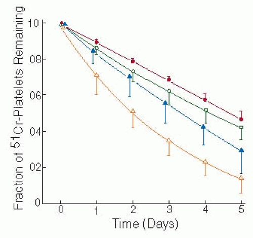


Table 60.1 Clinical implications of thrombocytopenia


Although the range of normal platelet counts in healthy individuals is 150,000-350,000/µL, the platelet counts of many individuals with borderline thrombocytopenia, that is, platelet counts of 100,000-150,000/µL, remain stable over time and most likely represent outliers of the normal platelet count distribution


The normal lifespan of circulating platelets is 7-10 d, but measured platelet lifespan decreases as the platelet count decreases


Most platelets are removed from the circulation because of senescence, but approximately 18% of platelet removal is random. It has been postulated that the function of randomly removed platelets is to support endothelial integrity


Approximately 30% of the normal platelet volume is present in the spleen. The amount of platelets sequestered in the spleen can increase with splenic enlargement


Platelets contain an endogenous apoptotic pathway that may be largely responsible for their senescence


Clinical experience suggests that a platelet count of 10,000/µL is a sufficient threshold for prophylactic platelet transfusions to minimize the risk of spontaneous bleeding in the absence of fever, infection, renal failure, and medications that impair hemostasis


Platelet counts of at least 50,000/µL are sufficient for most surgical procedures, but procedures involving the brain or eyes may require platelet counts >100,000/µL. Nonetheless, bleeding complications often do not correlate well with preprocedure platelet counts


Clinically relevant thrombocytopenia can result from decreased platelet production by bone marrow megakaryocytes, from accelerated platelet removal, or from platelet sequestration in an enlarged spleen


PLATELET KINETICS

Measurements of platelet survival in humans using autologous platelets labeled in vitro with either chromium-5117 or indium-11118,19 reveal that the lifespan of platelets in the circulation ranges from 7 to 10 days with a platelet turnover of 40,000 to 50,000/µL/day.20 111In complexed with 8-hydroxyquinolone (oxine) is currently the preferred radionuclide for these studies because of its shorter half-life, higher γ photon yield, and the greater affinity of the oxine complex for platelets.19,21 These features of111In-oxine have enabled the measurement of autologous platelet survival in patients with ITP who have platelet counts of <20,000/µL.22 Estimating platelet lifespan from the decrease in circulating platelet-associated radioactivity is accomplished by fitting the survival curve into one of several platelet survival models.23 The most commonly applied mathematical formulation is the multiple hit (or γ function) model developed by Murphy and Francis,24 based on the hypothesis that a platelet is repeatedly subjected to multiple external environmental insults or “hits” before it is removed from the circulation. However, the validity of this hypothesis has been questioned because of the observation that platelets contain an internal apoptotic pathway that may be largely responsible for their senescence25 (see below). Nonetheless, when the survival of autologous 51Cr-labeled platelets was compared in normal subjects and patients with stable thrombocytopenia due to bone marrow failure, platelet lifespan correlated with the platelet count, decreasing as the platelet count decreased (FIGURE 60.1).26 Based on a mathematical model in which a fixed platelet lifespan was reduced by a degree of random platelet destruction, the authors calculated that there was random destruction of approximately 7,100 platelets/µL/day.
This accounted for approximately 18% of the platelet turnover in normal individuals, while the remainder of platelet turnover occurred by senescent mechanisms. They also postulated that the shortened platelet lifespan they observed as platelet counts decreased resulted from an increase in the proportion of platelets removed by random processes and that the function of the randomly removed platelets is to support endothelial integrity.


FIGURE 60.1 Effect of platelet count on the lifespan of platelets labeled with 51Cr. Platelet lifespan was measured in normal individuals (closed circles, platelet lifespan 9.6 ± 0.6 days) and patients with bone marrow hypoplasia and stable platelet counts ranging from 50,000 to 100,000/µL (open circles, platelet lifespan 7.0 ± 1.5 days), platelet counts <50,000 (closed triangles, platelet lifespan 6.0 ± 1.7 days), and patients with more severe thrombocytopenia (platelet counts of 19,000 ± 6,000/µL) using labeled donor platelets (open triangles, platelet lifespan 3.4 ± 1.1 days). (Adapted from Hanson SR, Slichter SJ. Platelet kinetics in patients with bone marrow hypoplasia: evidence for a fixed platelet requirement. Blood 1985;66:1105-1109, with permission.)

Sixty to seventy percent of infused labeled platelets are recovered in the circulation of normal individuals, but this increases to nearly 100% when they are infused into individuals who have undergone splenectomy.27,28,29,30 Thus, there is a splenic platelet pool that amounts to approximately 30% of the platelet volume in normal individuals. The splenic pool is in dynamic equilibrium with the circulating platelet pool. For example, the splenic pool is depleted in parallel with the circulating platelet pool during platelet apheresis.31,32 Epinephrine infusions decrease splenic blood flow and the exchangeable splenic platelet pool and prolong the intrasplenic platelet transit time from its normal value of 10 to 12 minutes. This indicates that splenic blood flow is a major determinant of the size of the exchangeable splenic platelet pool.33 Moreover, because the intrasplenic transit time of platelets is 5 to 6 times greater than that of erythrocytes, it is likely that by virtue of their smaller size, platelets are required to traverse a complex pathway through the splenic cords.30


PLATELET SENESCENCE

Analyses of measurements of platelet survival suggest that the platelet lifespan is determined by a combination of random platelet removal and a nonrandom aging mechanism that appears to result in the removal of senescent platelets by the reticuloendothelial (RE) system. Although the factors responsible for platelet aging are not known with certainty, there is accumulating evidence that the intrinsic apoptotic pathway may act as an “internal timer” that regulates platelet senescence.25,34 Platelets contain several members of the antiapoptotic Bcl-2 family, as well as the proapoptotic proteins Bax and Bak.34,35 Mason et al.34 found that hypomorphic mutations of the antiapoptotic protein Bcl-xL caused thrombocytopenia in mice due to increased platelet clearance, whereas inactivating other antiapoptotic proteins, Bcl-2, Bcl-w, and Mcl-1, in platelets did not. Furthermore, treating mice with the BH3 mimetic ABT-737, which antagonizes Bcl-xL, caused acute thrombocytopenia. By contrast, absence of the proapoptotic proteins Bak and Bax was associated with a marked increase in platelet count and prolongation of the platelet lifespan.36 Since the half-life of Bcl-xL in platelets is shorter than Bak,34,37 it has been proposed that as the inhibitory effect of Bcl-xL declines, a threshold is reached, at which point uninhibited Bak is able to cause platelet apoptosis.25 Nonetheless, how platelet apoptosis could cause platelet removal by the RE system is still unclear. Apoptosis causes phosphatidylserine (PS) exposure on cell surfaces, which can be a signal for phagocytosis of the apoptotic cells. Although apoptosis causes PS exposure on the platelet surface, agonist stimulation also exposes PS as a component of platelet procoagulant activity by a pathway that is independent of the apoptotic mechanism.38 It remains to be established whether PS exposure plays a role in the clearance of senescent platelets, in addition to its role in supporting the formation of procoagulant complexes.

Only gold members can continue reading. Log In or Register to continue

Stay updated, free articles. Join our Telegram channel

Jun 21, 2016 | Posted by in HEMATOLOGY | Comments Off on Differential Diagnosis of Thrombocytopenia

Full access? Get Clinical Tree

Get Clinical Tree app for offline access