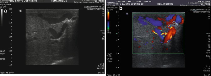
Fig. 9.1
Three-month-old girl with a soft-tissue mass. (a) Lesion is well limited with variable echogenicity. (b) Highly vascularized ≥5 vessels/cm2 are observed and (c) High-peak systolic Doppler shifts >2 KHz with low-resistive index mean (0.61)
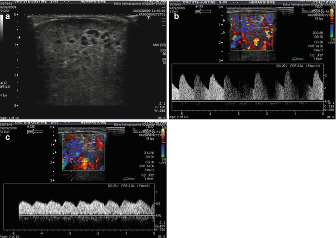
Fig. 9.2
Two-month-old female with intraparotid infantile hemangioma. (a) Ultrasound reveals a soft-tissue lesion with visible vessels. (b) Numerous arteries and veins are visible with high-systolic flow and (c) arterialization of the veins associated with arteriovenous fistula
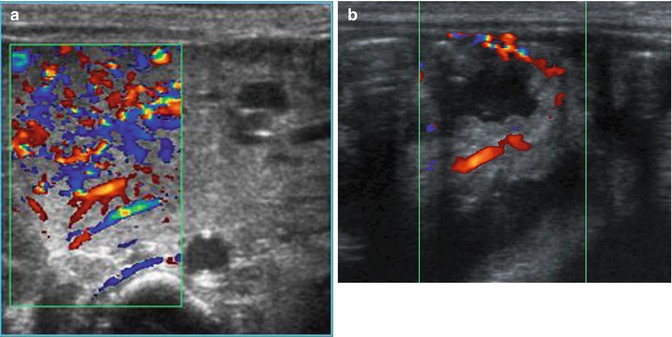
Fig. 9.3
Five-week-old female with severe intestinal bleeding required multiple transfusions. At physical exam, a segmental hemangioma of the leg was noticed. (a) Abdominal color ultrasound reveals a high-flow lesion in the head of pancreas and (b) in the small intestines. She was treated with octreotide, Cyklokapron, propanolol, and steroid with complete regression of the lesion
During the involuting phase, the lesion appears as a sonographically heterogeneous mass that becomes hyperechoic ultimately. The mass decreases in volume with a reduced number of vessels, but most of the time, the high Doppler shift persists in the remaining vessels.
On CT scan , during the proliferative phase, an infantile hemangioma shows a homogeneous mass with intense, persistent homogeneous enhancement, usually organized in a lobular pattern. During the involuting phase, CT scan shows a heterogeneous mass with less intense staining, as well as intralesional fibrofatty tissue.
MRI typically shows a well-defined, non-infiltrating lesion, with an intermediate signal intensity on T1-weighted sequences and increased signal intensity on T2-weighted sequences. Fast-flow vessels are identified by the presence of flow voids within and around the soft-tissue mass on spin-echo sequences and as high-signal intensity on gradient-recalled echo sequences. Perilesional edema should not be seen. The vessels with fast-flowing blood appear within and at the periphery of the mass. After gadolinium injection, high enhancement is observed. The MRI signal in involuting hemangiomas depends on the amount of fibrosis (hypointense on T1 and T2) and the quantity of fat (hyperintense on T1 and T2). The gadolinium enhancement decreases significantly with the involution [8–10] (Figs. 9.4, 9.5, and 9.6).
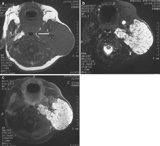
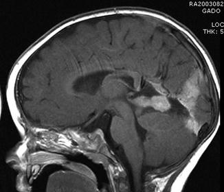
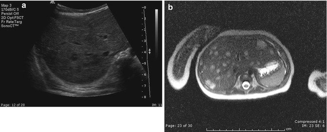
Fig. 9.4
Three-month-old child with left parotide infantile hemangioma. (a) Axial T1-weighted scan of the face shows a well-defined hypointense mass located in the parotid gland. Vessels are also identified by the presence of flow voids within the mass (arrow). (b) On axial T2-weighted scan with fat suppression, the mass is hyperintense. Flow void areas are still detected. No perilesional edema is visible, and (c) axial T1-weighted scan performed after fat suppression and gadolinium injection shows high enhancement of the lesion
Fig. 9.5
One-month-old male with hemiface infantile hemangioma. Sagittal brain contrast T1-weighted image with fat suppression shows an enhancement of the pineal, meningeal, and tentorial areas, confirming the IH diagnosis. One year later, the MR reveals the complete regression of these lesions
Fig. 9.6
Three-month-old male with multiple infantile skin hemangiomas. (a) Ultrasound reveals multiple hypoechoic lesions, and (b) axial T2-weighted image reveals multiple hyperintense lesions in the liver, confirming the IH diagnosis
Differential Diagnostic
Congenital hemangiomas
RICH and NICH share similar imaging findings. Most of these can display the same appearance as infantile hemangioma on US and CT or MR. Some differences like various-sized vascular aneurysms, intravascular thrombi (calcifications), more visible vessels, and venous ectasia are observed in these congenital hemangiomas. Microarteriovenous shunting is frequently identified and sometimes arterial aneurysms are present [11, 12] (Figs. 9.7 and 9.8).