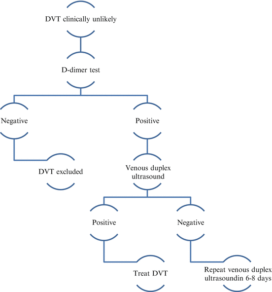
Figs. 1 and 2
Illustration of diagnostic approach for suspected DVT
3.1 The Wells Score
NICE recommend the use of the two-level DVT Wells score to estimate the clinical probability of DVT as shown in Table 1. Patients with a score of 2 points or more should be offered a venous duplex ultrasound scan carried out within 4 h of being requested. If a venous duplex ultrasound scan is not available within 4 h of being requested, a D-dimer test and interim 24 h dose of parental anticoagulation is suggested.
Clinical feature | Points |
|---|---|
Active cancer (treatment ongoing, within 6 months, or palliative) | 1 |
Paralysis, paresis or recent plaster immobilisation of the lower extremities | 1 |
Recently bedridden for 3 days or more or major surgery within 12 weeks requiring general or regional anaesthesia | 1 |
Localised tenderness along the distribution of the deep venous system | 1 |
Entire leg swollen | 1 |
Calf swelling at least 3 cm larger than asymptomatic leg | 1 |
Previously documented DVT | 1 |
An alternative diagnosis is at least as likely as DVT | −2 |
Clinical probability simplified score | |
DVT likely | 2 points or more |
DVT unlikely | 1 point or less |
3.2 Duplex Ultrasound
Venous duplex ultrasound has been widely adopted as the first line investigation for suspected DVT. It is able to identify DVT of the leg with a sensitivity of 96.5 % for proximal (above knee) DVT and 71.2 % for calf DVT, both with a specificity of 94 % (Goodacre et al. 2005).
A screening two-point ultrasound is often used in clinical trials to detect the possibility of DVT in asymptomatic patients. Compressibility of the deep veins at two points (the common femoral and the popliteal veins) can characterise the scan as normal (fully compressible), abnormal (non-compressible) or non-diagnostic (due to poor images). This two-point screening test has been shown to be sensitive and specific (Robinson et al. 1998). However it will not detect below knee or non-occlusive proximal (iliac) DVT. Abnormal and non-diagnostic examinations are usually followed up by a complete diagnostic venous duplex ultrasound which includes B-mode imaging for compressibility and visualisation of the thrombus, spectral display to determine flow direction and respiratory phasicity and colour-flow imaging to determine flow. Virtually all vascular labs use the inability to collapse a vein with probe pressure as the primary diagnostic criteria for DVT. A recent RCT showed that the two-point and whole leg strategies are equivalent in identifying DVT in symptomatic patients but despite this screening two-point ultrasound is not routinely used in clinical practice (Bernardi et al. 2008).
The venous duplex ultrasound can be repeated 6–8 days later in patients with persistent swellings, a positive D-dimer and an initial negative duplex ultrasound. Most DVT algorithms and duplex ultrasound protocols for DVT only include initial ultrasound evaluation of the proximal leg veins only even in symptomatic patients. This is largely based on out-dated perceptions that ultrasound examinations of the calf veins are inaccurate and such strategies are inefficient and not cost-effective. With the commonest site for DVT being the muscular calf vein, scanning the proximal veins will invariably miss a large proportion of DVT (Yoshimura et al. 2012; Zierler 2004).
The duplex examination can also be useful to help determine the cause of leg pain and swelling when a DVT is excluded. Intramuscular hematomas, Bakers cysts and varicose vein disease can mimic DVT and be identified on duplex ultrasound.
3.3 D-Dimer Test
In patients in whom DVT is suspected and with a low Wells score, a D-dimer test is suggested. The use of the D-dimer test to rule out DVT and remove the need for more expensive testing has increased in popularity over the last decade. D-dimers are degradation products produced as a result of plasmin on cross-linked fibrin indicating the development of a thrombus. There are however several other conditions that can elevate D-dimer results such as infection, myocardial infarction, malignancy, trauma and pregnancy which make its interpretation difficult (Schumann and Ewigman 2007).
D-dimer test can reliably exclude DVT with a 99 % negative predictive value (Wells et al. 2003). Patients with a high Wells score with a negative venous duplex ultrasound scan are recommended to have a D-dimer test. A negative proximal ultrasound scan and D-dimer can reliably exclude DVT. In practice where a D-dimer test will not provide a reliable result, duplex ultrasound remains as the most robust diagnostic tool.
3.4 Venography
Catheter-based contrast venography had traditionally been accepted as the reference diagnostic test for DVT before the widespread introduction of duplex ultrasound. Contrast venography was up until recently still widely used in clinical trials investigating anticoagulant therapies (Eriksson and Quinlan 2006). Because of its invasive nature, technical difficulty and cost it is not deemed suitable for routine clinical evaluation of suspected DVT (Zierler 2004). It is however still used for suspected pelvic and iliac vein DVT.
Magnetic resonance venography (MRV) is an alternative to catheter-based venography and has been shown to be highly accurate with sensitivies of 97 % and specificity of 100 % with excellent inter-observer variability for iliac, femoral and below knee DVT (Fraser et al. 2002; Spritzer et al. 2001). Compared with conventional catheter-based contrast venography, it is non-invasive and avoids ionising radiation. It is however expensive compared to venous duplex ultrasound and not available to patients with metal implants.
Computerised tomographic (CT) venography is similarly advantageous as MRV over duplex ultrasound but does involve ionising radiation and the use of intravenous contrast. It is reported to be 97 % sensitive and 100 % specific at determining lower limb DVT compared with duplex ultrasound (Frankel and Bundens 2014).
4 Management of Early DVT
4.1 Anticoagulation
The aim of treatment for DVT is to relieve the acute symptoms whilst reducing the risk of recurrent thrombosis and post-thrombotic syndrome. The initial anticoagulant regime for DVT can be a choice of either intravenous or subcutaneous un-fractionated heparin (UFH), low molecular weight Heparin (LMWH), Fondaparinux, Rivaroxaban and Apixaban. Vitamin K antagonists such as Warfarin can be initiated simultaneously with heparin to a target international normalised ratio (INR) of 2.0–3.0 In the UK LMWH in the form of Enoxaparin, Dalteparin or Tinzaparin are most frequently used in hospitalised patients whilst the target INR is reached. It is favoured because it can be administered by a single daily subcutaneous injection and is less likely to produce heparin related thrombocytopenia when compared to UFH. There are several LMWH on the market some of which are licensed for treatment and pharmacological VTE prophylaxis; prescribers must review local hospital guidance on which agent to use.
New oral anticoagulants such as Rivaroxaban and Apixaban are now recommended for use in the treatment of acute VTE in adult patients after review by NICE in 2012 and 2015 respectively. Both drugs have been shown to be clinically more effective and cheaper for patients requiring anticoagulation for longer than 12 months as they remove the need for regular monitoring and blood tests (Rivaroxaban for the treatment of deep vein thrombosis and prevention of recurrent deep vein thrombosis and pulmonary embolism). A recent meta-analysis of new oral anticoagulants (Dabigatran, Rivaroxaban, Apixaban and Edoxaban) showed that they significantly reduced the risk of stroke or systemic embolic events by 19 % when compared with warfarin (RR 0.81, 0.38–0.64, p < 0.0001) but was associated with greater risk of gastrointestinal bleeding (Ruff et al. 2014). No clinically relevant increases in major bleeding events were noted in the RE-COVER, RE-MEDY and the EINSTEIN trials that established the efficacy of dabigatran and rivaroxaban for the prevention of VTE (Sarrazin 2015).
Vitamin K antagonists include substances with a short (acenocoumarol), intermediate (warfarin, fluindione), or long (phenprocoumone) half-life. Because of the variable vitamin K content of food, a narrow therapeutic index, and several interactions with other drugs, treatment with vitamin K antagonists needs close monitoring. The safety their use can be increased by encouraging compliance, avoiding use of concurrent drugs with potential interactions, and restricting alcohol intake.
The duration of oral anticoagulant depends upon patient presentation, history of prior venous thromboembolism and condition of the patient after 6–12 months of therapy. The American College of Chest Physicians (ACCP) recommendations for duration are summarised in Table 2 but the optimal duration is still uncertain. Co-morbidities, family history, BMI and gender all need to be taken into account before stopping anticoagulation. Serial D-dimer testing to determine the risk of recurrent DVT has been shown to produce promising results in large cohort studies. Patients with abnormal D-dimer levels 1 month after discontinuation of anticoagulation were shown to have significantly higher incidence of recurrent VTE compared to patients with persistently negative D-dimers after stopping anticoagulation (Palareti et al. 2006, 2014).
Table 2


Recommended duration of anticoagulation (Kearon et al. 2008)
Stay updated, free articles. Join our Telegram channel

Full access? Get Clinical Tree


