Fig. 12.1
Basal cell carcinoma: cohesive clusters composed of small hyperchromatic epithelial cells
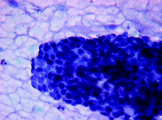
Fig. 12.2
Basal cell carcinoma: epithelial cells with scanty cytoplasm, indistinct cell borders and slight nuclear pleomorphism
A somewhat elongated, sometimes columnar, peripheral cells usually forming a palisade.
A slight nuclear pleomorphism.
Occasional mitotic figures may be seen.
Variants of basal cell carcinoma may show phagocytised melanin pigment (pigmented pattern), keratinised cell whorls within sheets of tumour cells (keratotic pattern) and elongated tumour cells or the presence of mucin. These findings reflect the histological variants of basal cell carcinoma and have no clinical significance. The differential diagnosis includes benign and malignant tumours of the sweat glands or of follicular apparatus.
BCC is the most common skin tumour and one which can be easily diagnosed by scrape specimen. Cytologic diagnosis has a high accuracy rate for margin control in BCC surgery. This application would especially be useful in areas with limited medical resources or in centres where Mohs micrographic surgery cannot be performed [8].
Squamous Cell Carcinoma (SCC)
Single cells due to loss of cohesion although solid cohesive fragments of tumour may be present in the less well-differentiated tumours
Bizarre cell shapes
Large irregular hyperchromatic nuclei
Necrosis
The presence of numerous neutrophils and sometimes eosinophils
The cancer cells with smaller nuclei and abundant cytoplasm may mimic normal squamous cells. In poorly differentiated tumours the cancer cells are smaller.
Keratinisation is a common and useful feature in diagnosis, especially when Papanicolaou staining is used (see Fig. 12.3). Some of the cells appear highly orangeophilic. Single-cell keratinisation is the most reliable indicator of squamous differentiation.
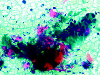
Fig. 12.3
Squamous cell carcinoma: small and loosely structured cluster composed of large keratinised (orangeophilic) and non-keratinised epithelial cells with bizarre shapes and irregular nuclei
The differential diagnosis includes:
Basal cell carcinomas: Contrary to BCC, squamous cancers do not form cohesive clusters of small cells. The cancer cells are much larger and dispersed and form only small, loosely structured clusters.
Regenerative squamous epithelium at the edge of an ulcer: The regenerative squamous cells are usually cohesive and nuclear atypia is not generalised or severe.
Solar keratosis and Bowen’s disease have some similar features but lack the extremes of cytoplasmic and nuclear pleomorphism found in squamous cell carcinoma.
Malignant Melanoma
Cellular smears.
Single-cell population (aggregates of cells are rare).
High nuclear/cytoplasmic ratio; large nuclei; prominent, often multiple large, irregularly shaped nucleoli and large intranuclear cytoplasmic inclusions.
Binucleated and multinucleated cells are frequent.
Cytoplasmic pigment.
Malignant melanoma is notorious for the great variability of its presentation and may mimic almost any malignant tumour. The common denominator of most (but not all) tumours is the presence of melanin pigment in tumour cells (see Fig. 12.4). Still, the diagnosis must be based on malignant features of the cells, because the melanin pigment phagocytised by macrophages may occur in a variety of skin disorders.
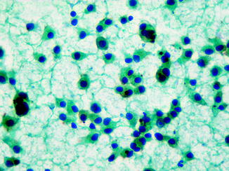
Fig. 12.4
Melanoma: cellular smear composed of a single-cell population with large nuclei, prominent nucleoli and occasional melanin pigment
Merkel Cell Carcinoma (Primary Neuroendocrine Carcinoma of Skin)
Cellular smears.
Dispersed tumour cells or loosely structured clusters, sometimes forming rosettes.
The cytoplasm is scanty and disintegrates easily with resulting debris and naked nuclei.
Granular nuclei.
Spherical, eosinophilic pink, cytoplasmic inclusions located near the nuclei or within nuclear indentations.
Paget’s Disease
Dispersed malignant cells
Large hyperchromatic nuclei with prominent nucleoli
Sebaceous Carcinoma
Moderately cellular smears
Clusters of irregular cells with cytoplasmic vacuoles rich in lipidic material
Central nuclei with prominent nucleoli
Smaller basaloid cells [9]
Dermatofibrosarcoma Protuberans
Clusters and dispersed uniform or slightly atypical spindle cells
Collagenous matrix
Storiform pattern [10]
Other Malignant Primary Tumours
Cytological findings of skin tumours, such as ectopic meningioma [11], Ewing’s sarcoma [12], malignant proliferating trichilemmal tumour [13], apocrine sweat gland carcinoma [14], Langerhans’ cell histiocytosis [15] and other uncommon lesions, are described by other workers.
Related posts:
 Epidemiology and Prevention of Cutaneous Tumors
Systemic Treatment of Primary Cutaneous Lymphomas
Photodynamic Therapy
Sunlight-Induced Skin Cancer in Companion Animals
Molecular Pathology of Melanocytic Skin Cancer
Cutaneous Squamous Cell Carcinoma: Focus on Biochemical and Molecular Characteristics
Epidemiology and Prevention of Cutaneous Tumors
Systemic Treatment of Primary Cutaneous Lymphomas
Photodynamic Therapy
Sunlight-Induced Skin Cancer in Companion Animals
Molecular Pathology of Melanocytic Skin Cancer
Cutaneous Squamous Cell Carcinoma: Focus on Biochemical and Molecular Characteristics
Stay updated, free articles. Join our Telegram channel

Full access? Get Clinical Tree


