Cystic Lesions
SIMPLE COLLOID CYSTS AND HEMORRHAGIC CYSTS
Cystic change within nodules of the thyroid gland is easily identified on ultrasound. Large cysts become the target for fine needle aspiration (FNA) as core biopsy is not suited for sampling of these lesions. Some cysts will not have any solid component and are typically of low clinical suspicion. However, many are ultrasonographically “complex cysts” with both cystic and solid components [1]. The cyst contents are often not of particular diagnostic value, and sampling of the solid component is mandated either through evacuation of the cyst contents followed by repeat aspiration of any residual solid mass or under ultrasound guidance [2, 3].
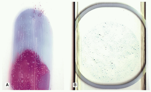
Simple colloid cysts of the thyroid are common due to excess colloid accumulation within inactive follicles. Aspiration of these cysts yields abundant colloid that is often watery in consistency and frequently lost in ThinPrep preparations but readily apparent on direct smears (Fig. 7.1). Although epithelium may be completely absent in these samples, usually some simple, bland, monolayered sheets of follicular epithelial are present and readily lead to a benign diagnosis. In the absence of follicular epithelium, but a tremendous abundance of colloid, it is accepted that a benign diagnosis can still be rendered.
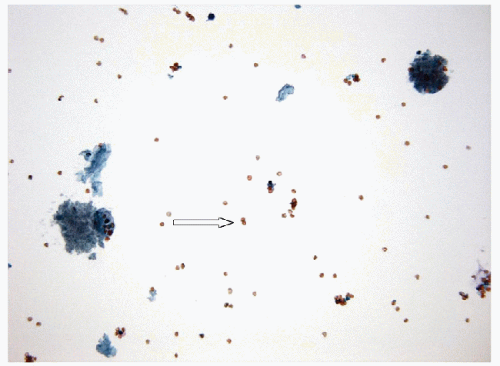
Hemorrhagic cysts, sometimes referred to as “degenerate cysts” or “degenerative changes,” are exceedingly common, much to the annoyance of both aspirators and pathologists alike. The majority of these hemorrhagic cysts presumably arise from ischemic involution of hyperplastic nodules due to overtaxing a tenuous vascular supply. Infarction generates a cystic cavity containing blood that undergoes lysis, leaving hemosiderin-laden macrophages, foamy macrophages with altered “old” erythrocytes that are characteristically resilient to common erythrolytic processes that readily lyze “young” circulating erythrocytes (Fig. 7.2) [4]. Follicular epithelium may be identified in the aspirate of some of these lesions, but these lesions are the genesis of one of the major controversies in the realm of FNA adequacy, as follicular epithelium is frequently absent.
It should be remembered that hemorrhagic cysts may have an iatrogenic origin due to hematoma formation resulting from previous FNA or
core biopsy and can be rapidly enlarging due to the rapid expansion of a hematoma and thereby frighten both the patient and clinician. However, the major differential diagnosis of concern with benign hemorrhagic cysts is that of a cystic papillary carcinoma [1]. Herein is the explanation of the need for follicular epithelium to evaluate the nature of the underlying lesion and the reason for the mandate to sample any solid components. Statistically, a hemorrhagic cyst is far more likely to be a benign lesion than a cystic papillary carcinoma but to establish the diagnosis pathologically one must carefully evaluate the epithelium for the presence or absence of diagnostic features of papillary carcinoma [5]. In case of doubt, or when the material is insufficient, molecular testing may add value [6].
core biopsy and can be rapidly enlarging due to the rapid expansion of a hematoma and thereby frighten both the patient and clinician. However, the major differential diagnosis of concern with benign hemorrhagic cysts is that of a cystic papillary carcinoma [1]. Herein is the explanation of the need for follicular epithelium to evaluate the nature of the underlying lesion and the reason for the mandate to sample any solid components. Statistically, a hemorrhagic cyst is far more likely to be a benign lesion than a cystic papillary carcinoma but to establish the diagnosis pathologically one must carefully evaluate the epithelium for the presence or absence of diagnostic features of papillary carcinoma [5]. In case of doubt, or when the material is insufficient, molecular testing may add value [6].
Hemorrhagic cysts also generate dilemmas due to the cytologic “atypia” that can be seen in the degenerate or regenerative epithelial cells [7]. Typically, these cells are large with preserved or even lowered nuclear/cytoplasmic ratio, but with large, rounded nuclei and often large nucleoli. The cells may resemble enlarged oncocytes. These cells typically form small simple flat sheets or are seen individually. It should be remembered that most thyroid carcinoma are well differentiated, and these suspect cells do not fit the description of the typical culprits. Thus, a degree of tolerance should be exercised when dealing with rare individual cells or small groups of these large “atypical” cells in a hemorrhagic cystic lesion.
THYROGLOSSAL DUCT CYSTS
These congenital lesions occur anywhere along the midline developmental tract of the thyroid from the base of the tongue to the mediastinum but are frequently in the region of the hyoid bone [8]. They are identified clinically as mass lesions and are biopsied to exclude malignancy [9, 10].
By FNA, these lesions are typically devoid of colloid or contain watery colloid appearing as a thin proteinaceous fluid. Cyst contents in the form of vacuolated macrophages are almost universal. In most aspirates of thyroglossal duct cysts, epithelial elements are completely lacking, and no specific diagnosis can be rendered. Follicular epithelial cells are usually absent, but when present, they are in the form of simple monolayered sheets with bland nuclear morphology, and when this occurs, it becomes impossible to determine, cytologically, if the thyroid gland (particularly the pyramidal lobe) or an extrathyroidal mass has been sampled. The most common epithelial elements found in FNA of thyroglossal duct cysts are squamous cells or ciliated respiratory epithelial cells. From the foregoing discussion, it is self-evident that a definitive diagnosis of thyroglossal duct cyst is not possible by cytologic features alone, and it is actually a clinicopathological correlation that suggests the diagnosis.
Stay updated, free articles. Join our Telegram channel

Full access? Get Clinical Tree


