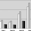
The current American Joint Commission for Cancer staging system for melanoma includes thickness, ulceration, and mitotic index as primary tumor factors for patients with stage I and II disease. Number and size of nodal metastases, presence of satellitosis and in-transit disease, and tumor ulceration status categorize patients with stage III disease. Presence and location of distant metastatic disease and increased lactate dehydrogenase level stratify prognosis in patients with stage IV disease. Factors predictive of sentinel lymph node positivity are also studied, particularly in patients with T1 melanomas, but are not always congruent with those predictive of survival.
Key points
- •
In the current (seventh) edition of the American Joint Commission for Cancer staging system, tumor thickness, ulceration status, and mitotic index categorize patients with stage I and II melanoma.
- •
Size (micro vs macro), number of nodal metastases, presence of satellitosis or in-transit disease, and primary tumor ulceration status categorize patients with stage III disease.
- •
For stage IV disease, location of distant metastases (skin/subcutaneous/nodal vs lung vs nonlung visceral) and increased lactate dehydrogenase levels categorize patients with M1 status.
- •
Other pathologic and clinical factors of the primary tumor have been reported with variable prognostic significance: presence of tumor-infiltrating lymphocytes, absence of regression, younger age, female gender, and extremity location have generally been associated with more favorable outcomes.
- •
Prognostic factors for sentinel lymph node positivity overlap but are not uniformly congruent with those for survival.
Introduction
The current (seventh) edition of the American Joint Commission on Cancer (AJCC) staging system for melanoma is now approaching its sixth year since publication and is based on 30,946 patients with stage I, II, and III melanoma and 7972 patients with stage IV melanoma from 17 major medical centers or independent cancer centers. This article reviews the notable changes in the current staging system compared with the last and discusses this in the context of clinical management of patients. Tumor and patient factors are discussed that do not feature in the staging system but that nonetheless carry important prognostic significance.
Melanoma staging system
The current AJCC staging system for melanoma is shown in Tables 1 and 2 .
| T Classification | Thickness (mm) | Additional Stratification |
| Tis | NA | NA |
| T1 | ≤1.00 | a: Without ulceration and mitoses <1/mm2 |
| b: With ulceration or mitoses ≥1/mm2 | ||
| T2 | 1.01–2.00 | a: Without ulceration |
| b: With ulceration | ||
| T3 | 2.01–4.00 | a: Without ulceration |
| b: With ulceration | ||
| T4 | >4.00 | a: Without ulceration |
| b: With ulceration | ||
| N Classification | Number of Metastatic Nodes | Metastatic Burden |
| N0 | 0 | NA |
| N1 | 1 | a: Micrometastasis (identified on SLN biopsy) |
| b: Macrometastasis (identified on clinical examination) | ||
| N2 | 2–3 | a: Micrometastasis (identified on SLN biopsy) |
| b: Macrometastasis (identified on clinical examination) | ||
| c: In-transit metastases/satellites without nodal metastases | ||
| N3 | 4+ metastatic nodes, matted nodes, or in-transit metastases/satellites with nodal metastases | — |
| M Classification | Site | Serum LDH Level |
| M0 | No distant metastases | NA |
| M1a | Distant skin, subcutaneous, or nodal metastasis | Normal |
| M1b | Lung metastases | Normal |
| M1c | All other visceral metastases | Normal |
| Any distant metastasis | Increased |
| Clinical Staging | T | N | M |
| 0 | Tis | N0 | M0 |
| IA | T1a | N0 | M0 |
| IB | T1b | N0 | M0 |
| T2a | N0 | M0 | |
| IIA | T2b | N0 | M0 |
| T3a | N0 | M0 | |
| IIB | T3b | N0 | M0 |
| T4a | N0 | M0 | |
| IIC | T4b | N0 | M0 |
| III | Any T | N > N0 | M0 |
| IV | Any T | Any N | M1 |
| Pathologic Staging | |||
| 0 | Tis | N0 | M0 |
| IA | T1a | N0 | M0 |
| IB | T1b | N0 | M0 |
| T2a | N0 | M0 | |
| IIA | T2b | N0 | M0 |
| T3a | N0 | M0 | |
| IIB | T3b | N0 | M0 |
| T4a | N0 | M0 | |
| IIC | T4b | N0 | M0 |
| IIIA | T1–4a | N1a | M0 |
| T1–4a | N2a | M0 | |
| IIIB | T1–4b | N1a | M0 |
| T1–4b | N2a | M0 | |
| T1–4a | N1b | M0 | |
| T1–4a | N2b | M0 | |
| T1–4a | N2c | M0 | |
| IIIC | T1–4b | N1b | M0 |
| T1–4b | N2b | M0 | |
| T1–4b | N2c | M0 | |
| Any T | N3 | M0 | |
| IV | Any T | Any N | M1 |
Stay updated, free articles. Join our Telegram channel

Full access? Get Clinical Tree


