COMPLICATIONS OF BRAIN IRRADIATION
Modern megavoltage radiotherapy produces minimal acute toxicity, including temporary alopecia, mild dermatitis, and a serous otitis media if the middle ear is included in the treatment field. These acute toxic effects typically are grade 2 or less. The focus of the treatment planning process is to minimize late toxic effects, which are uncommon but can be devastating. Late toxic effects occur predominantly in tissue with a slow turnover time and reflect a combination of direct cellular and indirect vascular injury.
The factors that predict a higher rate of complications include very young age, large total dose, large fraction size, large irradiated volume of normal tissue, and underlying medical conditions. In an unselected series of 134 patients who had undergone pituitary-hypothalamic irradiation over an 18-year period, 97% of whom had been treated to 45 to 50 Gy, complications attributable to radiotherapy occurred in 7 patients (2 second malignancies, 2 auditory deteriorations, and 3 vision deteriorations), underscoring the 5% or less risk from a carefully prescribed course of radiotherapy.64
ENDOCRINE COMPLICATIONS
Despite a low rate of cell proliferation in pituitary adenomas, the sequential assessment of hormone levels has demonstrated that the secretory functions of the pituitary gland are relatively susceptible to irradiation.65 The incidence of hypopituitarism was analyzed in a group of 165 patients who underwent cranial irradiation to total doses of 37.5 to 42.5 Gy in 2.25- to 2.65-Gy fractions.66 The analysis, which spanned a 10-year period, tested anterior pituitary function using insulin hypoglycemia or glucagon stimulation; thyrotropin-releasing hormone and luteinizing hormone–releasing hormone levels; and basal estimations of GH, prolactin, thyroid hormones, and testosterone or estradiol. Tests were repeated at 6- to 12-month intervals. Hyposecretion developed most rapidly for GH and least rapidly for thyroid-stimulating hormone; gonadotropins and
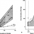
ACTH declined at an intermediate rate. The time required for 50% of patients with normal pituitary function to develop a hypofunctional pituitary was 1.2 years for GH, 3 years for luteinizing hormone and follicle-stimulating hormone (Fig. 22-1), and 3.2 years for ACTH. Almost two-thirds of patients exhibited the GH–luteinizing hormone/follicle-stimulating hormone–ACTH–thyroid-stimulating hormone sequence of hypopituitarism. In another report of 84 patients, radiotherapy resulted in local control of the majority of pituitary adenomas, but by 10 years of follow-up, the prevalence of hypopituitarism rose from 29% to 92%, suggesting that residual pituitary function is highly susceptible to long-term negation after radiotherapy. Patients should, therefore, be appropriately evaluated in follow-up and counseled regarding the almost universal need for hormone replacement after radiation.66
ACTH declined at an intermediate rate. The time required for 50% of patients with normal pituitary function to develop a hypofunctional pituitary was 1.2 years for GH, 3 years for luteinizing hormone and follicle-stimulating hormone (Fig. 22-1), and 3.2 years for ACTH. Almost two-thirds of patients exhibited the GH–luteinizing hormone/follicle-stimulating hormone–ACTH–thyroid-stimulating hormone sequence of hypopituitarism. In another report of 84 patients, radiotherapy resulted in local control of the majority of pituitary adenomas, but by 10 years of follow-up, the prevalence of hypopituitarism rose from 29% to 92%, suggesting that residual pituitary function is highly susceptible to long-term negation after radiotherapy. Patients should, therefore, be appropriately evaluated in follow-up and counseled regarding the almost universal need for hormone replacement after radiation.66
Related posts:


Stay updated, free articles. Join our Telegram channel

Full access? Get Clinical Tree


