Common Infections in Athletes—The Basics


Common Infections in Athletes—The Basics


Jessica Seidelman

Nicholas A. Turner


Introduction

Athletes are at increased risk for infections for several reasons. For example, athletes have frequent skin-to-skin contact and are often in crowded locker room environments. Infections can lead to adverse outcomes in athletes, including lost practice time and lost playing time. In extreme cases, infections can end athletic careers. While infections in athletes may not be entirely preventable, early recognition of infections can decrease the risk of adverse events.

This chapter summarizes common infections in athletes. We encourage athletes, parents, coaches, and team medical personnel to review the information herein. Essential information is included for each type of infection, including basic information and typical presentation. Infections highlighted in this chapter are presented because they are more common in athletes. In most instances, these infections are highlighted because they can be transmitted from athlete to athlete. As a result, this chapter also includes high-level information about strategies to prevent transmission in a locker room. Additional details on effective prevention strategies are provided throughout the remainder of the book.

Infections in this chapter are highlighted because (1) they are more common in athletes and/or (2) can be transmitted from athlete to athlete.


Localized Cutaneous Infections


Bacterial Skin Infections


DISEASE BASICS

Infections of the skin can be divided into two clinical categories: infections with pus (purulent) and infections without pus (nonpurulent).1 Purulent skin infections are associated with drainage of pus and/or abscesses, whereas nonpurulent skin infections have inflamed skin without
pus. These infections are typically caused by bacteria such as staphylococci or streptococci that are present on the skin. Infection occurs when there is a break in the skin that allows bacteria to enter.2

Streptococci are a group of gram-positive bacteria that cause several clinically important illnesses, including skin infections. They are broadly organized into two groups: pyogenic (beta-hemolytic) and alpha-hemolytic. For example, Streptococcus pyogenes (group A Strep) is a beta-hemolytic

Streptococcus that commonly causes skin infections and can even cause necrotizing soft-tissue infection. In general, streptococci lead to cellulitis and nonpurulent skin infections.

Staphylococcus aureus is another gram-positive bacterium that commonly causes skin infections.3 An estimated 30% of humans are colonized with S. aureus on their skin and in their nostrils.3,4 However, when the skin or mucosal barriers are disrupted, S. aureus causes significant infections. Methicillin-resistant Staphylococcus aureus (MRSA) are strains of S. aureus that are resistant to β-lactam antibiotics and therefore can be more difficult to treat. In general, S. aureus—and particularly MRSA—leads to purulent skin infections.

Skin + Pus = MRSA until proven otherwise.

Risk factors for developing a bacterial skin infection include (1) a recent injury to the skin through shaving or a cut, abrasion, or scrape suffered during athletic activities; (2) the presence of a viral or fungal infection such as herpes or athlete’s foot; or (3) chronic skin conditions such as eczema or psoriasis. Bacterial skin infections occur primarily in athletes with prolonged skin-to-skin contact, including wrestlers, rugby, judo, hockey, basketball, and football players. However, these infections can occur even in athletes with no risk factors and among athletes with little skin-to-skin contact.5 Recurrence of skin infections is common: 22% to 49% of patients with a skin infection report at least one prior episode.2

Risk factors for skin infections? Anything that disrupts the skin barrier.


TYPICAL PRESENTATION

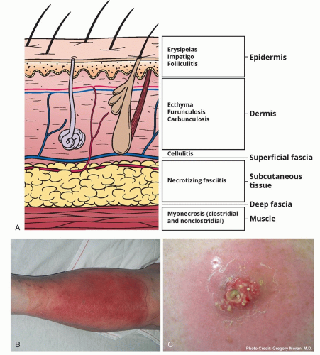
Bacterial skin infections can manifest in several clinical syndromes (Figure 1.1). One form of skin infection is called impetigo, which is characterized by well-defined, yellow crusted, scaling plaques. Erysipelas is a well-defined, erythematous plaque. Furunculosis occurs when multiple smaller boils (or pockets of pus) in the same area join together to form a large boil. Lastly, folliculitis presents as small pustules around hair follicles. Pseudomonas folliculitis or “hot tub folliculitis” can also occur in athletes who use hot tubs and whirlpools for rehabilitation.6 This infection typically presents as pruritic pustules that emerge on the skin that was submerged in the whirlpool. More specifically, the affected area typically involves skin covered by the bathing suit.6

The most common symptom of a bacterial skin infection is a dull ache or pain at the area of involvement. Other symptoms include swelling, warmth, or redness. These symptoms typically worsen with the redness spreading over a period of hours to days. The onset of skin infections can be gradual or sudden. Itching is not a common symptom. Fever and chills can accompany bacterial skin infections but are not always present. That is, bacterial skin infections may occur in the absence of fever.


TRANSMISSION

S. aureus can spread through contact with pus or drainage from an infected wound, skin-to-skin contact with an infected person, or indirectly through a contaminated environment. These routes of transmission are common in the athletic setting due to the number of athletes
sharing the training facility and frequent, close interaction. Outbreaks of S. aureus skin infections have occurred in football, wresting, rugby, and basketball athletes, among others.7,8,9,10 In one outbreak among fencers, sharing of equipment was linked to transmission of MRSA.8


Figure 1.1 A, Skin infections and skin structures involved. B, Cellulitis. C, Abscess. (B, Reproduced with permission from: Berg D, Worzala K. Atlas of Adult Physical Diagnosis. Philadelphia: Lippincott Williams & Wilkins; 2006. Copyright © 2006 Lippincott Williams & Wilkins. C, Photo by Gregory Moran, MD.)


PREVENTION MEASURES

Treatment typically includes antibiotic therapy and drainage of pus if present (typically via an incision and drainage or “I&D”). Skin lesions should be properly covered until they are healed.
Implementing therapy, covering the infection, and promptly removing infected athletes from contact with other athletes (ie, “isolation”) can help decrease the number of missed practices and risk of transmission.5

If pus is present, it needs to be drained.

In general, athletes should not share equipment or personal items such as towels, razors, knee/elbow pads, or other athletic gear.11 Athletes with skin infections should also avoid common areas (eg, weight rooms) and the hydrotherapy pools until the infection is controlled and can be covered. If the infected area cannot be covered, athletes should not participate in team activities such as practice and competition until the lesions heal.

For the prevention of Pseudomonas folliculitis, athletic trainers should ensure adequate chlorine levels and proper pH (7.0-7.4) in pools and whirlpools.5 In addition, if trainers suspect that a pool has been exposed to an infected athlete, the pool should be drained and cleaned.5 See Chapter 4 for more details about routine maintenance of hydrotherapy pools.


Herpes Gladiatorum


DISEASE BASICS

Herpes simplex virus (HSV) is classified into two broad types, HSV-1 and HSV-2. Overall, HSV infection is extremely common. Seroprevalence studies indicate that >50% of US adults have been exposed to HSV-1 and >15% exposed to HSV-2.12

Herpes gladiatorum is a viral skin infection caused by HSV. Nearly all reported outbreaks of herpes gladiatorum involve HSV-1, which is the same virus responsible for nearly 80% of herpes labialis cases (cold sores). HSV-2 causes the majority of genital herpes infections, but neither virus is strictly limited to one body site.13 Since the virus gains entry to the body via small breaks in the skin, athletes involved in contact sports are at increased risk.13 While the majority of outbreaks have been described among wrestlers, transmission between rugby players has also been described (informally called “scrum pox”).13,14,15,16 The same mechanisms of transmission—frequent abrasions and skin-to-skin contact between athletes—put athletes at risk.

HSV most commonly causes cold sores. Herpes gladiatorum occurs when HSV enters breaks in skin.


TYPICAL PRESENTATION

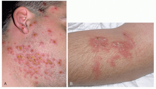
Herpes gladiatorum may be confused with other bacterial skin infections, including impetigo or folliculitis, leading to delayed diagnosis and ongoing transmission. The incubation period for herpes gladiatorum ranges from 4 to 11 days after exposure. Some people experience numbness at the site of inoculation prior to the development of skin lesions. Eventually a papulovesicular rash (small, fluid-filled spots) develops, usually in a localized distribution corresponding to areas of direct skin-to-skin contact (Figure 1.2).17 The rash is often painful and may be accompanied by a low-grade fever and malaise. After 7 to 10 days, the lesions crust over and resolve. Rarely, scarring or postinflammatory hyperpigmentation persists at the site. After the initial infection resolves, the virus persists in a latent state within sensory neurons. As a result, herpes infections can recur. Recurrence may be triggered by stress, other illnesses, sun exposure, or trauma.


Figure 1.2 Herpes gladiatorum involving face (A) and more localized involvement of arm (B). (Part A, Reprinted with permission from Craft N, Fox LP, Goldsmith LA, et al. VisualDx: Essential Adult Dermatology. Philadelphia, PA: Wolters Kluwer Health; 2010. Part B, Reprinted with permission from McDonagh DO, Micheli LJ, Frontera WR, et al. FIMS Sports Medicine Manual. Philadelphia, PA: Wolters Kluwer Health; 2011.)

Ocular involvement is a rare but serious complication of herpetic skin infection. Herpes keratitis (infection of the cornea) can occasionally result in blindness. Prompt evaluation by an ophthalmologist is warranted for cases involving the eye.


TRANSMISSION

HSV is transmitted by direct physical contact and may enter the skin through small abrasions or breaks. Viral shedding is most active when lesions are present; however, shedding may also occur for a few days before any visible rash develops. Outbreaks related to contact sports such as rugby or wrestling are common.13,14 Epidemiologic studies have yet to demonstrate a clear role for indirect transmission through contamination of training mats or shared gym equipment, but disinfection of equipment is still strongly recommended.18


PREVENTION MEASURES

Athletes with active herpes skin lesions should be excluded from competition until the skin lesions have dried and fully crusted over. Antiviral agents (acyclovir, valacyclovir, or famciclovir) inhibit replication of the herpes virus and may be prescribed to shorten the course of infection. For individuals with frequently recurrent herpes simplex outbreaks, the same antiviral agents may be prescribed for suppression. The determination of when an athlete is no longer contagious is best made by a physician after examining the lesions. In general, all lesions should be fully crusted over and no new lesions occurring before clearance to return to contact sports.

Although the role for indirect transmission of herpes simplex remains unclear, HSV can survive on various surfaces. Consequently, some facilities disinfect surfaces as a precaution, particularly in the setting of outbreaks.


Tinea


DISEASE BASICS

Tinea refers to superficial fungal skin infections caused by dermatophytes. The dermatophytes contain three genera of fungi: Microsporum, Epidermophyton, and Trichophyton.19 Dermatophyte infections are common and can affect nearly any body site. In one recent US survey, tinea accounted for >25% of skin infections among athletes.20 Athlete’s foot refers to one of the most common sites of involvement. Warm, damp conditions produced by occlusive footwear place football and soccer players at particularly high risk.21 Transmission via direct contact at other body sites has also been reported, mostly among contact sports participants such as wrestlers.22 Compared to bacterial skin and soft-tissue infections, tinea infections are less invasive and severe but usually require longer courses of therapy.


TYPICAL PRESENTATION

As tinea can affect multiple body sites, specific names have been given to each site of infection: tinea capitis involves the scalp, tinea unguium: the nails (also called onychomycosis), tinea pedis: the feet, tinea corporis: the trunk, tinea faciei/barbae: the face/beard, and tinea cruris: the inguinal region (groin).19

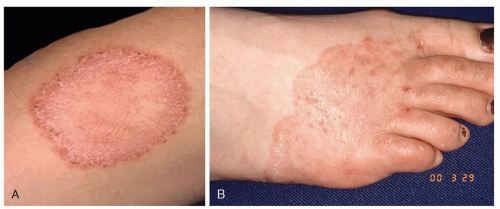
While appearance may vary by site, most tinea infections present as a localized scaly, erythematous rash (Figure 1.3). Most are pruritic. The leading or surrounding edge may be slightly elevated, referred to by the common name “ringworm.” Distribution often favors moist flexural regions (skin creases) of the body. Onychomycosis, or involvement of the nails, results in thickened, brittle, sometimes discolored nails.

The diagnosis of tinea is often made by appearance alone. In cases where uncertainty exists, a Wood lamp examination can confirm infection due to Microsporum, which fluoresces green.19 A skin scraping can also be performed for KOH prep and dermatophyte culture. For onychomycosis, fungal culture of nail clippings can confirm the diagnosis.

Treatment varies by body site. Topical therapy is effective for tinea pedis, tinea cruris, and tinea corporis. Allylamines (eg, terbinafine) and azoles (eg, clotrimazole, econazole, miconazole) are first-line therapies and readily available over the counter.23 Successful
treatment usually requires at least 2 to 6 weeks, with many providers recommending continuation for 1 to 2 weeks after visual resolution of the lesion to minimize risk of recurrence. Tinea capitis and onychomycosis respond poorly to topical therapies. Oral, systemic therapies such as griseofulvin or terbinafine are commonly used for tinea capitis. Treatment duration ranges from 6 to 12 weeks. Onychomycosis usually requires 3 to 6 months of oral terbinafine or occasionally itraconazole.


Figure 1.3 Tinea corporis (A) and tinea pedis (B). (Part A, Reprinted with permission from Gru AA, Wick M. Pediatric Dermatopathology and Dermatology. 1st ed. Philadelphia, PA: Wolters Kluwer Health; 2018. Part B, Reprinted with permission from Chung EK, Atkinson-McEvoy LR, Lai NL, Terry M. Visual Diagnosis and Treatment in Pediatrics. 3rd ed. Philadelphia, PA: Wolters Kluwer Health; 2014.)


TRANSMISSION

Damp conditions produced by occlusive footwear likely account for the increased risk of tinea pedis seen among soccer and football players.21 Tinea can also be spread by direct skin-to-skin contact, as outbreaks have been reported among athletes.18 Finally, tinea pedis may be acquired or spread via shower floors in shared bathing facilities.24

Tinea pedis (athlete’s foot) can be acquired from shower and locker room floors.


PREVENTION MEASURES

As tinea often favors moist body sites, removal of sweaty clothing, shoes, and socks promptly after activity may help reduce risk. Similarly, complete drying after showering may help to reduce susceptibility to tinea in flexural surfaces. Wearing sandals in the hydrotherapy and shower rooms can help athletes avoid exposure.


Central Nervous System Infections


Aseptic Meningitis


DISEASE BASICS

Aseptic meningitis refers to inflammation of the lining around the brain (meninges) without any bacterial growth on cerebrospinal fluid (CSF) culture. Because aseptic meningitis may closely resemble bacterial meningitis (see below), diagnosis is not always straightforward and often requires lumbar puncture (spinal tap) to rule out potentially life-threatening causes of meningitis. The majority of cases are caused by viral infection, particularly the enteroviruses. However, certain medications (especially nonsteroidal anti-inflammatory agents or NSAIDs) can cause aseptic meningitis as well.25,26,27 For particular regions of the United States, tickborne diseases such as Rocky Mountain spotted fever (caused by Rickettsia) can also cause an aseptic meningitis (see Table 1.1). Fortunately, if other more serious causes can be ruled out, aseptic meningitis is generally self-resolving and requires only supportive care.

Diagnosis of meningitis typically requires a lumbar puncture (“spinal tap”).

Outbreaks of aseptic meningitis have been reported among athletes.28,29 Close contact among athletes and sharing of water bottles are presumed to put athletes at increased risk of enteroviral transmission.30 The tendency for enteroviral transmission to peak in summer may also correspond with training camps and other preseason activities.31


Table 1.1 Causes of Aseptic Meningitis


Type of Aseptic Meningitis


Example Causes


Infectious causes


Common:


Enterovirus


Herpes simplex virus


Varicella zoster virus


Less common/rare:


Arboviruses


St Louis encephalitis virus


Eastern equine encephalitis virus


West Nile virus


Coxsackievirus


Epstein-Barr virus


HSV


HIV


Measles


Mumps


Influenza


Rocky Mountain spotted fever (Rickettsia)


Ehrlichia


Leptospirosis


Syphilis


Tuberculosis


Mycoplasma


Neuroborreliosis


Medication-related


Nonsteroidal anti-inflammatories (NSAIDs)


Azathioprine


Carbamazepine


IVIg (immune globulin)


Autoimmune


Sarcoidosis


Lupus


Behçet disease


Adapted from Hasbun R. The acute aseptic meningitis syndrome. Curr Infect Dis Rep. 2000;2(4):345-351 and Kupila L, Vuorinen T, Vainionpää R, Hukkanen V, Marttila R, Kotilainen P. Etiology of aseptic meningitis and encephalitis in an adult population. Neurology. 2006;66(1):75-80.


TYPICAL PRESENTATION

Aseptic meningitis can closely resemble bacterial meningitis. Headache, fever, neck stiffness, and photophobia are common. Presence of sore throat/pharyngitis or rash may hint at a viral cause, but the presence of these symptoms cannot reliably rule out other causes. Enterovirus has a strong seasonal trend, with the majority of cases occurring in summer.26 Lumbar puncture is usually required to rule out bacterial meningitis. Care is supportive, and recovery is spontaneous once more serious causes (eg, bacterial meningitis) are ruled out.


TRANSMISSION

While medication-related aseptic meningitis is not communicable, enterovirus is transmissible through a variety of mechanisms. Enterovirus has been detected in respiratory secretions, blister fluids from rashes, and saliva. Enterovirus is also capable of survival on surfaces touched by infected individuals, so indirect transmission via touching contaminated surfaces can occur as well. Poor hand hygiene and sharing of water bottles have been implicated in some of the reported outbreaks among team athletes.29

Viruses that cause aseptic meningitis can be spread by sharing water bottles.


PREVENTION MEASURES

There are no specific vaccinations for enterovirus, the most common viral cause of aseptic meningitis. Based on modes of transmission and a high propensity for survival on surfaces, good hand hygiene and avoidance of sharing water bottles with infected individuals can reduce risk of infection.


Bacterial Meningitis


DISEASE BASICS

Bacterial meningitis is a serious infection that can be caused by a variety of pathogens. Streptococcus pneumoniae, Haemophilus influenzae, and Neisseria meningitidis are three of the most common causes in adults.32 While any can be lethal, N. meningitidis is the most feared due to its particularly high mortality rate, extremely rapid progression, and tendency to occur in outbreaks among young adults. Risk for meningococcal meningitis occurs in two peaks: one in the first year of life and a second peak between the ages of 15 and 24 years. This second peak is the most likely to affect young athletes.

Bacterial meningitis is a medical emergency requiring rapid treatment with antibiotics. Although vaccination against N. meningitidis is now recommended for all adolescents and required by most colleges, awareness of the disease remains relevant for any sport involving young athletes staying in close quarters.

Bacterial meningitis is a medical emergency.


TYPICAL PRESENTATION

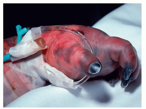
Meningitis classically presents with fever, headache, neck stiffness, and confusion—although fewer than half of patients will have all of these signs at the time of first presentation.33 Some patients with N. meningitidis will develop a characteristic rash; in severe cases, it can lead to gangrene of the extremities as sepsis progresses (Figure 1.4). Lumbar puncture should be undertaken without delay in order to confirm the diagnosis, unless the patient has evidence of high intracranial pressure or a focal brain lesion. Final diagnosis is confirmed by culture, but polymerase chain reaction (PCR) testing is increasingly used to provide rapid preliminary identification of pathogens. Empiric antibiotics (vancomycin plus ceftriaxone for most otherwise healthy adults) should be started immediately, ideally after blood and CSF cultures are obtained, as mortality climbs by the hour without treatment.


Figure 1.4 Gangrene of extremities in severe meningococcal sepsis. (Reprinted with permission from Kline-Tilford AM, Haut C. Lippincott Certification Review: Pediatric Acute Care Nurse Practitioner. 1st ed. Philadelphia, PA: Wolters Kluwer Health; 2015.)


TRANSMISSION

N. meningitidis may colonize the nose and upper throat of otherwise healthy individuals. Colonization converts to infection when these bacteria attach and invade through mucosal surfaces, leading to meningitis. Since N. meningitidis is found in saliva and respiratory secretions, it can be spread by kissing, coughing, or sharing of cups/eating utensils. Outbreaks are well reported in dense settings such as college dormitories.


PREVENTION MEASURES

Vaccination is highly effective, currently recommended for all adolescents, and generally required prior to attending college in the United States. There are several different vaccines available. The most widely used meningococcal conjugate vaccine (MCV4) protects against four common strains, but not meningitis B. MCV4 is recommended for all first-year college students residing on campus, all military recruits, individuals living with human immunodeficiency virus (HIV), those with asplenia or a complement deficiency, or those who may contract meningitis through travel or exposure to an outbreak. Because meningitis B is still a relatively common cause of outbreaks in the United States and abroad, separate vaccination with N. meningitidis group a polysaccharide antigen is recommended for individuals who have had their spleen removed, those with complement deficiency, or those with exposure to meningitis B through either occupation or an outbreak.

Antibiotic prophylaxis can be given to unvaccinated close contacts of patients with N. meningitidis to reduce risk of disease.


Gastrointestinal Infections


Hepatitis A


DISEASE BASICS

Hepatitis A is an acute, self-limiting viral infection involving the liver. Hepatitis A is typically acquired through contaminated food. Although overall incidence declined significantly following introduction of an effective vaccine in 1995, hepatitis A continues to occur in outbreaks
spanning multiple US states.34,35 Although several recent outbreaks predominantly occurred among urban homeless populations, transmission via infected food handlers or contaminated water is well-documented.36 Consequently, transmission via contaminated foods at restaurants or banquets poses a potential threat to traveling sports teams.

Outbreaks of hepatitis A are increasingly common.


TYPICAL PRESENTATION

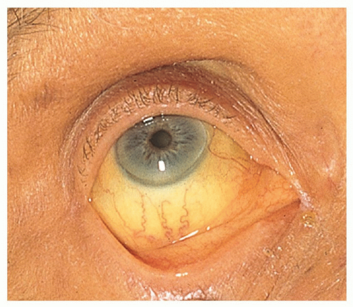
Disease severity varies with age. Childhood hepatitis A infection is usually asymptomatic. In contrast, adults are much more likely to experience fever, fatigue, jaundice (Figure 1.5), and right upper quadrant abdominal pain. Most adults recover spontaneously within 2 months, but some experience prolonged or waxing/waning disease occasionally lasting up to 6 months. Laboratory testing generally reveals significant elevations of the transaminases (aspartate aminotransferase [AST] and alanine aminotransferase [ALT], often in the thousands) and bilirubin. Diagnosis is confirmed by the presence of anti-HAV IgM antibodies.

There are no antiviral treatments for hepatitis A. Care is supportive and includes avoidance of alcohol or other hepatotoxins during convalescence.


TRANSMISSION

Transmission occurs via fecal-oral route (contaminated food/water), sexual contact, and illicit drug use. In developed countries with widespread access to safe drinking water, hepatitis A is most commonly acquired either through consumption of contaminated food or water during international travel or through consumption of contaminated foods. Virus may be shed for 1 to 2 weeks prior to symptomatic disease.


PREVENTION MEASURES

Receipt of hepatitis A vaccine leads to immunity. If exposure occurs prior to vaccination, the vaccine can still be given to reduce risk of disease if given within 2 weeks of exposure.
Preexposure vaccination is recommended for adults traveling to certain regions with high rates of hepatitis A or for individuals with known liver disease. For the prevention of foodborne outbreaks, cooking to temperatures >185°F for >1 minute inactivates the virus.


Figure 1.5 Scleral icterus/jaundice. (Used with permission from Bickley LS. Bates’ Guide to Physical Examination and History Taking. 12th ed. Philadelphia, PA: Wolters Kluwer Health; 2016.)


Hepatitis B


DISEASE BASICS

Hepatitis B virus (HBV) causes an infection of the liver. HBV is a global health issue; more than 250 million people are infected with HBV in the world.37 Approximately 80% of HBV infection in adults are clinically silent. While 20% of patients are symptomatic, fewer than 0.5% develop fulminant hepatitis. Almost 95% of adults with HBV infection achieve viral clearance, while the remaining 5% become chronic hepatitis B carriers.38,39


TYPICAL PRESENTATION

Symptoms from acute hepatitis B begin an average of 90 days after exposure (range 60-120 days).40,41 Symptoms can include fever, abdominal pain, fatigue, dark urine, loss of appetite, clay-colored bowel movements, nausea, vomiting, joint pains, muscle aches, or jaundice (yellowing of the eyes).42 Acute hepatitis B symptoms typically last for a few weeks but can persist up to 6 months.40,41 In contrast, chronic hepatitis B infection is typically asymptomatic. Symptoms from chronic hepatitis B typically do not occur until a person has been infected with the virus for 10 to 30 years.43


TRANSMISSION

Hepatitis B is typically acquired as a result of exposure to blood or blood products, intravenous drug use, sexual transmission, tattooing, body piercing, acupuncture, and needlestick injuries.44 During competition and training, athletes may be exposed to HBV through bleeding wounds and mucous membranes. The risk of transmission following high-risk exposures to blood infected with hepatitis B is approximately 33% (one in three). HBV is more likely to be transmitted than HIV because of its higher concentration in the blood and its stability in the environment.45 In addition, HBV can remain stable in the environment for up to 7 days.45 Therefore, HBV can theoretically be spread through inanimate objects contaminated with infected blood. However, HBV is not spread through food, water, sharing eating utensils, hugging, kissing, hand-holding, coughing, or sneezing.

Hepatitis B is highly contagious. Exposure to blood infected with hepatitis B leads to transmission in one out of three exposures. In contrast, hepatitis C is transmitted in approximately 1 of 30 exposures, and HIV is transmitted in 1 of 300 exposures.

Athletes may have a higher risk of hepatitis B due to injuries with bleeding and close contact with teammates. The estimates for risk of HBV transmission during athletic activity are wide ranging. The risk is estimated to be between one transmission in every 10,000 games and one transmission in every 4.25 million games.46 An outbreak of hepatitis B among sumo wrestlers occurred in 1980.47 This outbreak was caused by an asymptomatic wrestler. Another case series summarized an outbreak among 11 of 65 American football players who developed hepatitis B over a period of 19 months.48 This outbreak was also caused by an asymptomatic carrier that likely occurred as a result of contact with open wounds of the carrier during sporting activity.
Why vaccinate against hepatitis B? Transmission of hepatitis B can happen during athletic activities.


PREVENTION MEASURES

Vaccination is highly effective in reducing the risk of acquiring HBV to near zero, is long lasting, and is safe.49 Hepatitis B vaccination has been recommended by the ACIP for all children since 1991. The National Collegiate Athletic Association (NCAA) has recommended HBV vaccination for all student athletes since 1994.50

Theoretically, bloodborne infections may be transmitted through sharing a water container, as bleeding around the mouth is common in contact sport.51 Therefore, ideally, athletes should use squeeze water bottles to reduce the risk of oral and blood contamination.

Any blood spills (including dried blood) can be infectious.52 Therefore, blood on any surfaces or equipment should be cleaned with a blood spill kit while wearing gloves.

Jun 20, 2021 | Posted by in INFECTIOUS DISEASE | Comments Off on Common Infections in Athletes—The Basics

Full access? Get Clinical Tree

Get Clinical Tree app for offline access