(1)
Research Oncology, Guy’s Hospital, London, United Kingdom
Abstract
Breast cancer is rare in men and most males referred to breast clinics will have benign conditions, frequently true or pseudo–gynaecomastia. For both genders, individuals with breast problems should have triple assessment (clinical evaluation, imaging and biopsy as indicated) in a specialist breast clinic. Ultrasound is the best initial imaging method for assessing the male breast and will differentiate between simple gynaecomastia and MBC in the majority of cases. Mammography is not required in all symptomatic males but should be added when there is a strong suspicion of breast cancer or the findings on clinical and ultrasound assessment are equivocal. Core biopsy is the method of choice for tissue sampling of masses and other suspicious findings in men. Before treatment all men with proven breast cancer should have bilateral mammography and ultrasound assessment of the axilla if these have not already been performed.
One man among a thousand I found. Ecclesiastes
Presentation
As would be expected, the commonest presenting complaint in MBC is a lump and in his series Treves reported that 72% had a mass with the next most common symptom being ulceration, present in 10% [1]. In a series of 257 cases from Denmark only 13% had a breast mass as a single symptom [2]. Pain is a presenting feature in up to 10% of men in the large series which have been summarised in Table 2.1 [1–5]. In Ribeiro’s large series from Manchester, 81% complained of a lump and 10% had nipple retraction [3]. The largest national series of 489 Frenchmen with breast cancer reported that 83% had lumps, 7% had nipple retraction and 2% had nipple discharge [4]. In a Moroccan series the majority of the men had advanced disease and 98% complained of a lump with 2% having Paget’s disease of the nipple [5].
Table 2.1
Presenting features of MBC in large series
Author | n | Lump | Pain | Ulcer | Nipple retraction | Nipple discharge | Paget’s | Other | Asymp-tomatic |
|---|---|---|---|---|---|---|---|---|---|
Treves 1955 [1] | 146 | 105 (72%) | 19 (8%) | 14 (10%) | 11 (8%) | 4 (3%) | 2 (1%) | 13 (9%) | 5 |
Schieke 1973 [2] | 257 | 182 (71%) | 19 (8%) | 17 (7%) | 10 (4%) | 9 (4%) | 2 (1%) | 13 (5%) | 5 (2%) |
Ribeiro 1985 [3] | 301 | 244 (81%) | 12 (4%) | 18 (6%) | 30 10% | 9 (3%) | |||
Borgen 1992 [6] | 104 | 77 (74%) | 18 (17%) | 16 (15%) | |||||
Goss 1999 [7] | 229 | 196 (86%) | 22 (10%) | 18 (8%) | 60 (26%) | 20 (9%) | 81 (40%) | 3 (1%) | |
Cutuli 2010 [4] | 489 | 403 (83%) | 33 (7%) | 11 (2%) | 42 (8%) | ||||
Bourhafour 2011 [5] | 127 | 124 (98%) | 3 (2%) |
Clinical Evaluation
The principles of clinical evaluation of men with breast symptoms are similar to those applied to females but with certain important differences. In terms of history-taking, after eliciting the presenting sign(s) and duration, a family history of FBC and occasionally MBC should be sought, together with ovarian or prostatic cancer. For the reproductive history those who are in a heterosexual partnership and have not had children should be asked whether this was out of choice. Prior testicular damage or undiagnosed Klinefelter’s syndrome may be responsible for male infertility with an associated increase in risk of MBC. Many of these patients will be retired but their prior occupation should be sought since some such as blast furnace workers may have testicular malfunction due to a prolonged high ambient temperature.
Another important aspect of the history is use of regular medications since several of these may cause gynaecomastia. Risk factors for gynaecomastia are largely similar to those for MBC, including, increased aromatisation of androgens to estrogens in obesity, liver disease, testicular failure and testicular tumours, chronic renal failure and HIV. Details of drugs that have been implicated in increased risk of gynaecomastia are given in Table 2.2.
Definite cause | Probable association |
|---|---|
Spironolactone | Risperidone |
Cimetidine | Verapamil |
Ketoconazole | Nifedipine |
Human growth hormone (hGH) | Omeprazole |
Estrogens | Alkylating agents |
Human chorionic gonadotrophin (HCG) | Anti-HIV Efavirenz |
Antiandrogens | Anabolic steroids |
Gonadotrophin releasing analogues (GnRH) | Alcohol |
5 alpha reductase inhibitors | Opioids |
After inspection and palpation of the breasts, axillae and neck with the patient in the supine position, he is then asked to turn half on his side so that the examination can be repeated both facing towards and away from the examiner. Following the breast examination, the abdomen is palpated to determine whether hepatomegaly is present together with any signs of hepatic dysfunction. As a final part of the routine male examination the testes should be examined for signs of atrophy or tumour. If there is nipple discharge this should be tested for the presence of occult blood.
Many of these men will have gynaecomastia of variable extent. It is important to distinguish between pseudo-gynaecomastia which is lipomastia in the obese, without associated increase in glandular tissue, and true gynaecomastia in which glandular hypertrophy is present. Simon described three grades of gynaecomastia [8].
Grade I | Minor but visible breast enlargement |
Grade IIa | Moderate breast enlargement without skin redundancy |
Grade IIb | Minor breast enlargement with minor skin redundancy |
Grade III | Gross breast enlargement with skin redundancy and ptosis |
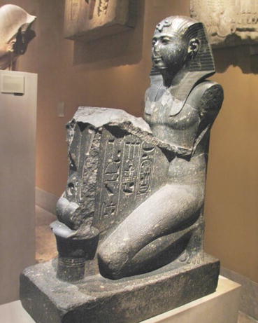
In 1958 Treves reported a series of 406 males with breast hypertrophy [9]. He pointed out that gynaecomastia is an ancient disease with statues of Pharaoh Seti I (1303–1290 BC) showing breast enlargement which would now be classified as grade III gynaecomastia Fig. 2.1.


Fig. 2.1
Pharaoh Seti I
Approximately 25% of men with gynaecomastia have developed the condition as a result of the medications that they are taking or the drugs that they are abusing. An evidence based review has categorised drugs into those that were definitely responsible and others with a possible association [10]. Spironolactone is a major offender producing gynaecomastia in 10% of men treated for severe cardiac failure [11]. Both spironolactone and cimetidine bind to androgen receptors producing an effective androgen blockade. For the majority of the agents there are effective alternatives which may reverse or reduce the gynaecomastia. The results are summarised in Table 2.2.
Ambrogetti reported a large series of 748 consecutive males patients referred for breast screening in Florence [12]. All had a clinical examination and mammography with sensitivities of 85% and 89% respectively. The average age was 50.5 years and cancers were found in 20 men (0.27%) of whom 17 were >60 years. Following biopsy 92 benign lesions were diagnosed of which 74 (80%) were gynaecomastia cases. The combination of palpation and mammography had 100% sensitivity. The authors concluded that the diagnostic protocol used in females appeared to be fully effective in men. Hence triple assessment (clinical evaluation, imaging and cytology/core biopsy) should be the standard management for a man with a breast mass.
Imaging
1975 Kalisher & Peyster described xerographic manifestations of male breast disease, and in particular the distinguishing features of unilateral gynecomastia and MBC [13]. Gynaecomastia was characterised by increased ducts, ductal hyperplasia, stromal proliferation around small ducts and fatty replacement. MBC was usually central and dense, with irregular spiculated margins and sometimes skin changes or axillary lymphadenopathy.
Ouimet-Oliva reported radiological findings in 171 symptomatic men of whom 20 had MBC and 150 were diagnosed with benign lesions [14] (Table 2.3). They suggested a triad of diagnostic signs of MBC, namely a small mass, which was well defined and located eccentrically to the nipple. Dershaw et al. reviewed the mammograms taken on 23 men with proven breast cancer and found that the commonest radiological sign was an uncalcified subareolar mass present in 72% [15]. Two men had a mass with associated microcalcification but not of the typical appearance seen in women with breast cancer. No evidence of the cancer was seen in 3 (13%) including one man with gynaecomastia which obscured the lesion.
Table 2.3
Imaging investigation of symptomatic males
Author | Mammos | U/S | Biopsy | Benign | Cancer |
|---|---|---|---|---|---|
Ouimet-Oliva 1978 [13] | 171 | – | 150 | 20 | |
Cooper 1994 [15] | 263 | – | 20 | 14 | 6 |
Chantra 1995 [16] | 118 | – | 3 | ||
Ambrogetti 1996 [11] | 748 | 110 | 92 | 18 | |
Gunhan-Bilgen 2002 [18] | 236 | 236 | 43 | 29 | 14 |
Chen 2006 [19] Centre A Centre B Centre C | 339 119 261 | 120 119 261 | 15 24 27 | 13 20 19 | 2 4 8 |
Patterson 2006 [20] | 164 | 68 | 6 | ||
Muñoz Carrasco 2010 [21] | 518 | 423 | 103 | 84 | 19 |
Adibelli 2010 [22] | 164 | 164 | 147 | 17 | |
Taylor 2013 [23] | 679 | 364 | 25 | ||
Tangerud 2015 [24] | 539 | 483 | FNAC 261 Core 4 | 257 | 8 |
Cooper reported that in a series of 263 symptomatic males, 66 (25%) had diffuse breast enlargement, 88 (33%) had pain and 20 (8%) had pain and swelling [16]. Mammographic findings were gynaecomastia in 213 (81%), solitary mass in 7 (3%) and multiple masses in 1 case. Chantra et al. carried out 118 mammograms on males during a 40 month period and found bilateral gynaecomastia in 66 (56%), unilateral gynaecomastia in 30 (25%), pseudogynaecomastia in 11 (10%), lipomas in 6 (5%), normal breast tissue in 2 (1%) and cancer in 3 (2%) [17].
In a large series of 748 consecutive symptomatic males seen in Florence, malignancy was diagnosed in 20 of whom 18 had invasive MBC, 1 had DCIS and the other a myxosarcoma [11]. Sensitivity was 85% for palpation, 89% for mammography, 94% for cytology and 100% for US with respective specificities of 95%, 94%, 96% and 98%. The combination of palpation and mammography achieved 100% sensitivity.
Applebaum et al. examined the mammographic findings in a series of 97 males with a histological diagnosis of breast disease which was gynaecomastia in 65 cases [25]. Of these 61 (94%) were diagnosed by mammographic signs as being nodular, dendritic or diffuse. Nodular gynaecomastia manifested as a fan–shaped density which radiates from the nipple. The dendritic variety comprised a retroareolar soft tissue density with extension into the surrounding fat. Diffuse gynaecomastia showed a mixed density similar to the adult female breast. There were 12 MBCs of which 3 (25%) had an associated DCIS component. On mammography the cancers were usually retroareolar masses sometimes eccentric and occasionally located peripherally. Margins could be well or ill-defined sometimes with spiculation and the shape was variously round, oval, irregular or lobulated. Additional signs were microcalcification in 3 (25%), nipple retraction in 7 (58%)and skin thickening in 7 (58%). The authors concluded, although there were mammographic features of MBC which could be recognised, nevertheless there was considerable overlap between signs of benign and malignant disease.
Hanavadi et al. were prompted to ask “Is mammography overused in male patients”? [26] Because no protocols existed for the appropriate use of mammography in assessment of symptomatic males that it was likely that the investigation would be overused. They carried out an audit of all 220 male patients referred to the breast clinic at Cardiff University Department of Surgery between January 2001 and December 2003. Mammography was carried out in 134 (61%), usually before the patient was seen by a clinician. A total of four cases of MBC were diagnosed and in every case the diagnosis was suspected on clinical examination and subsequently confirmed histologically. It was concluded that mammography was unnecessary for most males and did not have a role in routine imaging.
Gunhan-Bilgen et al. described their experience of investigating 236 Turkish males both in terms of mammography and ultrasound [17]. There was a range of final diagnoses: gynaecomastia (206), MBC (14), fat necrosis (5), lipoma (3), subareolar abscess (2), skin cyst (2), haematoma (1), myeloma (1), and metastatic carcinoma (2). Of those with gynaecomastia 73 (35%) were dendritic, 71 (34%) nodular and 62 (31%) diffuse glandular. The 13 MBC manifested as a non-calcified mass in 12 (86%) and with calcification in 1 (7%). Gynaecomastia completely obscured the cancer in one case and partially in two others. The ultrasound findings were of irregular margins in 12 (86%) and well-defined margins in 2 (14%). This illustrated the value of ultrasound particularly in males where the cancer was obscured by gynaecomastia. Further evidence of the value of combined mammography and ultrasound was provided by Jackson et al. who reviewed the mammographic and ultrasonic findings in 41 men with breast enlargement [27]. Of the men 29 (71%) had both mammography and ultrasound, 9 (22%) had ultrasound alone and 3 (7%) had mammography without ultrasound. There were five cases with equivocal or suspicious findings on mammography but of the two that showed suspicious changes on ultrasound one proved to have MBC and the other had gynaecomastia on core biopsy.
Stay updated, free articles. Join our Telegram channel

Full access? Get Clinical Tree


