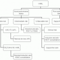
The initial histologic diagnosis provides a useful basis for further evaluation. Almost all initial light microscopic diagnoses fall into one of five categories: poorly differentiated (anaplastic) neoplasm, poorly differentiated carcinoma (PDC), adenocarcinoma, squamous cell carcinoma, and neuroendocrine carcinoma. Occasional sarcomas or melanomas are also diagnosed without an obvious primary site, and management of these patients should follow established guidelines.
Histologic Subtypes
Poorly Differentiated Neoplasm
The diagnosis of poorly differentiated neoplasm using light microscopy implies the inability of the pathologist to distinguish between the major categories of malignant disease, including carcinoma, lymphoma, and various mesenchymal tumors. Approximately 5% of all CUP patients present with this histologic diagnosis, but few remain without undefined lineage after specialized pathologic study.
1,2,3 In this group of patients, establishing a precise diagnosis is extremely important because various highly treatable cancers are poorly differentiated. In adults, the most frequent
poorly differentiated neoplasm for which specific, highly effective therapy is available is non-Hodgkin lymphoma. When additional pathologic evaluation is performed, either using immunoperoxidase staining or electron microscopy, 34% to 66% of poorly differentiated neoplasms are found to be intermediate- or high-grade non-Hodgkin lymphoma.
1,2 In children or young adults, various highly treatable malignancies can be identified, including various sarcomas, neuroblastoma, Ewing tumor, and germ cell tumors.
IHC staining, electron microscopy, and genetic analysis are helpful in the differential diagnosis. Initial communication with the pathologist is important because one common cause of a nonspecific light microscopic diagnosis is a small or poorly preserved biopsy specimen. Fine-needle aspiration biopsy
frequently provides an inadequate amount of tissue for optimal evaluation of poorly differentiated tumors because histology is difficult to evaluate and the ability to perform special studies is limited. A larger biopsy specimen is recommended and is often adequate to make a more specific diagnosis. When the tumor type is unclear after examination of an adequate biopsy specimen, additional pathologic studies are required.
Poorly Differentiated Carcinoma
Approximately 20% of patients with carcinoma of unknown primary site have PDC, and an additional 10% have poorly differentiated adenocarcinoma (PDA). It is now recognized that some of these patients have neoplasms that are highly sensitive to chemotherapy and a small percentage is curable with appropriate therapy. Careful initial evaluation is therefore critical in this patient group to ensure that patients with highly responsive neoplasms are identified and treated.
Histologic features that can differentiate chemotherapyresponsive tumors from nonresponsive tumors have not been identified.
4 Therefore, all PDCs should have additional pathologic study using IHC staining (see
“Immunohistochemical Staining”).
Electron microscopy should be reserved for tumors in which IHC is not contributory because this technique is less widely available and requires specific tissue preparations. Lymphoma can be reliably differentiated from carcinoma in most instances. In addition, sarcoma, melanoma, mesothelioma, and neuroendocrine tumors can occasionally be defined by subcellular features.
Identification of tumor-specific cytogenetic abnormalities may be useful in patients with PDC. Specific chromosomal abnormalities include t(11;22) in patients with peripheral neuroepitheliomas or Ewing tumor
5,6 and an isochrome of the short arm of chromosome 12 (i12p) in most patients with germ cell tumors.
7 The clinical relevance of molecular and cytogenetic studies has been demonstrated in young patients with PDC or adenocarcinoma involving primarily midline structures or with elevated serum levels of human chorionic gonadotropin (HCG) or α-fetoprotein (AFP).
8 In a group of 40 such patients, specific diagnoses were suggested by genetic studies in 17 (42%): germ cell tumor, 12; melanoma, 2; lymphoma, 1; peripheral neuroepithelioma, 1; and desmoplastic small cell tumor, 1. In the 12 patients with germ cell tumors, cisplatin-based therapy produced a 75% overall response rate, with 45% complete responses. Similar therapy yielded responses in only 17% of patients without specific abnormalities, with no complete responses.
Adenocarcinoma
Well-differentiated and moderately well-differentiated adenocarcinomas are the most common tumors identified using light microscopy and account for approximately 60% of CUP diagnoses (about 50,000 patients annually in the United States). As with most types of adenocarcinomas of known primary site, the incidence of adenocarcinoma of unknown primary site increases with advancing age. Most patients have multiple metastases. Common metastatic sites include the liver, lungs, bones, and lymph nodes.
The diagnosis of adenocarcinoma is usually made without difficulty on the basis of light microscopic features and is based on the formation of glandular structures by neoplastic cells. However, adenocarcinomas from many sites share these histologic features, and therefore, the site of tumor origin is usually impossible to pinpoint. Certain histologic features can suggest specific primary sites, but even these are usually not specific enough to make a definitive diagnosis. Examples include papillary features, typically seen in ovarian or thyroid cancers, and signet ring cells, typically associated with gastric adenocarcinoma.
The diagnosis of PDA should be interpreted with caution because patients with this diagnosis may be distinct from patients with well-differentiated adenocarcinoma in both tumor biology and responsiveness to systemic therapy. Criteria for the diagnosis of PDA may differ slightly among pathologists because there is clearly a spectrum of differentiation, ranging from very well-differentiated adenocarcinoma to completely anaplastic carcinoma that fails to show any differentiation. Minimal glandular formation or positive mucin staining in an otherwise PDC often results in the diagnosis of PDA. Additional pathologic study in these patients is therefore appropriate, as described for patients with PDC.
The identification of relatively cell-specific antigens by IHC staining has improved the ability to predict the site of origin in patients with adenocarcinoma of unknown primary site.
3,9 Panels of IHC stains are most useful and should be directed by clinical features (e.g., sites of metastases and gender). Molecular tumor profiling assays also appear relatively accurate and often provide additional diagnostic information (see sections on
“Immunohistochemical Staining” and
“Molecular Tumor Profiling and CUP Classification”).
Squamous Carcinoma
Squamous carcinoma of unknown primary site represents approximately 5% of patients with CUP. The diagnosis of squamous carcinoma is usually made definitively by examination of histology. Additional pathologic evaluation is usually not necessary. Effective treatment is available for the majority of these patients, and appropriate clinical evaluation is important.
Neuroendocrine Carcinoma
Neuroendocrine tumors account for approximately 3% of all CUP. Improved pathologic methods for diagnosing neuroendocrine tumors have resulted in the recognition of an increased incidence and wider spectrum of these neoplasms. Neuroendocrine carcinomas with widely differing histologic and clinical features are represented, and accurate categorization is important in planning treatment.
Two subgroups of neuroendocrine carcinoma can be routinely recognized by histologic features. Well-differentiated (low-grade) neuroendocrine tumors share histologic features with carcinoids and islet cell tumors and frequently secrete bioactive substances. A second histologic group (variously
described as small-cell carcinoma, atypical carcinoid, or poorly differentiated neuroendocrine carcinoma) has typical neuroendocrine features and an aggressive biology.
A third group of neuroendocrine carcinomas appears histologically as a poorly differentiated neoplasm or PDC. Accurate identification of these tumors requires IHC staining, and occasionally electron microscopy.
Immunohistochemical Staining
A large number of relatively sensitive and specific IHC stains are now widely available and frequently aid in the classification of poorly differentiated tumors. Immunoperoxidase reagents consist of either monoclonal or polyclonal antibodies directed at various specific cell components or products, including enzymes, normal tissue components, hormones, oncofetal antigens, and other tumor markers. Most stains can be performed using formalin-fixed, paraffinized tissue. Many new antibodies are being developed against various rather cell-specific proteins, making this area of diagnostic pathology a dynamic and evolving field.
Table 37-1 summarizes the IHC stains that are important in the diagnosis of specific tumor types. In the evaluation of poorly differentiated neoplasms, several important issues can usually be resolved by IHC staining. First, and most important, the leukocyte common antigen (LCA) stain can be used to identify non-Hodgkin lymphomas with a high level of accuracy.
10 Poorly differentiated neoplasms diagnosed as non-Hodgkin lymphoma on the basis of IHC staining can be treated effectively with combination chemotherapy.
2 Second, neuroendocrine tumors can be identified by staining for chromogranin and/or synaptophysin.
11 Third, unsuspected melanomas and sarcomas can be diagnosed with reasonable accuracy.
12,13 Finally, staining for germ cell tumors (HCG, AFP, OCT4, and PLAP) is suggestive in an appropriate clinical situation.
14
The ability of IHC staining to identify the origin of various adenocarcinomas has improved, but in most cases the staining results must be interpreted in the context of clinical and histologic features. An exception is the PSA stain, which is very specific for prostate carcinoma.
15 Stains suggestive of other primary sites are summarized in
Table 37-1; specificity is improved using panels of stains.
3,16,17,18Several problems are associated with the IHC stains. Technical expertise is required to perform these tests accurately and reproducibly, and proper interpretation requires an experienced pathologist. None of the stains is entirely specific. For example, some carcinomas stain with vimentin, some sarcomas
stain with cytokeratins, and a wide variety of carcinomas do not always stain in the expected patterns.
3,16 The typical staining patterns (
Table 37-1) often overlap with the staining patterns of other adenocarcinomas, forcing the pathologist to consider two or three possible primary sites. However, the clinical setting should be used to direct the selection of IHC stains and may narrow the spectrum of possibilities if staining patterns are not completely specific. For example, a CK20
+/CK7
– staining pattern provides strong evidence for the colon as a primary site in a patient with mucin-positive adenocarcinoma and metastases limited to the liver. Conversely, IHC findings may direct additional diagnostic procedures; in the above example, a colonoscopy should be performed and may result in the identification of a primary site.
In many cases, a single primary site cannot be identified with certainty even after histologic examination, IHC staining, and correlation with clinical features. Additional pathologic evaluation with either electron microscopy or a search for specific chromosomal abnormalities is useful in a few situations. In addition, molecular tumor profiling is a new technique that promises to be of importance in identifying the tissue of origin in patients with CUP.
Molecular Tumor Profiling and CUP Classification
Gene expression or molecular profiling of human neoplasms was first made possible by the development of DNA microarray analysis techniques.
19,20 A pivotal study in cancer classification and diagnosis was reported by Golub et al.,
21 who demonstrated for the first time that patterns of gene expression alone could discriminate acute myeloid leukemia from acute lymphoblastic leukemia. Other investigators demonstrated that numerous cancer types could be classified accurately by measuring the differential expression of specific gene sets.
22,23,24,25 The basis of molecular profiling in recognizing specific cancer types is the identification of the genes responsible for the synthesis of proteins required for specific normal cellular functions (e.g., milk production in breast luminal duct cells, albumin production in hepatocytes, etc.). Cancer cells retain some normal cell-type specific functional characteristics, so their origin can usually be predicted from their gene-expression profile.
25 Molecular profiling assays designed to determine the tissue of origin, therefore, measure gene expression dynamics in relation to cell lineage, rather than tumor-specific markers.
Patients with CUP have a clinically undefined primary tumor site, and are ideal candidates for classification by molecular profiling.
26 Molecular profiling may identify the specific type of cancer present, and when used in concert with the clinical and pathologic features may be useful in predicting the site of tumor origin. Primary site identification in CUP would probably improve the therapeutic outcome by allowing site-specific therapy to be administered, rather than a single empiric regimen to all patients. In addition to defining the precise tumor type, molecular tumor profiling may aid in unraveling various gene-specific cancer-activated or overexpressed cellular pathways and in identifying new targets for therapy.
27,28Molecular profiling assays have been validated in patients with metastatic tumors of known primary site. When applied to biopsy specimens from a metastatic site, various molecular assays have correctly predicted the primary site in 76% to 89% of patients.
23,29,30,31,32,33 Correct identification of the primary tumor type in CUP is difficult to validate because the primary tumor site is unknown and rarely becomes apparent during the subsequent clinical course of these patients. In several retrospective studies using archived biopsy specimens, molecular profiling assays predicted a tissue of origin in most cases.
29,34,35,36,37,38 Although these predictions were generally consistent with clinical features, IHC staining patterns, and response to empiric therapy, no confirmation of accuracy was possible.
More direct evidence regarding the accuracy of molecular tumor profiling is now available from a study of CUP patients who had a primary site identified later during their clinical course (latent primary).
39 Twenty such patients who had primary sites identified 2 to 54 months (median 10 months) after the initial diagnosis of CUP were identified. Since publication of the initial report, four additional patients have been identified. The initial diagnostic biopsies were evaluated by a molecular profiling assay using RT-PCR methodology (Cancer Type ID; BioTheranostics, Inc.) capable of identifying 32 tumor types. In 18 of 24 biopsies (75%), the primary tumor was accurately predicted (matched the latent primary site identified), providing direct validation of the accuracy of a molecular profiling assay in identifying the tissue of origin in CUP patients.
Developing evidence therefore indicates that molecular tumor profiling can accurately predict the tissue of origin in a majority of patients with CUP and is an important new diagnostic tool. Although its exact role in the diagnostic evaluation awaits definition, molecular tumor profiling is probably more accurate than IHC staining in many patients with CUP. However, there is little published data regarding the impact of diagnoses made by molecular profiling on results of treatment. Until such data exist, these patients should still be considered to have CUP when planning management. In some cases, consideration of treatment based on the predicted primary site is now appropriate (see
“Treatment” section).


