Acute Myeloid Leukemia


Acute Myeloid Leukemia


Radwan F. Khozouz

Carl E. Freter


Acute myeloid leukemia (AML) accounts for 90% of acute leukemias diagnosed in adults, while comprising only a minority (15% to 20%) of childhood acute leukemias. According to SEER Cancer Statistics Review 1975 to 2007 published in 2010, approximately 12,330 new cases of AML are expected in 2010 (6,590 in men and 5,740 in women), resulting in a combined incidence of 3.5/100,000 (4.3 in men vs. 2.9 in women). Approximately 8,950 deaths are expected in 2010 from AML, accounting for a 2.8/100,000 death rate (3.6 in men vs. 2.8 in women).

Multiple etiologic agents have been implicated, including environmental, chemical and radiation exposure, genetic disorders (Down syndrome, Bloom syndrome, Fanconi anemia, and ataxia telangiectasia), preexisting marrow disorders (myelodysplastic syndrome [MDS], myeloproliferative disorders, aplastic anemia, and paroxysmal nocturnal hemoglobinuria), and preceding cytotoxic chemotherapy (alkylating agents and topoisomerase II inhibitors). AML results from mutations that either result in survival and a proliferation advantage, or impairment of differentiation.1 Tables 44-1 and 44-2 depict the classification of AML according to the current WHO 2008 and FAB classification, respectively.

In view of the different approach to the management of acute promyelocytic leukemia (APL) and its unique features, natural history, and complications associated with treatment, it will be discussed separately.


KARYOTYPE AND GENETIC MUTATIONS AS RISK DETERMINANTS IN AML

Karyotype is considered the strongest prognostic factor for response to induction therapy and survival. Classically, three large risk categories have been recognized. The favorable-risk group includes APL with t(15;17) and core-binding factor (CBF) AML involving t(8;21), inv(16), and t(16;16). The high risk group in AML includes complex cytogenetics defined as the presence of three or more chromosomal abnormalities in the absence of recurring chromosomal translocations or inversions, t(6;9), t(3;3), -5, del(5q) or -7. Patients with normal karyotype are grouped into a single intermediate prognosis group.1,2

Recent studies have shown that AML with normal cytogenetics constitutes a large and genetically diverse group, and the presence of specific genetic mutations translates into various risk categories that mandate different approaches to treatment and management. Genetic mutations involving FLT3, nucleophosmin gene 1 (NPM1), c-KIT, CCAAT/Enhancer Binding Protein Alpha (CEBPA), WT1, and MN1 genes are among the most studied and will be discussed shortly in this section. Table 44-3 summarizes the cytogenetic and mutational risk stratification in AML.


FMS-Like Tyrosine Kinase 3

FMS-like tyrosine kinase 3 (FLT3) is a transmembrane tyrosine kinase receptor that is involved in cell proliferation upon activation. Mutations in FLT3 are reported in 10% to 30% of cytogenetically normal AML. Two types of FLT3 mutations have been recognized. The most common has activating internal tandem duplications (FLT3-ITD), which along with point mutations in the activating loop, result in ligand-independent activation of FLT3. The presence of FLT3-ITD confers a worse prognosis and inferior outcomes, secondary to higher relapse rates. Current data suggests that internal tandem duplication mutations of FLT3 are stronger predictors of poor prognosis than point mutations.3,4,5 Clinical trials with FLT3 tyrosine kinase inhibitors, sorafenib, and sunitinib have shown evidence of activity, albeit moderate and transient.6,7 Other FLT3 inhibitors are currently in development.


Table 44-1 WHO 2008 Classification of AML


AML with recurrent genetic abnormalities


AML with t(8;21), inv(16), t(16;16), t(15;17), t(9;11), t(6;9), inv(3), t(3;3), t(1;22)


AML with myelodysplasia-related changes


t-AML


AML not otherwise specified


AML with minimal differentiation (FAB M0), without maturation (FAB M1), with maturation (FAB M2), myelomonocytic (FAB M4), monocytic (FAB M6), megakaryocytic (FAB M7), basophilic, acute panmyelosis with myelofibrosis


Myeloid sarcoma


Myeloid proliferations related to Down


Blastic plasmacytoid dendritic cell neoplasm


AML of ambiguous lineage


Nucleophosmin Gene 1

Through its association with nucleolar ribonucleoprotein and ribosome function, NPM1 is involved in the regulation of cell proliferation and differentiation. Mutations in NPM1 are seen in up to 50% of cases of cytogenetically normal AML. Current studies indicate that presence of NPM1 mutations in cytogentically normal AML confer a favorable prognosis comparable to that in the good-risk cytogenetics AML. The combined presence of NPM1 mutation and FLT3-ITD results in significantly poorer outcome compared with patients with NPM1 and wildtype FLT3.8,9,10,11,12


c-KIT Gene

KIT is a class of receptor tyrosine kinase that is involved in several pathways of cell differentiation and proliferation. KIT mutations, seen in 20% to 30% of AML, result in ligandindependent activation of downstream pathways, which translates clinically into higher relapse rates and shorter survival.13,14 Studies attempting to incorporate tyrosine kinase inhibitors into treatment regimens have not shown significant activity. A study evaluating the benefit of adding imatinib to low-dose cytarabine as induction regimen in elderly c-KIT positive AML revealed no added benefit to the combination.15


CCAAT/Enhancer Binding Protein Alpha Gene

The CEBPA gene product is involved in myeloid lineage differentiation, and mutations in the CEBPA gene occur in 10% to 20% of cytogenetically normal AML. Studies have shown that the presence of CEBPA mutations confers a favorable outcome.16,17,18 Data from recent studies, however, suggests that the favorable effect conferred by CEBPA mutations is limited to cases with two copies of the mutant allele, whereas single allele mutations have outcomes comparable with wild-type CEBPA genes.19,20,21 Hypermethylation of the CEBPA gene also appears to negate the benefit conferred by CEBPA mutations.22


Table 44-2 FAB Classification of AML


FAB Classification


AML


Characteristic Cytogenetics


M0


With minimal differentiation


None


M1


Without maturation


None


M2


With maturation


t(8;21)


M3


Promyelocytic


t(15;17)


M4/M4eo


Myelomonocytic/M4 with eosinophilia


t(16;16), inv(16), del(16q)


M5


Monocytic


t(9;11), del(11q), t(11;19)


M6


Erythroid


None


M7


Megakaryocytic


T(1;22)


Table 44-3 Cytogenetic and Mutational Risk Stratification in AML


Favorable risk


t(8;21)


t(15;17)


t(16;16)


inv(16)


CN-AML-mutated NPM1 without FLT3-ITD


CN-AML with mutated CEBPA


Intermediate risk


t(9;11)


t(8;21) with c-KIT mutations


t(16;16) with c-KIT mutations


inv(16) with c-KIT mutations


CN-AML-mutated NPM1 with FLT3-ITD


CN-AML wild-type NPM1 with FLT3-ITD


CN-AML wild-type NPM1 without FLT3-ITD


Adverse risk


Complex karyotype


inv(3)


t(3;3)


t(6;9)


del(5q)


del(7q)


Monosomal karyotype, -5, -7


Isocitrate Dehydrogenase 1 and 2 Genes

Isocitrate dehydrogenase (IDH) gene 1 and 2 products are involved in NADPH production through the Krebs cycle. Mutations of IDH 1 and 2 genes are found in 20% to 30% of AML with normal cytogenetics and appear to confer a negative outcome. In a study evaluating the prognostic significance of IDH1/IDH2 mutations, the presence of IDH1 mutations in otherwise low-risk, cytogenetically normal young AML subjects, with NPM1 mutations and negative FLT3-ITD, was associated with shorter 5-year disease-free survival rates of 42% versus 59% and a trend toward shorter 5-year overall survival of 50% versus 63% when compared with wild-type IDH1. A similar picture was seen with IDH2 mutations, where a lower
complete remission (CR) rate of 38% was seen compared with 75% in patients with wild-type IDH2.23


Wilms Tumor 1 Gene

Despite early studies showing Wilms tumor 1 (WT1) gene to exhibit a tumor-suppressor function, more recent data suggests that the WT1 gene exhibits a poorly understood dual function of an oncogene or a tumor suppressor, depending on the tissue of origin.24 Mutations in WT1 gene are reported in 10% to 20% of cytogenetically normal AML cases.25 The bulk of currently available data suggests that mutations in WT1 gene in cytogenetically normal AML constitute a negative prognostic factor.26,27,28


Meningioma Gene 1

The meningioma gene 1 (MN1) product is involved in transcriptional regulation, and mutations involving the MN1 gene have been reported to confer negative outcomes.29,30


TREATMENT OF AML IN YOUNGER ADULTS

Induction chemotherapy constitutes the first step in the treatment of AML. The goal of induction is to rapidly restore normal bone marrow function by reducing the total leukemia cell population to below the cytologically dectectable level. A residual leukemia burden, however, will result in a rapid relapse if no postinduction therapy is administered. In view of the vast difference in treatment outcomes and prognosis between younger and older adults (over the age of 60), these two groups will be discussed separately below.


Induction Therapy of AML in Younger Adults

The most common induction regimen for AML is a combination of a 7-day continuous infusion of cytarabine with three daily infusions of an anthracycline (7 + 3 regimen). The most common anthracycline used is daunorubicin. Studies attempting to substitute or incorporate other agents to induction or to increase the cytarabine dose have resulted in worsening toxicity and no added benefit.31,32 In a study evaluating anthracycline dose intensification for young adults, two different daunorubicin doses were compared; 45 versus 90 mg per m2. Higher rates of CR and overall survival were noted in the higher daunorubicin group with no increase in adverse effects.33 This data supports the use of more intense induction regimens in young adults. There is currently no standard anthracycline agent for induction. In a study comparing idarubicin with daunorubicin for induction, the idarubicin-allocated arm exhibited higher rates of CRs and overall survival compared with the daunorubicin arm. However, the use of nonequitoxic dose of daunorubicin might have resulted in the favorable outcome in the idarubicin-treated arm.34

Response assessment with bone marrow aspiration and biopsy, cytogenetics and FISH or PCR-based studies for known leukemic mutations, is commonly performed between 21 and 28 days after induction with a “classical” 7 + 3 regimen. Earlier response assessment at 7 to 14 days after induction is indicated in the case of new or investigational agent use.1


Postremission Therapy of AML in Younger Adults

Postremission therapy includes consolidation chemotherapy with or without hematopoietic stem cell transplantation (HSCT). Selection of the appropriate treatment regimen largely depends on the risk of relapse, which is mainly dictated by karyotype, although increasingly other risk factors are being incorporated into medical decision making as described previously1


Favorable-Risk AML

A landmark study of intensive postremission consolidation chemotherapy showed that high-dose cytarabine (HDAC) administered at 3 g per m2 every 12 hours for six doses on days 1, 3, and 5 monthly for four cycles, resulted in significant improvement in disease-free survival rates in younger AML patients. A 4-year disease-free survival rate of 44% was seen in patients younger than 60 years, compared with <16% in those older than 60 years.35 A study examining the potential benefit of sequential multiagent chemotherapy versus HDAC, as postremission consolidation in young patients in first CR, revealed no benefit to multiagent consolidation.36 A systemic review and meta-analysis of prospective trials evaluating the benefit of allogeneic HSCT in AML patients in first CR showed significant benefit in terms of relapse-free and overall survival, confined only to the intermediate and high-risk cytogenetic groups. Any benefit of allogenetic HSCT provided in the favorable cytogenetics group was offset by high transplant-related mortality.37,38 Recent data suggests that AML patients harboring mutations in RAS proto-oncogene appear to benefit from HDAC consolidation more than AML cases with wild-type RAS.39 Based on current recommendations, four monthly cycles of HDAC appear appropriate in young AML patients in first CR with a favorable cytogenetic profile.1


Intermediate-Risk AML

Available options for patients in the intermediate-risk group include HDAC consolidation as administered to the favorablerisk group. This could be further intensified with autologous HSCT.40,41 Allogeneic HSCT also constitutes a viable option.1 Intermediate-risk patients, despite having higher transplantrelated mortality, appear to benefit from allogeneic HSCT in terms of decreased relapse and longer disease-free survival rates.37,42

Based on the most recent recommendations, patients with AML harboring unfavorable features; positive FLT3-ITD and c-KIT mutations, and wild-type NPM1, but otherwise exhibiting favorable cytogenetics should be approached as intermediaterisk groups. While patients with normal cytogenetics AML harboring NPM1, mutations and wild-type FLT3 (negative for FLT3-ITD) should be approached as favorable-risk groups.1,3


Unfavorable-Risk AML

Allogeneic HSCT constitutes the backbone of the management of high-risk AML patients after attainment of first CR. This group of patients exhibits poor survival when treated with consolidation chemotherapy alone, with only 15% of patients
in CR after 5 years.37,38,42,43 In contrast to autologous HSCT, which depends on the effect of a pretransplantation intensive chemotherapy regimen to reduce leukemia cell burden, allogeneic HSCT provides an additional graft-versus-leukemia (GVL) effect that has been shown to decrease relapse rates. The benefits of GVL effect are clearly demonstrated as increased relapse rate of AML patients receiving syngeneic transplants, and the ability of donor lymphocyte infusions to induce partial remissions in patients who relapse after receiving allogeneic HSCT.44,45 Figure 44.1 depicts the treatment algorithm for AML patients younger than 60 years.


TREATMENT OF AML IN OLDER ADULTS

Survival rates for AML decrease as a function of aging. In addition to decreased functional reserves and comorbidities, age is associated with an increased incidence of unfavorable cytogenetics and multidrug resistance (Fig. 44.2).46,47 The decision to treat an elderly patient should take into consideration multiple factors, such as age, functional status, comorbidities, and patients’ personal desires and expectations. Treatment should not be denied based on age alone.48,49


Induction Therapy of AML in Older Adults

The classical induction regimen (7 + 3) continues to be a viable option in fit elderly patients. In a study comparing two different daunorubicin doses as part of 7 + 3 induction regimen in elderly AML patients, the group assigned to daunorubicin at 90 mg per m2 compared with 45 mg per m2 exhibited higher CR rates with no significant increase in toxicity. Subgroup analysis revealed that patients 60 to 65 years of age and with core-binding factor leukemias; t(8;21), t(16;16), and inv(16) exhibited longer event-free and overall survivals.50 Elderly AML patients harboring favorable translocations, t(8;21) and inv(16), were shown to exhibit better response compared with historical controls.51 Currently, there is no clear evidence to recommend one anthracycline agent over another if used at equitoxic doses.52 Hypomethylating agents (azacitidine and decitabine) are well tolerated and constitute a viable alternative in frail elderly patients unable to tolerate intensive standard induction. Available data suggests that patients with chromosome 5 and 7 abnormalities might derive the most benefit from hypomethylating agents.53,54,55,56 In a study comparing cytarabine at a low daily dose (LDAC) compared with hydroxyurea in elderly AML patients unfit for intensive treatment, LDAC was associated with higher CR rates of 18% versus 1% for hydroxyurea, and hence is an option for frail elderly patients.57


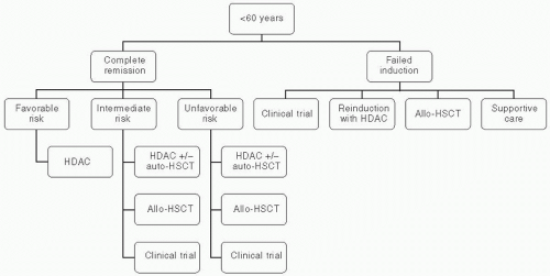
Figure 44.1 Treatment algorithm for AML patients younger than 60 years.


Postremission Therapy of AML in Older Adults

No standard postremission regimen exists for elderly patients. Studies have shown that HDAC consolidation results in extremely poor outcomes and increased toxicity in patients over the age of 60 years.35 In a study randomizing elderly AML patients after attainment of first CR, patients who were randomized to receive six monthly courses of outpatient chemotherapy, with 1 day of anthracycline and five daily subcutaneous cytarabine injections, exhibited higher rates of disease-free and overall survival, and lower rate of death compared with a second intensive induction course.58

Allogeneic HSCT is preferred for the treatment of high-risk younger adults with unfavorable cytogenetics secondary to the associated GVL effect. To gain the benefits of GVL effect while avoiding high transplant-related morbidity and mortality associated with allogeneic HSCT, reduced-intensity conditioning (RIC) allogeneic HSCT were devised. Several studies
employing RIC allogeneic HSCT showed that development of graft-versus-host-disease (GVHD) was associated with lower rates of relapse.59,60 A large study evaluating the effect of age on the outcomes of RIC allogeneic HSCT in elderly AML patients in first CR revealed no significant effect of age on nontransplant mortality, relapse rates, and disease-free and overall survival. This study supports the notion that available modalities of treatment should be considered thoroughly in elderly patients. Chronologic age alone should not be used as the sole discriminant to deny potentially effective therapy.61 Patients’ preferences, performance status, and comorbidities should be integrated into the medical decision making regarding further postinduction therapy in elderly adults. Figure 44.3 depicts the treatment algorithm for AML patients older than 60 years.


Figure 44.2 Percentage of different cytogenetic risk groups as a function of age. (From Appelbaum FR, Gundacker H, Head DR, et al. Age and acute myeloid leukemia. Blood. 2006;107(9):3481-3485.)


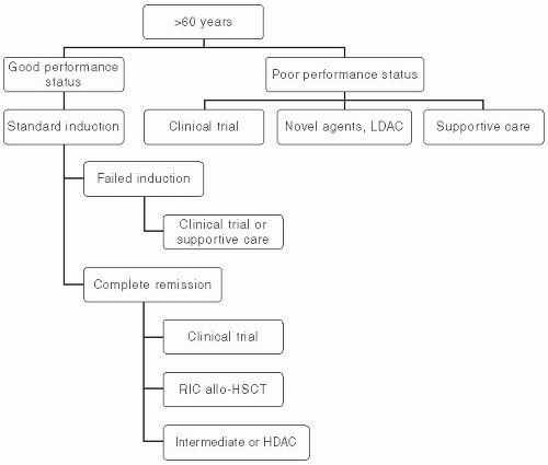
Figure 44.3 Treatment algorithm for AML patients older than 60 years.


TREATMENT OF AML IN RELAPSE

The duration of first CR has prognostic significance in relapsed AML. Remission durations of <1 year are associated with poor response to reinduction with standard regimens, which translates into poor overall survival. This group of patients should be considered for investigational agents, whereas those who relapse more than 1 year after induction could be considered candidates for reinduction with standard regimens.62 Patients with early relapse or those with resistant disease should be considered for allogeneic HSCT or investigational agents. In a retrospective study examining the outcomes of relapsed AML and salvage therapies, which included allogeneic HSCT, standard and HDAC induction second remissions were not durable, achieved in a minority of patients, and translated into poor survival rates. First remission duration of <12 months and age >60 years was among the factors associated with worse outcomes.63

Gemtuzumab ozogamicin, an anti-CD33 antibody linked to a cytotoxic agent calicheamicin, when used as a single agent in relapsed AML results in CR rates of 30%.64,65 It has been granted accelerated approval by FDA in May 2000 for the treatment of CD33-positive AML in first relapse in patients older
than 60, or those who are not candidates for intensive therapy.66 Multiple postmarketing reports described severe hepatotoxicity, including hepatic sinusoidal obstruction syndrome (hepatic veno-occlusive disease). Toxicity was noted both among recipients of allogeneic HSCT and those who were treated with gemtuzumab as a single agent.67,68,69 Gemtuzumab was subsequently withdrawn from the market in June 2010.


Chemotherapy for Relapsed A ML

Multiple studies have evaluated different combinations of cytotoxic agents for relapsed AML. Remissions are not durable and are often achieved at a cost of excessive toxicity. Intermediate and HDAC either as single agent, or in combination with mitoxantrone, etoposide in combination with mitoxantrone or cyclophosphamide, have been tried with minimal encouraging results.70,71,72,73,74 A combination of fludarabine, cytarabine, and granulocyte colony-stimulating factor (G-CSF) resulted in CR rates of 30% to 50% in primary refractory or relapsed AML. The combination was well tolerated and was not associated with significant toxicity. Median survival was considerably shorter with early relapsed AML compared with late relapses.75,76


Hematopoietic Stem Cell Transplantation

Autologous HSCT is likely to be of no benefit as first-line therapy in low-risk AML patients; however, it could be a viable option at relapse. Relapses are common secondary to loss of GVL effect, and there is no available data to show any survival benefit to autologous HSCT in AML.77

Myeloablative allogeneic HSCT are associated with significant transplant-related morbidity and mortality, especially in patients with relapsed AML. Thus, multiple studies have evaluated the role of RIC regimens followed by allogeneic HSCT. Overall survival rates of 30% to 40% have been achieved with RIC allogeneic HSCT. It was well tolerated by elderly patients with no excessive mortality.61,78,79

For patients who relapse after allogeneic HSCT few options are available. A second RIC allogeneic HSCT can be attempted in medically fit patients.80,81 Reduced immunosuppression and donor lymphocyte infusion are potentially viable options, however, this intervention is associated with worsening GVHD. CR rates and 2-year overall survival rates of <20% have been reported.82,83 Figure 44.4 depicts the treatment algorithm for relapsed AML.


Figure 44.4 Treatment algorithm for relapsed AML


TREATMENT OF APL IN ADULTS

APL, classified as AML-M3 in the French-American-British classification system, and currently as AML with t(15;17) in the WHO classification, is characterized by its responsiveness to alltrans-retinoic acid (ATRA) and relatively good prognosis with currently available aggressive treatment regimens. Minor genetic variants of APL with t(11;17) and t(5;17) that might have different sensitivities to ATRA have also been described. Contrasting with the treatment of other subtypes of AML, treatment of APL involves induction, consolidation, and maintenance phases that may span up to 2 years.84 APL is considered an oncologic emergency secondary to the high mortality rate attributed to hemorrhagic complications arising from APL-associated disseminated intravascular coagulation (DIC). Treatment with ATRA, which acts on malignant promyelocytes to resume terminal differentiation to mature neutrophils, should be started on the first suspicion of APL and continued until APL is ruled out by cytogenetic studies or FISH.84,85 With appropriate treatment, 10-year survival rates of 77% have been reported.86

Therapy-related APL (t-APL) deserves special attention, as it has been reported after treatment with topoisomerase II inhibitors for breast cancers and non-Hodgkin lymphomas. Of interest are the recent reports of t-APL after the use of mitoxantrone for the treatment of patients with multiple sclerosis.87 The latency period is short, <3 years, and preceding MDS has not been described. The prognosis of t-APL appears to be favorable, however, prior exposure to anthracyclines might limit treatment options to ATRA and/or arsenic trioxide (ATO) containing regimens.84


Induction Therapy of APL

Monotherapy with ATRA results in high rates of CR, however, remissions are not durable. Studies have shown that a combination of ATRA and chemotherapy administered simultaneously for induction is superior to either ATRA or chemotherapy alone in terms of CR and relapse rates, and longer disease-free survival.88,89,90,91 ATRA has shown beneficial effects in decreasing the markers and duration of coagulopathy associated with APL.88,92 Currently, there is no evidence supporting the use of ATRA in non-APL variants of AML.93

Only gold members can continue reading. Log In or Register to continue

Stay updated, free articles. Join our Telegram channel

Jun 19, 2016 | Posted by in ONCOLOGY | Comments Off on Acute Myeloid Leukemia

Full access? Get Clinical Tree

Get Clinical Tree app for offline access