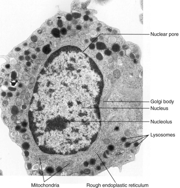
After completion of this chapter, the reader will be able to: 1. Describe the general function and chemical composition of cellular membranes. 2. List and describe the components of the nucleus, including staining qualities visible by light microscopy. 3. Correlate the nuclear structures to the activities of the cell. 4. Name and describe the ultrastructural appearance of the cytoplasmic organelles found in the cell and staining qualities by light microscopy, if appropriate. 5. Correlate the cytoplasmic structures to the activities of the cell. 6. Describe the function of the hematopoietic inductive microenvironment. 7. Associate stages in the cell cycle with activities of the cell. 8. Correlate regulatory proteins with the stage of the cell cycle. 9. Discuss the function of checkpoints in the cell cycle and where in the cycle they occur. The numerous multichannel instruments available to assist in the clinical diagnostic process have revolutionized the study of hematology. The technologies of light scatter, electrical impedance, and conductivity have added parameters and scatter plots whose significance is yet to be fully realized and clinically applied, but morphologic examination of the peripheral blood film by light microscopy remains the hallmark for clinical evaluation of patients with hematologic abnormalities. The study of cells under the microscope was greatly enhanced when Paul Ehrlich (1854-1915) developed staining techniques to better differentiate the various normal and abnormal cells present in human blood. The development of the electron microscope revolutionized the ability to study and understand the internal components of the cell.1 Cells are structural units that constitute living organisms (Figures 6-1 and 6-2). Many cells have specialized functions and contain the components necessary to perform and perpetuate these functions. Regardless of shape, size, or function, most cells have three basic parts: unit membranes, the cytoplasm, and the nucleus. Each of these basic parts has components or subdivisions that assist in their varied functions. Table 6-1 summarizes the cellular components and functions, which are explained in more detail later. TABLE 6-1 Summary of Cellular Components and Functions The cell membrane serves as a semipermeable outer boundary separating the cellular components from their surrounding environment. The cell membrane serves three basic functions: (1) it restricts and facilitates the interchange of substances with the environment by selective permeability, endocytosis, exocytosis, and locomotion; (2) it detects hormonal signals facilitating cell-to-cell recognition; and (3) it is the location of surface markers for cell identity.2 Monoclonal antibodies are used to identify a cell’s surface markers. The nomenclature uses the letters CD (cluster designation) and a number following the CD. This common terminology assists in unifying classification in clinical practice, research efforts, and the literature (see Chapter 33). Many components found within the cell (e.g., the mitochondria, Golgi apparatus, nucleus, and endoplasmic reticulum) have similarly constructed membrane systems. The red blood cell membrane has been widely studied and serves as an example of a cell membrane (see Figure 9-2). Most proteins present in the cell membrane are called glycoproteins and are found floating in the lipid bilayers.2,3 Two types of proteins, integral and peripheral, have been described in the cell membrane. Integral proteins may traverse the entirety of the lipid bilayers and penetrate the outside of the membrane or only the cytoplasmic side of the membrane. These transmembrane proteins are thought to serve as a communication and transport system between the cell’s interior and the external environment. Peripheral proteins are found only on the inner cytoplasmic side of the membrane and form the cell’s cytoskeleton. Peripheral proteins also are attached to the cytoplasmic ends of integral proteins to form a reticular network for maintaining structural integrity and holding the integral proteins in a fixed position. Membrane carbohydrates occur in combination with proteins (glycoproteins) and lipids (glycolipids). The carbohydrate portion usually extends beyond the outer cell surface, giving the cell a carbohydrate coat often called the glycocalyx. These carbohydrate moieties function in cell-to-cell recognition and provide a negative surface charge, surface receptor sites, and cell adhesion capabilities.2 The function of the red blood cell membrane is discussed in detail in Chapter 9.
Cellular Structure and Function
Cell Organization
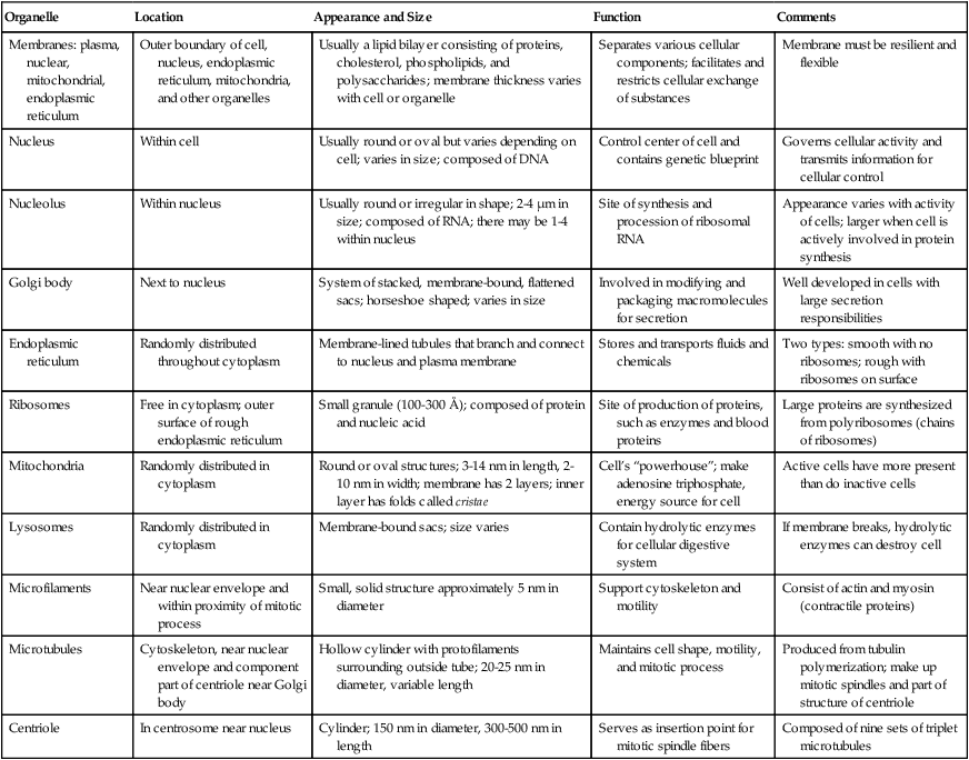
Organelle
Location
Appearance and Size
Function
Comments
Membranes: plasma, nuclear, mitochondrial, endoplasmic reticulum
Outer boundary of cell, nucleus, endoplasmic reticulum, mitochondria, and other organelles
Usually a lipid bilayer consisting of proteins, cholesterol, phospholipids, and polysaccharides; membrane thickness varies with cell or organelle
Separates various cellular components; facilitates and restricts cellular exchange of substances
Membrane must be resilient and flexible
Nucleus
Within cell
Usually round or oval but varies depending on cell; varies in size; composed of DNA
Control center of cell and contains genetic blueprint
Governs cellular activity and transmits information for cellular control
Nucleolus
Within nucleus
Usually round or irregular in shape; 2-4 µm in size; composed of RNA; there may be 1-4 within nucleus
Site of synthesis and procession of ribosomal RNA
Appearance varies with activity of cells; larger when cell is actively involved in protein synthesis
Golgi body
Next to nucleus
System of stacked, membrane-bound, flattened sacs; horseshoe shaped; varies in size
Involved in modifying and packaging macromolecules for secretion
Well developed in cells with large secretion responsibilities
Endoplasmic reticulum
Randomly distributed throughout cytoplasm
Membrane-lined tubules that branch and connect to nucleus and plasma membrane
Stores and transports fluids and chemicals
Two types: smooth with no ribosomes; rough with ribosomes on surface
Ribosomes
Free in cytoplasm; outer surface of rough endoplasmic reticulum
Small granule (100-300 Å); composed of protein and nucleic acid
Site of production of proteins, such as enzymes and blood proteins
Large proteins are synthesized from polyribosomes (chains of ribosomes)
Mitochondria
Randomly distributed in cytoplasm
Round or oval structures; 3-14 nm in length, 2-10 nm in width; membrane has 2 layers; inner layer has folds called cristae
Cell’s “powerhouse”; make adenosine triphosphate, energy source for cell
Active cells have more present than do inactive cells
Lysosomes
Randomly distributed in cytoplasm
Membrane-bound sacs; size varies
Contain hydrolytic enzymes for cellular digestive system
If membrane breaks, hydrolytic enzymes can destroy cell
Microfilaments
Near nuclear envelope and within proximity of mitotic process
Small, solid structure approximately 5 nm in diameter
Support cytoskeleton and motility
Consist of actin and myosin (contractile proteins)
Microtubules
Cytoskeleton, near nuclear envelope and component part of centriole near Golgi body
Hollow cylinder with protofilaments surrounding outside tube; 20-25 nm in diameter, variable length
Maintains cell shape, motility, and mitotic process
Produced from tubulin polymerization; make up mitotic spindles and part of structure of centriole
Centriole
In centrosome near nucleus
Cylinder; 150 nm in diameter, 300-500 nm in length
Serves as insertion point for mitotic spindle fibers
Composed of nine sets of triplet microtubules
Cell Membrane
Membrane Proteins
Membrane Carbohydrates
Cellular Structure and Function