Bones
Age Changes
Bone structure changes throughout life owing to ongoing bone resorption by osteoclasts and bone growth by osteoblasts. With increasing age, the balance is lost, leading to a net increase in bone resorption. This in turn leads to gradual and progressive loss of bone from the age of 35 onwards. This process affects trabecular bone more than cortical bone. In osteoporosis, the molecular composition is similar to normal bone, but the microarchitecture is disorganized so that the bone is fragile and at increased risk of fractures.
Bone loss per year is 0.2% of the total from the age of 35. This rate of loss increases to 1% after the menopause in women. Therefore, on average, by the age of 80 a woman will have lost 30% of her bone mass, whilst a man of the same age will have lost 10%.
The shape of long bones changes with increasing age; the internal cavity increases in diameter, the outer cortical layer becomes thinner and the total bone diameter becomes expanded. These changes result in weaker bones.
Osteoporosis
Risk Factors
- Age > 65.
- Female sex.
- Family history, especially maternal hip fracture.
- Failure to maximize by early adulthood because of poor nutrition or oestrogen deficits, e.g. secondary to anorexia nervosa.
- Hormonal changes at the menopause: the fall in oestrogen causes a marked acceleration of bone loss. Fractures secondary to osteoporosis affect one in two post-menopausal women.
- Previous fragility fractures.
- Low body weight (BMI < 19 kg/m2).
- Physical inactivity, e.g. following a stroke.
- Use of corticosteroids for 3 or more months.
- Other drugs especially antiepileptics, aromatase inhibitors for breast carcinoma, anti-androgen treatment and caffeine.
- Alcohol intake > 3 units/day.
- Smoking.
- Endocrine disorders, e.g. thyrotoxicosis, Cushing’s disease, hyperparathyroidism and hypopituitarism.
- Chronic disease: CKD, liver disease and COPD.
- Malabsorption.
- Falls are not a cause of osteoporosis but are strongly predictive of osteoporotic fractures.
- Older people often have vitamin D deficiency and secondary hyperparathyroidism, and these are likely to contribute.
- The reduction of insulin-like growth factor production is also being investigated as a risk factor.
 hypogonadotrophic hypogonadism;
 steroids;
 alcohol;
 hyperparathyroidism;
 malabsorption, e.g. secondary to Crohn’s disease, gastric surgery and coeliac disease.
Clinical Features
Osteoporosis is usually asymptomatic until there has been a fracture:
- Often the first presentation is a Colles’ wrist fracture in women aged 50–65.
- Vertebral fractures may present as severe mid-thoracic or low back pain often with no history of trauma.
- Loss of height and dorsal kyphosis secondary to multiple vertebral fractures. A loss of > 4 cm suggests at least one vertebral fracture.
- Contact between ribs and iliac crests.
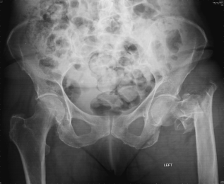
- Hip fractures: rising incidence with increasing age. See Figure 6.1 showing a comminuted fracture of the left hip. The incidence is rising faster than expected from demographic changes. There are now 57,000 cases per annum in the UK – 75% of which are aged over 75.
- Other fractures associated with osteoporosis include neck of humerus, pelvis and distal tibia/fibula.
Investigations
The WHO Fracture Risk Assessment Tool (FRAX) collates clinical information and risk factors to stratify the patient’s risk and determine whether a dual-energy X-ray absorptiometry (DXA) scan would give extra information.
Bone Mineral Density
The DXA scanner measures bone density usually at the proximal femur and lumbar vertebrae (because these are clinically important fragility fractures). Osteoporosis is defined as a bone mineral density (BMD) of greater than 2.5 standard deviations below that of a normal pre-menopausal woman and is expressed as a T score (i.e. T –2.5 SD).
Ultrasound of the Calcaneum
This has the advantage of being inexpensive and portable so that it can be used in the primary-care setting, but it has not been fully validated. It is probably most useful for risk stratification; if the result suggests low bone density, the patient should have DXA scan.
Excluding Causes of Secondary Osteoporosis
If serum calcium is raised, check PTH level to exclude primary hyperparathyroidism (see later).
- Thyroid function tests to exclude thyrotoxicosis (see Chapter 12).
- Liver and renal function tests.
- Bone function tests.
- Testosterone: in men with suspected hypogonadotrophic hypogonadism.
- ESR, serum immunoglobulins and urinary Bence–Jones protein to exclude myeloma.
- Dexamethasone suppression test to exclude Cushing’s disease.
Complications
- Fractures, as above.
- Pain and reduced mobility.
- Deformity: kyphosis, loss of height, abdominal protrusion. Cord compression is very uncommon in this context.
- Loss of independence and increased risk of being admitted to a care home.
- Use of resources: 25% of UK orthopaedic beds are occupied by patients with fractured hips. The average cost is £25,000 per patient. Fractures in osteoporotic bones now account for over 1 million bed-days annually in the NHS. The cost of treatment of these patients is now more than £5 million per week.
Prevention
- Adequate nutrition.
- Calcium and vitamin D.
- Hormone replacement therapy is most useful in women in early menopause.
- Exercise: should be regular and weight-bearing, e.g. brisk walking, Tai Chi.
- Stop smoking and moderate alcohol intake.
- Prophylactic use of bisphosphonates: if treating with steroids at > 7.5 mg prednisolone for three or more months.
Treatments
Calcium and Vitamin D
The debate regarding the role of calcium and vitamin D in the treatment of osteoporosis rages on. Questions surround the efficacy of both elements and the optimum dose. It has been shown that treatment with calcium and vitamin D prevented hip fractures in older people living in care homes. There is controversy about the role in other populations; research is ongoing. A large meta-analysis of women given calcium and vitamin D demonstrated a modest increase in cardiovascular events. The Scientific Advisory Committee on Nutrition is undertaking a new review of this controversial area.
What is certain is that all the trials demonstrating the efficacy of bisphosphonates, strontium and teriparatide have co-prescribed calcium and vitamin D preparations.
Bisphosphonates
Bisphosphonates, e.g. risedronate and alendronate, work by binding to hydroxyapatite in the bone, which inhibits recruitment of osteoclasts and apoptosis, thus inhibiting bone resorption. Effects are seen in the first 12–18 months of use. Both are shown to increase bone mass of the spine and the hip and to reduce fractures and are indicated in women and men. Weekly preparations are available. This is an advantage because bisphosphonates have to be taken on an empty stomach to ensure absorption. Thus, patients only miss their early morning cup of tea once a week! The patient also has to remain upright for 30 min to reduce the risk of oesophageal ulceration. Ibandronate has the added advantage of being a monthly oral preparation or a 3-monthly injection.
Intravenous zoledronate is given annually. The infusion is usually given in hospital because a few patients developed atrial fibrillation in the drug trials. More common side-effects include flu-like symptoms, fever, headache, diarrhoea and vomiting. Zoledronate is more potent than the other bisphosphonates.
Osteonecrosis of the jaw is a rare complication that seems more likely in patients on high-dose bisphosphonates for carcinoma; however, it is sensible to warn patients to inform their dentists if they are planning any invasive dental treatment.
There is also concern that bisphosphonates may be the cause of atypical subtrochanteric fractures below the neck of femur which produce a characteristic ‘beak’ appearance on the radiograph of the fracture site. Current advice is to reassess the patient’s risk-benefit after 5 years of treatment and consider stopping the bisphosphonate. Bisphosphonates are not recommended if the creatinine clearance is less than 35 ml/h.
Strontium Ranelate
This is thought to increase bone growth and reduce bone loss, although the precise mode of action is unknown. It comes in a sachet to be mixed with water and usually is taken at bedtime. It is an alternative for women over 75 years old who do not tolerate bisphosphonates. Side-effects include diarrhoea, nausea and increased thromboembolic risk.
Teriparatide, Recombinant 1-34 PTH
This is given daily by subcutaneous injection for 18 months. Given intermittently, PTH is anabolic and stimulates osteoblast activity (contrary to the effect of endogenous PTH which is catabolic). Use of teriparatide is restricted to patients with severe disease who have not tolerated or responded to other treatments because it is extremely expensive. Side-effects include gastrointestinal upset. It is contraindicated in renal impairment.
Selective Oestrogen Modulators (SERMs)
SERMS, e.g. raloxifene, act as oestrogen agonists in the bone and liver, but as oestrogen antagonists in the breast and uterus, and therefore increase bone density without increasing the risk of breast or uterine cancer. It reduces the risk of vertebral fractures only. HRT is now only used second line because of the high risk of cancer and heart disease in older women.
Synthetic Analogues of Calcitonin
Given as intramuscular injections, synthetic analogues of calcitonin also inhibit bone resorption. They are not as effective as other agents, so are used as second line. However, they may be a useful adjunct in pain control following an acute vertebral wedge fracture.
Denosumab
A monoclonal antibody to RANK ligand (receptor activator of nuclear factor kappa-B), denosumab is given by subcutaneous injections 6 monthly to post-menopausal women. The mechanism of action is shown in Figure 6.3.
Figure 6.3 The mechanism of action of denusomab. Osteoblasts build bone and have a role in bone resorption by producing RANK ligand (RANKL) and osteoprotegerin. RANKL binds to its receptor RANK and mediates osteoclast differentiation, activation and survival. The protein osteoprotegerin binds to RANKL and functions as an endogenous inhibitor of this pathway. Denosumab is a monoclonal antibody to RANKL and functions like osteoprotegerin, mopping up RANKL and reducing osteoclast activity and decreasing bone resorption.
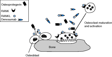
Side-effects include cellulitis, eczema and a few reports of osteonecrosis of the jaw. Denosumab must not be given without correcting calcium and vitamin D levels. If denosumab is discontinued, the patient must be treated with another preparation, e.g. IV zoledronate, or there is profound rebound bone-remodelling.
Analgesia
This is essential for all osteoporotic fractures.
Secondary Osteoporosis
Treat causes of secondary osteoporosis.
Internal Fixation of Hip Fractures
The most appropriate type of fixation depends on the fracture site.
Balloon Kyphoplasty
This is a treatment for vertebral collapse; it restores the vertebral body height, reduces pain rapidly and thus enables the patient to mobilize early.
Concordance
Concordance with calcium and vitamin D preparations and bisphosphonates is extremely poor even 3 months after they are first prescribed. Explaining the rationale to patients may improve this. Different preparations of vitamin D and calcium are available including soluble and capsule forms as well as the familiar chalky tablets, so it is worth trying to find the most palatable for an individual. It may be worth considering monthly or yearly bisphosphonates or denosumab injections; as expensive as these treatments are, they are cheaper than hospital admissions following fractures.
Patient Education
Encourage patients to stop smoking, reduce their alcohol intake and increase their weight-bearing activity.
Prevention of Further Fractures
- Treatment of osteoporosis.
- Exercise programmes.
- Falls prevention strategies (see Chapter 5).
- The benefit of hip protector pads is controversial (see Chapter 5).
Osteomalacia
This is reduced calcification of the osteoid matrix due to vitamin D deficiency. The amount of bone is normal, but it is soft and weak compared with normal bone.
Incidence
Incidence is uncertain and depends on the population studied:
- Admissions to Scottish departments of geriatric medicine, 4%.
- Post-mortem study of elderly patients, 12%.
- Biopsies on fractured neck of femur patients, 25%.
Causes
Reduced Vitamin D Availability
- Deficient diet.
- Reduced sun exposure: most common in Muslim and Hindu cultures (where women cover their heads) and in institutionalized elderly people. Ultraviolet light converts 7-dehydrocholesterol to previtamin D3 and then to vitamin D3 (cholecalciferol).
- Ten to fifteen minutes’ exposure to sunlight is sufficient to replenish the body’s stores.
- Vitamin D binding protein carries D2 and D3 to the liver which converts it to 25-hydroxyvitamin D. Then 1-alpha-hydroxylase in the proximal tubule of the kidney converts it to 1, 25-dihydroxycholecalciferol.
- Malabsorption of vitamin D in food and medications occurs in coeliac disease, diverticular disease of the small bowel and post-gastrectomy.
Impaired Vitamin D Metabolism
- Chronic kidney disease (reduced activity of 1-hydroxylation).
- Drugs that induce liver enzymes, e.g. phenytoin and carbamazepine.
Clinical Features
- Pain in the axial skeleton (spine, shoulders, ribs and pelvis).
- Muscle weakness.
- Waddling gait and difficulty standing from sitting secondary to osteomalacic myopathy.
- Fragility fractures.
Investigations
- X-rays may show insufficiency fractures, Looser’s zones.
- Bone scintigram may show ‘hungry bones’, so-called because the bones take up the isotope so readily that they are very bright and the kidneys may not be visible.
- Blood tests: raised alkaline phosphatase, low corrected calcium and low phosphate.
- Serum 25-hydroxyvitamin D3 will also be low.
- Bone biopsy will clinch the diagnosis where there is doubt.
Treatment
- Oral vitamin supplements, e.g. Calcichew D3 Forte.
- In the case of abnormal metabolism, e.g. renal disease, give a hydroxylated preparation, i.e. alfacalcidol or calcitriol.
- Watch for hypercalcaemia.
Paget’s Disease of the Bone
This is a localized abnormality of bone that arises because of increased activity of the osteoclasts and osteoblasts. The net result is an increase in bone turnover, which produces bone that is expanded, but is paradoxically weaker than normal bone. Paget’s disease of the bone most frequently affects the pelvis, spine, skull and the femur, although any bone can be affected. A single bone is affected in 10% of cases. The adjacent bone may also be affected.
Incidence
The incidence increases with age. The prevalence is 5% of over 40 year olds, rising to 10% of over 80 year olds. There is a slight male preponderance. It is more common in northern Europe, Australia and New Zealand and much less common in Asian countries.
Aetiology
This is still unknown, but there is likely to be a link between environmental and genetic factors. There have been studies linking it to various viruses including measles and the respiratory syncytial virus. Some families possess a candidate gene on chromosome 18q2. There may be a link with HLA DQW 1 antigen. RANK ligand has also been implicated and upregulation may account for the localization of the abnormal bone.
Clinical Features
- Usually asymptomatic and is diagnosed incidentally on X-rays, or because of a raised alkaline phosphatase.
- Pain, which might be localized to the affected bone or secondary to nerve entrapment.
- Deformity, e.g. enlargement of the skull, anterior bowing of the tibia or lateral bowing of the femur.
Investigations
- Raised serum alkaline phosphatase.
- Raised urinary hydroxyproline suggests active disease.
- Serum calcium is raised in patients who are immobile.
- X-rays show affected bones to be enlarged, abnormally dense and distorted, e.g. cottonwool appearance of the skull, picture frame appearance of vertebrae.
- Bone scintigraphy.
Complications
- Fractures of abnormal bone, usually the femur or tibia.
- Secondary osteoarthritis of adjacent joints.
- Neurological: compression of the cranial nerves as they exit the skull, most commonly affects the eighth nerve, but can also affect the second and the fifth; or paraplegia if the spinal cord is compressed.
- Hydrocephalus.
- High-output heart failure caused by Paget’s disease of the bone is very rare.
- The development of malignant tumours is also rare, but examples include osteosarcoma (incidence is < 1%) and chondrosarcoma.
Treatment
- Aimed at treating pain and preventing deformities and fractures.
- Acute disease is treated with a bisphosphonate, usually risedronate, which reduces disease activity (mirrored by a fall in serum alkaline phosphatase and urinary hydroxyproline) and pain within days of starting treatment.
- Risedronate is usually given for 2–6 months.
- Treatment can be repeated if necessary.
- Intravenous pamidronate can be given if an oral preparation is not tolerated or if the disease is rapidly progressing.
Primary Hyperparathyroidism
Incidence
- Common, affecting 1 in 500 women and 1 in 2,000 men per year.
- Occurs worldwide.
- Fifty-five percent are women over 70 years of age.
- Should be considered in any patients with hypercalcaemia in the context of normal renal function and no history of malignancy.
Clinical Features
- The majority of elderly patients (up to 80%) will be asymptomatic and the problem will have been discovered on biochemical testing done for other reasons.
- Asymptomatic patients should simply be observed and their biochemistry monitored.
- However, 12% of cases with a raised calcium level will have had documented episodes of confusion and dehydration and will merit treatment unless there are other major co-morbidities.
- Check PTH level.
- Serum phosphate may be low.
- Primary hyperparathyroidism is usually due to an adenoma in a single parathyroid.
- Sestamibi scan: technetium99 is preferentially taken up by the overactive parathyroid gland; used to demonstrate anatomy prior to surgery.
- Minimally invasive parathyroidectomy is now available, with good results and short hospital stays.
Hypercalcaemia
There are many causes of hypercalcaemia. In practice, the following are the main groups affecting older people:
- Primary hyperparathyroidism, as above.
- Hypercalcaemia of malignancy may be due to bone metastases but also non-metastatic manifestation of malignant disease. PTH is suppressed in this context.
- Myeloma (see Chapter 14).
- Drug induced: thiazide diuretics, lithium and vitamin D.
- Chronic kidney disease (see Chapter 13).
- Sarcoidosis, hyperthyroidism and Addison’s disease are all rare causes in this age group.
Emergency treatment involves rehydration with intravenous normal saline. If the patient is fluid overloaded, loop diuretics are useful to facilitate urinary excretion of calcium. Intravenous pamidronate is an effective treatment especially in malignancy.
Joints
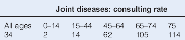
Many people accept joint problems as a part of growing older, but arthritis is not universal, and in old age it must be diagnosed accurately for appropriate management (Table 6.1).
Table 6.1 Patient consulting rate per 1,000 persons (by age in years) for patients with joint diseases
Osteoarthritis
- Osteoarthritis (OA) is the most common joint disorder and the incidence increases with increasing age.
- Eighty percent of 80 year olds will have some X-ray evidence of OA, but not necessarily symptoms.
- Small changes in management may produce big improvements in quality of life and symptom control.
- Long-standing, complicated and burnt-out rheumatoid arthritis may be difficult to differentiate from generalized OA in old age.
- Aetiology usually unknown, i.e. primary osteoarthritis.
- May be secondary to:
 Genetic predisposition.
Related posts:

Stay updated, free articles. Join our Telegram channel

Full access? Get Clinical Tree


