Asparaginase


Asparaginase


Bruce A. Chabner

Alison M. Friedmann


The growth of malignant as well as normal cells depends on the availability of specific nutrients and cofactors. Some of these nutrients can be synthesized within the cell, but others such as essential amino acids are required from external sources. Nutritional therapy for cancer has been directed at identifying differences between the host and malignant cells that might be exploited in treatment.1 Although some significant differences in glucose and lipid metabolism have been found, these attempts have been largely unsuccessful because of difficulties in producing a deficiency state by dietary means and a lack of clear differences in the nutritional requirements of rapidly proliferating host cells and the tumor. The most successful outcome has been the use of L-asparaginase in the treatment of childhood acute leukemia.

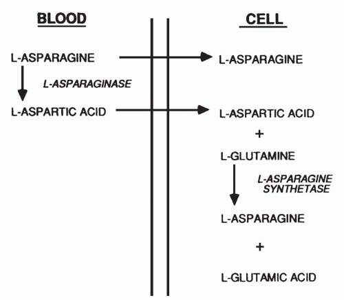
L-Asparagine is a nonessential amino acid synthesized by transamination of L-aspartic acid (Fig. 20-1). The amine group is donated by glutamine, and the reaction is catalyzed by the enzyme L-asparagine synthetase. This enzyme is constitutive in many tissues, which accounts for the modest toxicity of asparagine depletion from the plasma, but the capacity to synthesize asparagine is lacking in certain human malignancies, particularly those of lymphocytic derivation. In tumor cells lacking L-asparagine synthetase, such as L5178Y murine leukemia cells,2 the amino acid can be obtained only from a culture medium or, in vivo, from extracellular fluid and from plasma. There is evidence that mesenchymal stromal cells found in the bone marrow can produce and secrete sufficient L-asparagine to maintain acute lymphoblastic leukemia (ALL) cell growth.3

The enzyme L-ASP (L-asparagine aminohydrolase, EC 3.5.1.1), which catalyzes the hydrolysis of asparagine to aspartic acid and ammonia as end products, is found in many plants and microorganisms and in the plasma of certain animals. General interest in L-ASP as a therapeutic agent was the result of an unexplained observation by Kidd,4 who in 1953 reported that the growth of transplantable lymphomas in rodents was inhibited by guinea pig serum but not by rabbit, horse, or human serum. Ten years later, Broome5 demonstrated that the responsible factor was the enzyme L-ASP. Subsequently, highly purified preparations of L-ASP from Escherichia coli6 and Erwinia carotovora (also known as Erwinia chrysanthemi)7 have become standard components of remission induction, consolidation, and reinduction therapy in childhood and adult ALL.8 A chemically modified enzyme, pegaspargase, having a longer half-life and reduced immunogenicity, is approved for use in patients hypersensitive to the native E. coli enzyme and for first-line therapy of childhood ALL.9 The clinical and biochemical features of L-ASP chemotherapy have been summarized in comprehensive reviews.10 The key features of L-ASP pharmacology are listed in Table 20-1.


Properties and Mechanism of Action

L-ASP (L-Asp) purified from E. coli11 has been used most widely in both basic and clinical research, although L-ASP obtained from a number of other bacterial sources possesses antitumor activity. The purified bacterial enzyme has a molecular weight of 144,000 Da and is composed of four subunits, which associate as two dimers. These dimers undergo a transition to a fully active catalytic state with the binding of substrate.12 The gene coding for the Erwinia chrysanthemi13 enzyme has been cloned and sequenced and expressed in E. coli.14 Preparations of enzyme from different bacterial strains and by different purification methods show slight differences in enzyme characteristics. For the bacterial enzymes, the specific activity of purified enzyme is usually 300 to 400 μmol of substrate cleaved per minute per milligram of protein; the isoelectric point lies between pH 4.6 and 5.5 for the E. coli enzyme and is 8.6 for the Erwinia protein; and the Km (Michaelis-Menten constant) for asparagine is usually 1 × 10⁻⁵ mol/L.12,15 The E. coli enzyme contains 326 amino acids in each subunit16, and the Erwinia subunit has a molecular weight of 32,000 Da.17 (See Ref [9] for the amino acid sequence of the E. coli enzyme.) The sequence and crystal structure of the E. coli enzyme have been solved.16 The enzyme has only a 46% homology with the Erwinia chrysanthemi enzyme; the two enzymes lack antigenic cross-reactivity and differ in biochemical properties. For example, ammonia activates E. coli asparaginase, whereas oxygen represses its synthesis; neither affects the Erwinia enzyme.17

The E. coli and Erwinia enzymes are highly specific for L-asparagine as substrates and have less than 10% activity for the D-isomer, for N-acylated derivatives, or for L-asparagine in peptide linkage. In contrast, the enzyme from Saccharomyces cerevisiae has equal or greater activity with D-asparagine and with N-substituted substrates.18

The hydrolysis of L-asparagine proceeds according to a reaction mechanism that involves an initial displacement of the amino acid NH2 group during the formation of an enzyme-aspartyl intermediate, followed by hydrolytic cleavage of the latter bond to generate free L-aspartate and active enzyme. The reaction may be summarized as E + Asn ↔ NH3 E · Asp → E + Asp + NH3, where E · Asp represents the enzyme-aspartyl intermediate.18 A specific threonine in the enzyme catalyzes the nucleophilic attack and displacement of the amide group on the L-asparaginase molecule.16 The reaction is irreversibly inhibited by the L-asparagine analog 5-diazo-4-oxo-l-norvaline, which binds covalently to the enzyme’s active site.19


FIGURE 20-1 Sources of L-asparagine for peripheral tissues. The amino acid may be obtained directly from the circulating blood pool of L-asparagine or may be synthesized by transamination of L-aspartic acid, with L-glutamine acting as the NH2 donor in a reaction catalyzed by L-asparagine synthetase. The liver is a major source of L-asparagine found in plasma.


TABLE 20.1 Key features of Escherichia coli L-ASP pharmacology


L-Asparaginase (E. coli)


Pegaspargase


Mechanism of action


Depletion of the essential amino acid asparagines leads to inhibition of protein synthesis.


Same


Pharmacokinetics


Plasma half-life: 30 h


6 d


Blood levels proportional to dose


Dosage


1,000-25,000 IU/m2/dose, variable


2,500 IU/m2 q1-2wk


Elimination


Metabolic degradation, immune clearance


Same


Toxicities


Decreased protein synthesis


Same


Decreased procoagulant and anticoagulant clotting factors lead to thrombosis and (less commonly) hemorrhage.


Same


Hypoalbuminemia


Hyperglycemia


Hypersensitivity reactions


Anaphylaxis


Serum sickness


Cerebral dysfunction


Pancreatitis


Elevated hepatic enzymes


Drug interactions


Asparaginase blocks methotrexate action, “rescues” from methotrexate toxicity.


Same


Precautions


Use with caution in patients with hepatic dysfunction or pancreatitis.


If hypersensitive to pegaspargase, switch to Erwinia L-asparaginase.


If there is history of hypersensitivity to the drug, switch to pegaspargase or Erwinia asparaginase.


Cellular Pharmacology and Resistance

The enzyme L-ASP owes its antitumor effects to the rapid and complete depletion of circulating pools of L-asparagine. In clinical practice, hyperdiploid subtypes of ALL display marked sensitivity to treatment, for unexplained reasons.20 Similarly, intensive therapy with L-ASP appears to be important in the effective treatment of the less common T-cell variety of ALL, especially in adolescents.21 Plasma L-asparagine levels (usually in the range of 4 × 10⁻⁵ mol/L) are more than sufficient for L-asparagine-requiring tumor cells, which can grow at a normal rate in tissue culture medium containing 1 × 10⁻⁶ mol/L asparagine.22 Because the Km of the E. coli enzyme for L-asparagine is 1 × 10⁻⁵ mol/L, the hydrolysis of L-asparagine proceeds at less than maximal velocity once plasma levels fall below this concentration, and considerable excess L-ASP is required in plasma to degrade L-asparagine to sufficiently low concentrations to halt tumor growth. The critical enzyme concentration for maintaining depletion of L-asparagine appears to be at least 0.03 u/mL, but it is possibly 10-fold higher.23

The cellular effects of L-ASP result from inhibition of protein synthesis. Cytotoxicity correlates well with inhibition of protein synthesis. Inhibition of nucleic acid synthesis is also observed in sensitive cells but is believed to be secondary to the block in protein synthesis. Cells insensitive to asparagine depletion from growth medium in vitro are also insensitive to L-ASP and show little inhibition
of protein synthesis in the presence of the enzyme. These resistant cells have high endogenous activity of asparagine synthetase.24 The dependence on asparagine exhibited by sensitive cells may be related not only to the requirement for the amino acid itself as a constituent of protein but also to its role as a donor of the NH2 group in the synthesis of glycine.25 The mechanism of cell death may be the activation of programmed cell death, or apoptosis, as suggested by both in vitro and in vivo experiments.26


Resistance to L-Asparaginase

Resistance emerges rapidly when L-ASP is employed as a single agent, both in animal tumor systems and in humans. Early studies on cell culture24 and cells taken from resistant leukemia patients27 demonstrated elevated levels of asparagine synthetase (AspS), indicating the selection of cells that up-regulate the synthesis of asparagine in the presence of the enzyme. Subsequent studies showed that upregulation was associated with hypomethylation of the AspS gene.28 However, there is, as yet, no clear correlation of AspS expression and sensitivity to L-ASP in tissue culture studies or prospective clinical trials of human ALL.29 For example, AspS levels are high in the cells from patients with the (12:21) TEL/AML1 translocation, a type of ALL that exhibits high sensitivity to L-ASP both in vitro and in patients.29 Further, there is no evidence for greater AspS induction after treatment in resistant versus sensitive ALL cells. Definitive studies on the role of AspS are not yet available.30 Other possible mechanisms of resistance in ALL have been reported, including the development of neutralizing antibodies (referred to as “silent hypersensitivity”)31 and defective induction of apoptosis, a change that confers resistance to glucocorticoids as well.32 A 35-gene expression profile highly predictive of L-ASP resistance in vitro and in clinical outcomes has been reported but does not include AspS as a contributor.33


Chemical Modification

In an attempt to reduce the immunogenicity of L-ASP, to eliminate L-glutaminase activity from the molecule, and to prolong the enzyme’s plasma half-life, the E. coli asparaginase has been subjected to chemical modifications. Most bacterial L-ASP preparations contain significant L-glutaminase activity (3% to 5% of the L-ASP activity), activity linked to immunosuppression and cerebral dysfunction. Attempts to eliminate the L-glutaminase activity34 have met with limited success; the nitrated enzyme has little L-glutaminase but also has reduced L-ASP action.

A second objective has been to reduce immunogenicity. The E. coli enzyme, modified by conjugation with 5,000 D of monomethoxypolyethylene glycol (PEG), displays a similar decrease in immunogenicity and a 5- to 10-fold increase in plasma half-life and retains 50% of its initial activity.35 The PEG-asparaginase (pegaspargase) is active and nonimmunogenic in about 70% of patients hypersensitive to the native enzyme. A copolymer of asparaginase with albumin has markedly reduced immunoreactivity and “satisfactory” activity in mice.36 Pegaspargase, an effective alternative for patients hypersensitive to the E. coli enzyme, is increasingly employed in primary treatment regimens.37

Only gold members can continue reading. Log In or Register to continue

Stay updated, free articles. Join our Telegram channel

May 27, 2016 | Posted by in ONCOLOGY | Comments Off on Asparaginase

Full access? Get Clinical Tree

Get Clinical Tree app for offline access