Antimitotic Drugs


Antimitotic Drugs


Eric K. Rowinsky


Microtubules are highly strategic subcellular targets of anticancer therapies, and antimicrotubule agents are the mainstay constituents of both curative and palliative therapeutic regimens. Over the last several decades, an increasing number of structurally complex, naturally occurring alkaloids and synthetic compounds that disrupt microtubules have also been identified.1, 2, 3, 4, 5, 6, 7, 8 The natural product antimicrotubule agents are chemically diverse, and it is notable from an evolutionary standpoint that the microtubule seems to be a preferred, self-protective target of many marine and plant species alike. These organisms produce highly potent, compounds that bind to nearly identical sites on microtubules and induce almost identical toxic effects. Despite their early promise and diversity, only two antimicrotubule agents, the vinca alkaloids vincristine (VCR) and vinblastine (VBL), were widely used until the late 1980s. However, the identification of other classes of antimicrotubule agents with novel mechanisms of cytotoxic action and antitumor spectra, such as the taxanes, epothilones, semisynthetic vinca analogs, and estramustine phosphate, which primarily target the principal mitotic protein constituent tubulin, has resulted in a resurgence of interest in the microtubule as an important target in cancer chemotherapy. It has also been recognized that some tubulin targeting agents, particularly combretastatin and its analogs, can confer anticancer activity by preferentially targeting tubulin in vascular endothelial cells.9 In addition to discovery and development of novel agents targeting tubulin, recent efforts have been directed at understanding the anticancer effects conferred by targeting other related cellular constituents, such as kinesins and mitotic kinases, that play critical roles, along with the microtubule, in mitosis.10, 11, 12, 13


Microtubule Structure

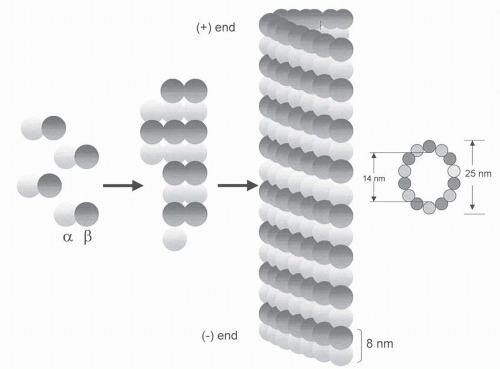
Microtubules are composed of tubulin, each of which is a heterodimer consisting of two tightly linked, closely related globular polypeptide alpha and beta subunits.1, 2, 3, 4, 5, 6, 7, 8,12 These protein subunits, α-tubulin and β-tubulin, each consist of approximately 450 amino acids with a molecular weight of 50 kDa and are encoded by small families of related genes.1,2,4,5,14, 15, 16 When tubulin molecules assemble into microtubules, they form linear “protofilaments” with the dimers aligned side by side around a hollow central core and the β subunit of one dimer in contact with the α-tubulin subunit of the next, as shown in Figure 13-1. Each microtubule is composed of 13 protofilaments. Typically, the protofilaments arrange themselves in an imperfect helix with one turn of the helix containing 13 tubulin dimers each from a different protofilament. These tubes can grow and shrink by the addition or removal of subunits from their ends. Because microtubules are composed of heterodimers, they are polarized, with α-tubulin exposed at the slow-growing minus end. The plus end terminates with β-tubulin, where the addition and removal of subunits are faster and net elongation occurs, whereas assembly is slow and net shortening occurs at the other negative end.

Microtubules, which are nucleated and organized by the microtubule organizing centers (MTOCs), most notably the centrioles and basal bodies, comprise an important element of the cellular cytoskeleton. Contained within the MTOC is another type of tubulin, γ-tubulin, which is distinct from the α– and β-subunits.17 γ-Tubulin combines with several other associated proteins to form a circular structure known as the γ-tubulin ring complex. This complex serves as a scaffold for the polymerization of α/β-tubulin heterodimers into microtubules. The complex essentially caps the minus end while microtubule growth continues away from the MTOC in the plus direction.

The functional diversity of microtubules is achieved through their association with various regulatory proteins, particularly the microtubule-associated proteins (MAPs), through variations in tubulin isotype composition, and through posttranslational tubulin modifications.16,18, 19, 20, 21, 22, 23, 24 The intrinsic dynamicity of microtubules is also influenced by the tubulin isotype composition, and the sensitivity of microtubules to both depolymerizing and polymerizing agents is also a function of the tubulin isotypes, their posttranslational modifications, and MAPs.19,21,22,24 There are at least six and seven isotypes of α– and β-tubulin, respectively, which are distinguished by their different C-terminal amino acid sequences, and are encoded by a large multigene family that has been highly conserved throughout evolution.16,18,19 Both α– and β-tubulin also undergo various posttranslational chemical modifications, including polyglutamylation, polyglycylation, phosphorylation, detyrosination, tyrosination, and acetylation, among others. These modifications produce structural and functional diversities that vary with the intracellular location of microtubules.16,21, 22, 23, 24

Modified regions of polymerized tubulin provide sites for the binding of MAPs, which regulate the dynamic behavior of cytoplasmic microtubules, generally by modifying their stability.7,20,22,23 The major classes of MAPs, which can be isolated from tubulin-rich brain tissues, include tau proteins (molecular weights, 40 to 60 kDa) and high-molecular weight proteins (200 to 300 kDa) whose members include MAP1, MAP1c (an adenosine triphosphatase [ATPase]), MAP2, MAP4, MCAK, Dis1/TOG, and XMAP215.7,20,22,23 Both classes of MAPs have two binding domains, one of which binds to microtubules. Because this domain also binds to free tubulin
molecules simultaneously, MAPs facilitate the initial nucleation step of tubulin polymerization. The other domain appears to be involved in linking the microtubule to other cellular components. Other MAPs that affect microtubule behavior include stathmin and catastrophin, which destabilizes microtubules; katanin that severs microtubules; cytoplasmic linker-associated proteins that facilitate interaction with the plasma membrane; and several types of motor proteins that transport vesicles along microtubules.7,10,20,22,23,25


FIGURE 13-1 Heterodimers of α-tubulin and β-tubulin assemble to form a short microtubule nucleus. Nucleation is followed by elongation of the microtubule at both ends to form a cylinder that is composed of tubulin heterodimers arranged head-to-tail in 13 proto-filaments. Each microtubule has an end where net addition of heterodimers takes place, called the plus (+) end, with β-tubulin facing the solvent, and a minus end (−) with α-tubulin facing the solvent.

Microtubule motor proteins, such as the dyneins (GTPases) and kinesins (ATPases), transform chemical energy into mechanical sliding force and move various solutes and subcellular organelles along microtubules.10,25 Motor proteins, which play critical roles in mitosis, premeiotic events, and organelle transport, are being evaluated as strategic targets for anticancer therapeutic development.10,25


Microtubule Function

The microtubules are primarily recognized as being principal components of the mitotic spindle apparatus that correctly separates, or segregates, the duplicate set of chromosomes into two daughter cells during cell division. The integrity of the mitotic spindle is required for cells to pass through various cell-cycle checkpoints, and errors in chromosome segregation trigger programmed cell death or apoptosis.6,7,12,26 The microtubules also play critical roles in many interphase functions such as maintaining cellular shape and serving as a scaffold for cellular organelles. Since they are capable of growing and shrinking, microtubules can generate force, and motor proteins allow organelles and other cellular constituents to move along the microtubule. The microtubules are also involved in intracellular transport, secretion, neurotransmission, and in relaying signals between the cell surface receptors and the nucleus.26, 27, 28, 29, 30

The unique functions of microtubules relate to their polymerization dynamics, which involve a constant shifting equilibrium between α-β-tubulin heterodimer subunits and microtubule polymers.1, 2, 3,5, 6, 7,15,22,30, 31, 32 Tubulin polymerization occurs by a nucleationelongation reaction, in which the slow formation of a short microtubule “nucleus” is followed by a rapid elongation of the microtubule by the reversible, noncovalent addition of α-β-tubulin heterodimers at it ends (Fig. 13-1). In essence, the microtubule polymer is in a dynamic equilibrium with the intracellular pool of α-β-tubulin heterodimers; free heterodimers are incorporated into the polymerized structure while heterodimers are simultaneously released into
the soluble tubulin pool. Microtubule assembly and disassembly occur simultaneously at both ends of the microtubule. The rates of these processes and net direction of microtubule growth are determined by the concentration of free tubulin, capping by a MTOC, and several chemical mediators that promote assembly (e.g., Mg2+, GTP) or disassembly (Ca2+).20,22,24,31, 32, 33, 34

Each tubulin molecule is associated with two molecules of GTP. Tubulin binds GTP with high affinity. The nucleotide bound to α-tubulin at the N-site is nonexchangeable, whereas the other bound to β-tubulin at the E site can be exchanged with free guanosine diphosphate (GDP). The assembly process uses energy provided by the hydrolysis of GTP. As tubulin-GTP is added to the ends of growing microtubules, the β-tubulin subunit hydrolyzes GTP to both GDP and Pi once it is incorporated into the microtubule. The Pi ultimately dissociates from the microtubule, leaving a microtubule core that consists of tubulin bound to GDP. The GDP nucleotide remains nonexchangeable until the tubulin subunit dissociates from the microtubule. During elongation of the microtubule, GTP hydrolysis lags behind subunit addition, thus forming a “GTP cap” that stabilizes the plus end and promotes further extension of the polymer. However, when the rate of subunit addition decreases, hydrolysis catches up and the GTP cap is lost, promoting rapid depolymerization of the microtubule. The switch from growth to shrinkage is known as catastrophe, and the switch from shrinkage to growth is known as rescue. Although tubulin polymerization and dissociation occur simultaneously at each end, the net changes in length at the more kinetically dynamic plus end are much larger over time than those at the minus end. If the polymerization reaction is followed in vitro, an initial lag phase occurs, after which microtubules form rapidly until a plateau phase is reached. In intact cells, the negative ends of microtubules are usually anchored to a specific nucleating site or the MTOC, which, in most cases, is the centrosome, whereas growth occurs in the direction of the free plus end.

At any instant, cytoplasmic microtubules are either rapidly growing or catastrophically dissociating. Microtubules undergo long periods of slow lengthening, short periods of rapid shortening, and periods of attenuated dynamics. During rapid polymerization, the high concentration of free tubulin results in net assembly until a plateau phase is reached, at which time the concentration of tubulin falls to a critical level and the rates of both polymerization and depolymerization are balanced. Two processes are principally responsible for the unique functionality and dynamics of microtubules in the living cell. The first, known as treadmilling, is the net growth at one end of the microtubule and the net shortening at the opposite end.32,35 Treadmilling plays a role in many microtubule functions, most notably in the formation of the mitotic spindle and the polar movement and segregation of the chromosomes during the anaphase stage of mitosis. In the second dynamic process, known as dynamic instability, the plus ends of microtubules switch spontaneously between states of slow sustained growth and rapid shortening.36,37 Dynamic instability is dependent on cycles of GTP hydrolysis and exchange. A microtubule can grow as long as it maintains a stabilizing “cap” of tubulin-GTP. Once the GTP cap is lost from the plus end, the end loses subunits more rapidly.38 Depolymerization occurs approximately 100-fold faster at a GDP cap than a GTP cap, and therefore, once rapid depolymerization occurs, the GTP cap is difficult to regain. On the other hand, the minus end is bound tightly to the MTOC, which interferes with both assembly and disassembly of the subunits. In essence, the capping process represents an adaptation that results in microtubule stability at the capped end. The rate of dynamic instability is accelerated during some processes, such as mitosis, which enables the mitotic spindle to “reach out” and probe the three-dimensional space around the spindle, thus facilitating the attachment of the mitotic spindles to the chromosomes, chromosome capture, and alignment. The rates and magnitudes of both dynamic instability and treadmilling are much slower in purified tubulin than in cells. These mechanisms can be altered by MAPs and other regulatory proteins, by variable expression of tubulin isotypes and by posttranslational tubulin modifications.2,22

In the nonmitotic phases of the cell cycle, microtubules radiate from the MTOC, which is located centrally near the nucleus and consists of a centrosome, a lattice of MAPs, γ-tubulin, and a pair of centrioles. The minus ends of the microtubules are positioned in or near the centrosome, whereas the plus ends extend out toward the cell periphery. The centrioles and other components of the centrosome are duplicated in interphase cells, but they remain together on one side of the nucleus until the beginning of mitosis. At that point, the two centrosomes then separate and move to opposite sides of the nucleus, forming the two poles of the mitotic spindle.

As the cell enters mitosis and as the nuclear envelop breaks down and releases the now condensed chromosomes, the microtubules of the interphase array disassemble and are replaced by a new population of mitotic spindle microtubules, which are much more dynamic and whose MTOCs are the newly duplicated centrioles at each of the two poles of the cell.39,40 During this process, the dynamics of microtubule assembly and disassembly also change dramatically. First, the rate of microtubule disassembly increases about 10-fold, resulting in overall depolymerization and shrinkage of microtubules. At the same time, the number of microtubules emanating from the centrosome increases by 5- to 10-fold.

Dynamic instability and treadmilling are vital to the assembly and function of the mitotic spindle. The high dynamaticity of mitotic spindle microtubules is required for the precise alignment of the chromosomes and their attachment to the spindle during metaphase, as well as chromosome separation during anaphase. These processes enable microtubules, which emanate from each of the two spindle poles, to make vast growing and shortening excursions, ultimately attaching the plus end of the microtubules to the kinetochore of the chromosomes. This attachment selectively “caps” or stabilizes this end of the mitotic spindle. If even a single chromosome is unable to achieve a bipolar attachment to the spindle, perhaps from drug-induced suppression of microtubule dynamics, the cell will not traverse beyond a prometaphase/metaphase-like state, eventually triggering apoptosis. Although mitotic spindles form in the presence of low concentrations of antimicrotubule agents, mitosis cannot progress beyond the mitotic cell-cycle checkpoint at the metaphase/anaphase transition or is delayed in this stage.2,6,22,38 Such perturbations in mitotic spindle dynamics may delay cell-cycle progression at critical mitotic checkpoints, ultimately triggering apoptosis.2,6,22,26,39,40 In the unperturbed normal state, oscillations of the duplicated chromosomes, dynamic instability, and microtubule treadmilling, in which there is addition of tubulin to the spindle at the kinetochore and loss of tubulin at the spindle poles, exert considerable tension on the chromosomes in metaphase.33 Both tension and oscillations are required for the proper function
of the mitotic spindle and progression from metaphase to anaphase. In the next mitotic stage, anaphase, microtubules that are attached to the chromosomes undergo shortening, while another subpopulation called interpolar microtubules lengthen, resulting in polar movement of the chromosomes. During an unperturbed mitosis, mitotic exit, often termed slippage, is triggered by the rapid degradation of cyclin B1. However, at this critical juncture, perturbations in microtubule dynamics, due to pharmacological disruption of treadmilling and dynamic instability of spindle microtubules, may reduce spindle tension and impede cell-cycle progression from metaphase to anaphase, as well as mitotic exit, ultimately triggering cell death.2,6,22,26,39,40


Vinca Alkaloids

The vinca alkaloids are naturally occurring or semisynthetic nitrogenous bases that are present in minute quantities in the pink periwinkle plant Catharanthus roseus G. Don (formerly Vinca rosea Linn). The early medicinal uses of C. roseus for controlling hemorrhage, scurvy, toothache, and diabetes, and for the healing of chronic wounds led to the screening of these compounds for their hypoglycemic activity, which turned out to be of little importance compared with their anticancer properties.41, 42, 43, 44, 45, 46, 47 Although many vinca alkaloids have been investigated clinically, only VCR, VBL, and vinorelbine (VRL) are approved for use in the United States. A third widely studied vinca alkaloid, vindesine (VDS, desacetyl VBL carboxyamide), a semisynthetic derivative and human metabolite of VBL, was introduced in the 1970s. It has been used in combination with other agents, particularly the platinating agents and/or mitomycin C (or both), to treat non-small cell lung cancer, but VDS is also active in other hematologic and solid malignancies.7,29,43,44 Although VDS demonstrated notable activity against several tumor types, particularly non-small cell lung cancer, it has been available only for investigational purposes in the United States and has not demonstrated a unique role in cancer therapeutics.43,44 The semisynthetic VBL derivative vinorelbine, VRL (5′-norhydro-VBL), which is structurally modified on its catharanthine nucleus, is approved in the United States as either a single agent or in combination with cisplatin to treat non-small cell lung cancer and has also been registered for advanced breast cancer in many other countries.7,29,45, 46, 47, 48 In addition to demonstrating broad antitumor activity as a single agent and some evidence that it may not be completely cross-resistant with VCR and VBL, VRL can be administered orally, in contrast to other available vinca alkaloids.49 More recently, vinflunine (VFL), which is a bifluorinated vinca alkaloid, has demonstrated notable clinical activity in bladder cancer, as well as preliminary activity in breast, lung, and other cancers, but despite these activities, unique roles for VDS and VFL relative to other vinca alkaloids have not yet been demonstrated.50, 51, 52, 53, 54 The key features of these vinca alkaloids are listed in Table 13-1.


TABLE 13.1 Key features of the vinca alkaloids


Vincristine sulfate


Vinblastine sulfate


Vindesine sulfate


Vinorelbine tartrate


Vinflunine ditartrate


Mechanism of action


Low concentrations inhibit microtubule dynamics (dynamic instability and treadmilling) High concentrations inhibit polymerization of tubulin


Standard dosage (mg/m2)


1-1.4 every 3 wk


6-8 every wk


3-4 every 1-2 wk


15-30 every 1-2 wk


320 every 3 wk


Pharmacokinetics and disposition


See Table 13-2


Principal toxicity


Peripheral neuropathy


Neutropenia


Neutropenia


Neutropenia


Neutropenia


Other toxicities


Constipation, SIADH, infusion site reactions, jaw pain


Thrombocytopenia, anemia, alopecia, peripheral neuropathy (mild), SIADH, constipation, diarrhea, nausea and vomiting, infusion site reactions, mucositis, alopecia, jaw pain


Peripheral neuropathy (moderate), alopecia, infusion site reactions, nausea and vomiting, diarrhea, SIADH, mucositis, alopecia, jaw pain


Peripheral neuropathy (moderate), anemia, thrombocytopenia, constipation, diarrhea, nausea and vomiting, fatigue, infusion site reactions, SIADH, mucositis, alopecia, jaw pain


Anemia, thrombocytopenia, fatigue, constipation, diarrhea, nausea and vomiting, mucositis, infusion site reactions, abdominal pain, peripheral neuropathy (mild), jaw pain


Precautions


Patients with abnormal liver function should be treated with caution. See section on dosage and schedule for specific dosing guidelines.


SIADH, syndrome of inappropriate antidiuretic hormone secretion.


Despite the minor structural differences between VCR and VBL, their antitumor and toxicologic profiles differ greatly. VCR is used more commonly in pediatric oncology than in adults with cancer, most likely owing to the higher level of sensitivity of pediatric malignancies and better tolerance of therapeutic VCR doses in children. VCR is an essential part of the combination chemotherapeutic regimens used for acute lymphocytic leukemia and plays an important role in the treatment of non-Hodgkin’s lymphoma. VCR-based combination regimens, particularly those in which VCR is administered with doxorubicin and dexamethasone (known as VAD) and occasionally other newer agents such as bortezomib, are used to treat multiple myeloma.29 VCR also plays a role in the treatment of Wilms’ tumor, neuroblastoma, medulloblastoma, osteosarcoma, Ewing’s sarcoma, and other types of pediatric sarcoma, and small cell lung cancer.29,41,42,44 The agent has also been used as a component of a combination regimen consisting of procarbazine, lomustine, and VCR (known as PCV) in the neoadjuvant, adjuvant, and advanced settings of several types of uncommon brain malignancies anaplastic including oligoastrocytoma and oligodendroglioma.55 In addition, VCR and VCR-loaded platelet transfusions are used occasionally to treatment refractory autoimmune thrombocytopenia, and VCR has been successfully used to treat other nonmalignant immunologically mediated disorders such as autoimmune hemolytic anemia, hemolytic uremic syndrome, thrombotic thrombocytopenia purpura, and steroid-dependent nephrotic syndrome.29,41,42,56 VBL has been an integral component of combination therapeutic regimens for germ-cell malignancies (with cisplatin and bleomycin, PVB) and Hodgkin’s disease (with doxorubicin [adriamycin], bleomycin, and DTIC, ABVD) and has been used in combination with other agents to treat Kaposi’s sarcoma and bladder, brain, and non-small cell lung and breast cancers.29 In addition to the clinically relevant antitumor activity of VRL in non-small cell and breast cancers, VRL has demonstrated activity in advanced ovarian carcinoma and lymphoma, but a unique role in the treatment of these cancers has not been defined.29,47 It has also been reported that VRL as single-agent treatment confers reasonable therapeutic benefit in elderly patients with advanced breast and lung cancers.29,46, 47, 48


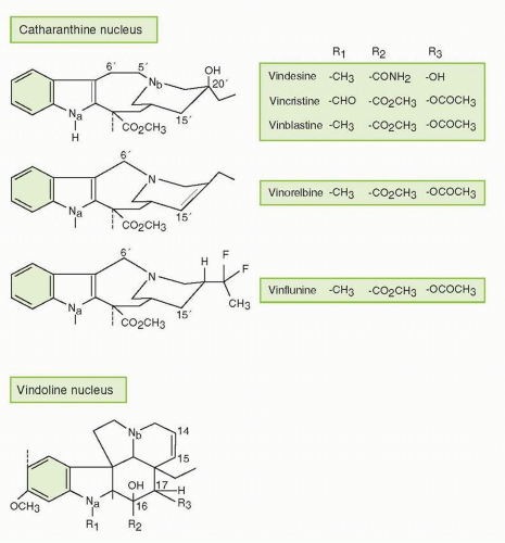
FIGURE 13-2 Structural modifications of the vindoline nucleus and catharanthine nucleus in various vinca alkaloids.


Structures

The vinca alkaloids have a large dimeric asymmetric structure composed of a dihydroindole nucleus (vindoline), which is the major alkaloid in the periwinkle, linked by a carbon-carbon bond to an indole nucleus (catharanthine), which is found in much lower quantities in the plant (Fig. 13-2). VCR and VBL are structurally identical except for the substituent (R1) attached to the nitrogen of the vindoline nucleus, where VCR possesses a formyl group and VBL has a methyl group. These small structural differences confer major clinical differences. VBL and VDS differ in two substituent (R2 and R3)
attached to the vindoline nucleus, whereas the catharanthine ring of VRL is modified. VFL is produced by a semisynthetic method that selectively introduces two fluorine atoms into the catharanthine ring at the 20′ position.


Mechanism of Action

The vinca alkaloids induce cytotoxicity by interacting with tubulin and disrupting microtubule function.2,3,5, 6, 7,29,30,41,42,47,50, 51, 52, 53,57, 58, 59 However, they affect other biochemical and biologic actions that may or may not be related to their effects on microtubules, such as the intracellular transport of amino acids; syntheses of RNA, DNA, and proteins; lipid metabolism; glutathione oxidation; cellular glycolysis; cellular release of hormones and pharmacological substances (e.g., antidiuretic hormone, histamine, epinephrine); calcium-calmodulin-regulated cAMP; and the integrity of the cellular membranes.7,27, 28, 29,41,47 Many of these effects occur only after treatment with superpharmacological concentrations that are not readily attained in vivo, whereas nanomolar concentrations, which are readily achieved in clinical practice, induce typical antimicrotubule effects. Although the vinca alkaloids preferentially disrupt proliferating cells and tissues, they also affect nonproliferating tissues that are rich in tubulin such as neurons and platelets.60

The cytotoxic actions of the vinca alkaloids are principally due to their effects on the mitotic spindle apparatus. In support of this mechanism, there is a strong relationship between cytotoxicity and the dissolution of the mitotic spindle.46 Furthermore, the accumulation of mitotic figures correlates with both drug concentration and duration of treatment. However, the vinca alkaloids and other antimicrotubule agents also perturb both malignant and nonmalignant cells in the nonmitotic cell-cycle phases, which is not surprising since microtubules are involved in many nonmitotic functions, including chemotaxis, migration, intracellular transport, movement of organelles, secretory processes, membrane trafficking, and transmission of growth factor signals from the cell surface receptor to the nucleus.15,61,62

The vinca alkaloids bind rapidly, avidly, and reversibly to sites on tubulin, known as the vinca domain, which appears to be the same binding sites for plant alkaloids such as maytansine, but distinct from those of the taxanes, GTP/GDP, and the site on the tubulin heterodimer shared with colchicine, podophyllotoxin, steganacin, combretastatin, and many synthetic compounds.2,3,5,7,30,31,41,51,53,54,57, 58, 59, 60, 61, 62, 63, 64, 65, 66, 67, 68, 69, 70 Unlike colchicine, the vinca alkaloids bind directly to microtubules without first forming a complex with soluble tubulin, and the vincas do not copolymerize with the tubulin lattice of the microtubule.2,3,5,65, 66, 67, 68

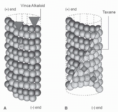
The vinca alkaloids bind to microtubules at two binding sites, each with different affinities including high affinity sites (Kd, 1 to 2 μmol) located at the ends of microtubules and low affinity sites (Kd, 0.25 to 0.3 mmol) sites located along the sides of microtubule surfaces (Fig. 13-3).3,6,65, 66, 67, 68 The binding of the vinca alkaloids to high-affinity sites is responsible for the substoichiometric and potent suppression of tubulin exchange that occurs at low vinca alkaloid concentrations (<1 μmol). This engagement disrupts treadmilling, dynamic instability, and other dynamic processes, but microtubule mass is not affected. Low concentrations of the vinca alkaloids strongly enhance dynamic instability at the minus end of microtubules, whereas dynamic instability is inhibited at the plus end. These actions increase the time that microtubules spend in a state of attenuated activity, neither growing nor shortening, the end result of which is a potent block at the metaphase/anaphase boundary in mitosis.2,3,5,55,56,65, 66, 67, 68


FIGURE 13-3 Antimitotic drugs bind to microtubules at different sites. A. A few molecules of the vinca alkaloids bound to high-affinity sites at the microtubule plus end are sufficient to suppress microtubule dynamics. B. A microtubule sliced away to show the interior surface. The taxanes binds along the interior surface of the microtubule, suppressing its dynamics.

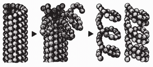
The binding of the vinca alkaloids at high stoichiometric concentrations (μmol) to low-affinity sites (Kd, 0.25 to 3.0 mmol) along the sides of microtubules is accompanied by reduced microtubule mass due to tubulin depolymerization. This type of binding induces tubulin to self-associate into nonmicrotubule tubulin polymers and ordered aggregates through a self-propagation pathway. Self-propagation occurs as vinca alkaloid binding progressively weakens the lateral interactions between protofilaments, induces conformation changes in tubulin, and exposes new sites. The exposure of new sites further increases the binding affinity of the vinca alkaloids and, in turn, results in the formation of vinca alkaloid-tubulin spiral aggregates, protofilaments, and paracrystalline structures, which ultimately results in the disintegration of the microtubules. MAPs stabilize the longitudinal interactions between dimers in the proto-filaments as they splay apart after binding to the vinca alkaloid, as illustrated in Figure 13-4.69 MAPS may also mediate the effects of the vinca alkaloids. For example, VBL increases the affinity of microtubules for the MAP stathmin, which destabilizes microtubules.71

The vinca alkaloids induce a block in mitosis at the metaphase/anaphase transition.2,3,6,7,27,57, 58, 59, 60, 61, 62, 63, 64, 65, 66, 67, 68, 69, 70 Following nuclear envelop breakdown, the vinca alkaloids block mitotic spindle formation and reduce tension at the kinetochores of the chromosomes. Although chromosomes may condense, they remain scattered in the cells. The chromosomes separate along their lengths but still remain attached at their centromeres.66,70 Following vinca alkaloid treatment, mitotic progress is delayed in a metaphase-like state with chromosomes “stuck” at the spindle poles, unable to move to the spindle equator.
The cell-cycle signal to the anaphase-promoting complex, which is required for the cell to transition from metaphase to anaphase, is blocked and the cells eventually undergo apoptosis. However, cyclin B concentrations may remain high and cell-cycle progression to interphase in the absence of anaphase or cytokinesis may occur, resulting in chromatin decondensation and formation of multilobed nuclei.2,3,6,64 Although treatment of cells with low concentrations of the vinca alkaloids may result in the disruption of spindle microtubule dynamics without microtubule depolymerization in mitotic cells, it can nonetheless lead to apoptosis, which involves the inactivation of antiapoptotic factors and induction of proapoptotic factors (see the sections on “Mechanism of Action” and “Mechanisms of Resistance” under “Taxanes”).2,3,6,7,22,26,51,52,54,70,72, 73, 74, 75, 76, 77, 78


FIGURE 13-4 Model of the vinca alkaloid-induced disassembly of microtubules containing MAP into spiraled protofilaments composed of one or two spirals. (Reprinted from Donoso JA, Haskins KM, Himes RH. Effect of microtubule proteins on the interaction of vincristine with microtubules and tubulin. Cancer Res 1979;39:1604, with permission.)

The processes that govern whether a cell dies during mitosis or exits mitosis in the presence of an antimitotic drug are not entirely clear. Several fates have been described for cells that exit mitosis in the presence of an antimitotic drug, including cell-cycle arrest, apoptosis, and cell-cycle progression. Although the molecular factors that govern these fates are not well understood, there is abundant evidence that p53 is involved, perhaps by restraining cell-cycle progression following exit from a prolonged mitotic block.6,26,73,74, 75, 76, 77 It is also becoming increasingly clear that the fate of the cell in response to drug is determined not only by events occurring during mitotic arrest but also by the consequences of events after mitotic exit, perhaps involving signaling pathways and cellular machinery that are principally operative during interphase including cdk1, cyclin B, p21 and various apoptosis proteins.6,26,73, 74, 75

The relationships between the antiproliferative actions of the vinca alkaloids and various relevant subcellular effects, such as mitotic arrest, mitotic spindle disruption, and microtubule depolymerization, have been characterized in a series of elegant studies.59 The antiproliferative effects of the vincas strongly related to the induction of both mitotic spindle disruption and metaphase arrest. These effects occur at the lowest effective drug concentrations with little or no microtubule depolymerization or disorganization of the mitotic spindle apparatus. With increasing drug concentrations, the organization of microtubules and chromosomes in arrested mitotic spindles deteriorates in a manner that is common to all vinca derivatives. The cumulative body of evidence data indicates that the antiproliferative effects of the vinca alkaloids at their lowest effective concentrations are caused by alterations in the dynamics of tubulin addition and loss at the ends of mitotic spindle microtubules rather than by depolymerization of the microtubules. Similar effects have been demonstrated with nocodazole, podophyllotoxin, and the taxanes.72,76,78,79

In addition to their direct cytotoxic effects on tumor cells, the vinca alkaloids and other antimicrotubule agents disrupt malignant angiogenesis with surprising potency.75,80, 81, 82, 83 In vitro, 0.1 to 1.0 pmol/L VBL blocks endothelial proliferation, chemotaxis, and spreading on fibronectin, all essential steps in angiogenesis.80,82 However, the relative contribution of these antiangiogenic effects to the antitumor activity of the vinca alkaloids in the clinic is unclear.

Additionally, the vinca alkaloids possess radiosensitizing properties in vitro related to their ability to induce cell-cycle block in the G2/M phase.84,85


Mechanistic and Functional Differences

With regard to the disruptive effects of the vinca alkaloids on microtubule dynamics, the naturally occurring vinca alkaloids VCR and VBL, the semisynthetic analog VRL, and the bifluorinated analog VFL impart similar actions, but they have distinguishing features as well.54,75,86,87 The vinca alkaloids differ in their tubulin-binding affinities: VCR > VBL > VRL > VFL.51,54 Consequently, the interaction between VFL and tubulin appears to be the most reversible, as shown by a greater reversibility of centrosome segregation after drug “wash-out” compared with the other vinca alkaloids, and this difference may be responsible, in part, for the differential effects of VFL on microtubule dynamics.51,54 However, the affinity for tubulin does not relate to the antitumor activity of the vinca alkaloid class. In fact, the relative tubulin-binding affinity may even be inversely correlated with their antitumor potencies in vivo.51 Although VFL has a lower overall tubulin-binding affinity and a lower potential to induce vinca alkaloid-tubulin spiral polymers than VCR, VFL is more active than VCR in a variety of murine and human tumor models.51,54,88,89 Moreover, the effects of VFL and VRL on microtubule dynamics differ from those induced by VBL and VCR in that they not only decrease the microtubule growth rate; increase the mean duration of a growth event; and increase the percentage of time that microtubules spend time growing; but VFL and VRL decrease the time spent in attenuation to a much greater extent. In contrast, VBL and VCR decrease the shortening rate and increase the time microtubules spend in an attenuated state to a much greater extent than VRL and VFL.51,86 Suppression of microtubule treadmilling also occurs with VFL treatment, but to a lesser degree compared with VRL and VBL.51,86


The explanation for the differential effects of the various vinca alkaloids on normal tissues and tumors is not clear. VCR, the most potent of the analogs in humans and the most neurotoxic, has the greatest affinity for tubulin.51,89 In contrast, VFL’s lower affinity for tubulin binding, as well as its greater potential intracellular sequestration, may contribute to its reduced incidence of peripheral neuropathy.51 Peripheral neurotoxicity, possibly due to drug-induced microtubule loss, steroid hindrance of MAPs, and/or altered microtubule dynamics in axonal processes, is a common adverse effect of first-generation vinca alkaloids.90 Although the vinca alkaloids may demonstrate similar potencies against preparations of tubulin derived from any given tissue, the differential sensitivities of various tissues to the vinca alkaloids are likely due to several factors.59,69,87,90, 91, 92, 93, 94, 95, 96 One possible factor is tubulin isotype composition, which is highly variable amongst tissues. Intracellular drug accumulation and tubulin binding vary according to tubulin isotype composition.18,19,97 Neurons are enriched in α-β-tubulin classes II and III, and the relatively high drug binding affinities for these isotypes may explain, in part, why the vinca alkaloids produce neurotoxicity.88,89,97 The variable potencies of the vinca alkaloids with regard to the induction of tubulin spirals also appear to relate to their relative neurotoxic potencies.88,89,97 In addition, the differences in the type and concentration of MAPs and posttranslational tubulin modifications between various tissues, which may influence drug interactions with tubulin, as well as differences in the cellular permeability and retention of the various vinca alkaloids, may affect the formation and stability of complexes formed between the vinca alkaloids and tubulin.27,59,86,89,91,92,98, 99, 100 For example, the higher cellular retention of VCR compared with VBL in cultured leukemia cells may explain why VCR is more potent than VBL after brief treatment periods, whereas these effects of the vinca alkaloids differ to a lesser degree with more prolonged exposure times.91,96,100, 101, 102, 103 Additionally, the vinca alkaloids directly inhibit palmitoylation of tubulin, and tubulin palmitoylation may relate to drug sensitivity.104 The intracellular concentration of GTP concentrations may also influence the type of interactions between the vinca alkaloids and tubulin, and variable vinca alkaloid retention among tumors may relate to GTP hydrolysis.101, 102, 103 Other factors that may explain why various tissues are differentially sensitive to the vinca alkaloid include differences in cellular pharmacology and pharmacokinetics, which is discussed in the next section.


Cellular Pharmacology

Although the vinca alkaloids are rapidly taken up into cells and accumulate intracellularly, steady-state intracellular/extracellular concentration ratios range from 5- to 500-fold depending on the cell type.91,93,99,103,105 In murine leukemia cells, the intracellular concentrations of VCR are 5- to 20-fold higher than the extracellular concentrations, and this ratio has been reported to range from 150- to 500-fold for other vinca alkaloids in both human and murine leukemia cell lines.96,105,106 In isolated human hepatocytes, VRL is more rapidly taken up and metabolized than other vinca alkaloids.96,105, 106, 107, 108 Although the vinca alkaloids are retained in cells for long periods and thus may have protracted cellular effects, there are marked differences in cellular retention amongst agents in this class.109, 110, 111, 112, 113, 114 Overall, the most important determinant of drug accumulation and retention is lipophilicity, although a number of other factors undoubtedly play a role.105,106 Drug uptake and retention also appear to be determined by tissue-specific and drug-specific factors, as illustrated by studies indicating that the accumulation and retention of VRL in neurons are less than other vinca alkaloids (see the section on “Mechanistic and Functional Differences” under “Vinca Alkaloids”).57,102 Recent studies using cellulose-purified tubulin from porcine brain have demonstrated the following pattern of binding affinities: VCR > VBL > VRL > VFL, which mirrors the relative potential of the vinca alkaloids to induce tubulin spirals.51,98 As discussed in the last section, the differential tissue uptake of the vinca alkaloid appears to be related to the tissue composition of tubulin isotypes, each possessing different binding characteristics, uptake kinetics, efflux pumps, and intracellular reservoirs for drug accumulation. Although the binding affinity for tubulin appears to be less for VFL than for the other vinca alkaloids, its intracellular accumulation has been demonstrated to be higher than for VBL, VCR, and VRL, which may be due to the sequestration of VFL in, as of yet, undefined intracellular compartments and its slower release over time.51,75,98

Temperature-independent, nonsaturable mechanisms, analogous to simple diffusion, seem to account for most transport, and temperature-dependent saturable processes are less important.27,30,74,93,105,106 Although both drug concentration and treatment duration are important determinants of drug accumulation and cytotoxicity, the duration of exposure above a critical threshold concentration is perhaps the most important determinant of vinca alkaloid cytotoxicity.96,107 Cytotoxicity is directly related to the extracellular concentration of drug when the duration of treatment is kept constant; for prolonged exposure to VCR, the concentration yielding 50% inhibition ranges from 1 to 5 nmol/L.107


Mechanisms of Resistance

Resistance to the vinca alkaloids develops rapidly in vitro with continuous exposure to these agents. Two types of mechanisms of resistance to the vinca alkaloids have been well characterized. The first mechanism is pleiotropic or multidrug resistance (MDR), which can be either innate (primary) or acquired. Although a large number of proteins mediate MDR, the best-characterized ones are the ATP-binding cassette (ABC) transporters, which transport a variety of substrates across cellular compartments and are encoded by a large transporter gene family.108 These intracellular and extracellular membrane-spanning proteins transport endobiotics and xenobiotics across membranes and confer resistance to the vinca alkaloids, taxanes, and other structurally bulky, natural product chemotherapeutic agents in vitro. The most extensively studied ABC transporters with respect to conferring resistance to the vinca alkaloids are the permeability glycoprotein (Pgp), or the MDR1 encoded gene product MDR1 (ABC Subfamily B1; ABCB1), and the multidrug resistance protein (MRP) (ABC Subfamily C2; ABCB1).108, 109, 110, 111, 112, 113, 114, 115, 116, 117, 118

MDR1 is a 170-kDa Pgp energy-dependent transmembrane transport pump that regulates the efflux of a large range of amphipathic hydrophobic substances, resulting in decreased drug accumulation. Pgp forms a channel in the membrane through which drugs are transported, and drug resistance is proportional to the amount of Pgp. Pgp is constitutively overexpressed by various normal tissues, including renal tubular epithelium, colonic mucosa, adrenal medulla, and other epithelial tissues. The efflux protein is also commonly
overexpressed by several human cancers, particularly those derived from tissues in which it is constitutively expressed (e.g., kidney and colon cancers). In the clinical setting, Pgp overexpression has been documented following treatment of patients with a variety of malignancies including lymphoma, leukemia, and multiple myeloma.

MDR1 confers varying degrees of cross-resistance to other structurally bulky natural products, such as the taxanes, anthracyclines, epipodophyllotoxins, actinomycin D (dactinomycin), and colchicine.119, 120, 121, 122, 123, 124 These cells may have homogeneously stained chromosomal regions or double-minute chromosomes, which indicates the presence of an amplified gene that codes for Pgp.110,111 The specific Pgp associated with resistance to the vinca alkaloids shows slight antigenic amino acid sequence differences and a different peptide map after digestion than does Pgp from cells selected for resistance to colchicine or paclitaxel.116,122 In fact, two forms of the protein are produced by a single clone of VCR-resistant cells, and these forms undergo posttranslational modifications, particularly N-glycosylation and phosphorylation, which results in further structural diversity. This diversity may explain the greater degree of resistance for the specific agent used to induce resistance compared with other MDR substrates, and it also may explain the variable patterns of resistance among cells with the MDR phenotype. The composition of membrane gangliosides in cancer cells resistant to the vinca alkaloids has also been shown to differ from that of wild-type cells.111 Although VFL is a substrate for Pgp, Pgp overexpression appears to be less involved in conferring resistance to VFL compared with other vinca alkaloids in various types of Pgpoverexpressing human cancers.118,125 The clinical ramifications of this resistance mechanism are not known, but VCR resistance, as assessed ex vivo, correlates with Pgp overexpression, particularly in childhood acute lymphoblastic leukemia (ALL).112

Resistance to the vinca alkaloids is also conferred by MRP1, which is a 190-kDa membrane-spanning protein that shares 15% amino acid homology with MDR1.116, 117, 118, 119, 120, 121, 122, 123, 124, 125, 126, 127, 128 The expression of MRP1, a member of the ABC protein family distantly related to Pgp, is found in many types of cancer and has been implicated as being responsible for the MDR phenotype in cancers of the lung, colon, breast, bladder, and prostate, as well as leukemia.116, 117, 118, 119, 120, 121, 122, 123, 124, 125, 126, 127, 128 Transient increase in MRP1 expression also correlates with resistance to MDR substrate drugs in cell lines transfected with MRP1.123,126 Amplification of MRP1 has been identified in several laboratoryderived cancer cell lines with elevated levels of MRP1 protein, as well as increased energy-dependent drug efflux.116, 117, 118,128 MRP1 has been shown to transport glutathione conjugates of several types of compounds, including alkylating agents, as well as etoposide and doxorubicin, but it only confers resistance to the latter agents. The MRP1 profile also encompasses resistance to methotrexate but confers a low level of resistance, if any, to the taxanes and colchicine.112,113,116,118,126, 127, 128 Also, MRP1 and other ABC transporters do not confer a significant resistance to VFL.118,125,129 Expression of MRP in transfected or selected cell lines is principally localized to the plasma membrane and endoplasmic reticulum, suggesting that MRP1 mediates resistance by affecting drug sequestration and/or vesicular transport.118,127 Several other ABC transporters have also been characterized in vitro, including several that enhance cellular resistance to the vinca alkaloids; however, their roles in conferring inherent or acquired resistance to the vinca alkaloids in the clinic are even less clear than those of MDR1 and MRP1.

Another important feature of MDR1 and MRP in vitro is that drug resistance may be reversed, in part, after treatment with various agents that have distinctly different structural and functional characteristics, such as the calcium-channel blockers, calmodulin inhibitors, detergents, progestational and antiestrogenic agents, antibiotics, antihypertensives, antiarrhythmics, antimalarials, and immunosuppressives.130, 131, 132 These agents bind directly to Pgp, thereby blocking the efflux of the cytotoxic drugs and increasing intracellular drug concentrations. Therefore, the role of MDR modulators has been a source of great contemporary interest, but the interpretation of clinical studies of resistance modulation has been confounded by the fact that MDR modulators, particularly MDR1 reversal agents, also enhance drug uptake in normal cells, decrease biliary elimination and drug clearance, and lead to enhanced toxicity.130, 131, 132 Overall, strategies aimed at reversing resistance to the vinca alkaloids in the clinic with pharmacologic modulators of both MDR1 and MRP1 have been disappointing, most likely due to the fact that many other proteins besides MDR1 and MRP1 occur in association with the MDR phenotype.92 Nevertheless, by characterizing the genetics and role of the ABC transporters in normal organ function and in the disposition of chemotherapeutic agents, there is a great deal to learn about how genetic polymorphisms in these proteins impact pharmacokinetics and drug toxicity.

The second well-characterized mechanism of vinca alkaloid resistance relates to tubulin isotypes. Mammalian cells have six α– and seven β-tubulin isotypes, whose expression may influence microtubule dynamics. Structural alterations in α– or β-tubulin due to either genetic mutations and consequential amino acid substitutions or posttranslational modifications, particularly phosphorylation and acetylation, have been identified in cancer cells with acquired resistance to the vinca alkaloids.18,19,57,69,117,118,133, 134, 135, 136, 137, 138, 139, 140, 141, 142 These alterations result in α– and β-tubulins that confer hyperstability to microtubule polymers and are collaterally sensitive to the taxanes and similar tubulin-stabilizing natural products (see “Mechanisms of Resistance” under “Taxanes”). Although the means by which tubulin alterations confer resistance to the vinca alkaloids are not entirely clear, this phenomenon is not apparently due to decreased binding affinity of the altered tubulins for drug.138, 139, 140, 141 Instead, alterations in α– and β-tubulins promote resistance to agents that inhibit microtubule assembly by increasing microtubule stability, perhaps by promoting longitudinal interdimer and intradimer interactions and/or lateral interactions between protofilaments.142

Decreased expression of class III β-tubulin, which increases the rate of microtubule assembly, in contrast to overexpression of class III β-tubulin, which is associated with rapid disassembly, is associated vinca alkaloid resistance in vitro.117,118,137,142, 143, 144, 145, 146, 147 Additionally, knockdown of either class II or IVb β-tubulin with siRNA hypersensitizes lung cancer cell lines to the effects of the vinca alkaloids, with the effects more pronounced following knockdown of class IVb β tubulin.118,137,147 A high level of class III β-tubulin expression also appears to independently predict a poor response and reduced survival in patients with non-small cell lung and breast cancer, following treatment with VRL or the taxanes.117,118,147, 148, 149, 150 These data suggest that class III β-tubulin may alter the dynamic
behavior of microtubules, as well as their sensitivity to inhibitors, but the overexpression of both class III and IVa β-tubulin appears to be more germane to conferring resistance to the taxanes and other microtubule-stabilizing agents (see the section on “Mechanisms of Resistance” under “Taxanes”). The expression of class II β-tubulin, which appears to be linked to p53 suppressor function, may also relate to vinca alkaloid resistance.117,151

Increased expression of MAPs, particularly MAP4, which promote microtubule assembly and hyperstability, perhaps by mechanisms similar to those linked with alterations in α– and β-tubulins, has also been associated with vinca alkaloid resistance.152,153

Various alterations in the principal components of the apoptotic pathway may confer resistance to the vinca alkaloids. Although much more work has been done in this area with the taxanes and various other microtubule-stabilizing agents (see the section on “Mechanisms of Resistance” under “Taxanes”), VFL resistance has been associated with down-regulation of the antiapoptotic factor Bcl-2, but this potential mechanism appears to be a cell type-dependent phenomenon.154 There is also experimental evidence suggesting that VBL and other antimicrotubule agents induce proapoptotic effects by phosphorylating the antiapoptotic factor Bcl-xL, thereby reducing the formation of Bax, whereas some VBL-resistance mutants that undergo reduced apoptosis have defects in BcL-xL phosphorylation.117,118,155


Clinical Pharmacology


Analytical Assays

The clinical pharmacology of the vinca alkaloids, which were largely studied decades ago, reflects the fact that sensitive, specific, and reliable analytic assays, capable of measuring the minute plasma concentrations that result from the administration of milligram quantity doses of these agents, were not available. Instead, pharmacologic studies were performed initially with radiolabeled drugs; however, interpretation of the results has been confounded by the chemical instability of these agents and the inability of such methods to differentiate metabolites from parent compound. Several vinca alkaloids, particularly VCR and VBL, may undergo spontaneous degradation under mild conditions, forming degradative products that can be separated using high-pressure liquid chromatography (HPLC).156 Radioisotopic methods coupled to HPLC were later used for improved separation of the various chemical moieties. Radioimmunoassay (RIA) and enzyme-linked immunosorbent assay (ELISA) methods, which can detect picomolar drug concentrations, have been developed157, 158, 159, 160, 161, 162; however, these methods also cannot reliably distinguish parent compounds from related derivatives. More recent technical advances in extraction and chromatographic detection (electrochemical and fluorescence) have made HPLC and gas chromatography the most feasible means of separating the vinca alkaloids from their metabolites. Tandem mass spectrometry used in conjunction with HPLC has enhanced the sensitivity of chromatographic methods.163, 164, 165, 166


TABLE 13.2 Pharmacokinetic parameters of the vinca alkaloids


Vincristine sulfate


Vinblastine sulfate


Vindesine sulfate


Vinorelbine tartrate


Vinflunine ditartrate


Standard adult dosage range (mg/m2/wk)


1.0-1.4 mg/m2/wk


6-8 mg/m2/wk


3-4 mg/m2/wk


15-30 mg/m2/wk


320 mg/m2 every 3 wk


Pharmacokinetic behavior


Triexponential


Triexponential


Triexponential


Triexponential


Triexponential


Elimination half-lives


α (min)


<5


<5


<5


<5


<5


β (min)


50-155


53-99


55-99


49-168


γ (h)


23-85


20-64


20-24


18-49


38.7


Clearance (L/h/kg)


0.16


0.74


0.25


0.4-1.29


0.64


Primary mechanism of disposition


Hepatic metabolism and biliary excretion


Hepatic metabolism and biliary excretion


Hepatic metabolism and biliary excretion


Hepatic metabolism and biliary excretion


Hepatic metabolism and biliary excretion


Pharmacokinetics


General

The vinca alkaloids are most commonly administered intravenously as a bolus injection or brief infusion. Table 13-2 summarizes the pharmacokinetic characteristics of the vinca alkaloids, which generally exhibit triphasic distribution.29 Pharmacokinetic characteristics include large volumes of distribution, triphasic clearance from plasma after intravenous administration with high early systemic clearance rates followed by long terminal half-lives (t1/2), and extensive hepatic metabolism and biliary/fecal elimination of principal metabolites. At conventional adult dosages, peak plasma concentrations (Cpeak), which persist for only a few minutes, range from 100 to 500 nmol, and plasma levels remain above 1 to 2 nmol for long durations.29,94,95,160, 161, 162,167, 168, 169, 170 There is also large interindividual and intraindividual variability in their pharmacologic behavior, which has been attributed to many factors, including differences in protein and tissue binding, hepatic metabolism, and biliary clearance. In comparative studies of the vincas, VCR had the longest terminal t1/2
and the lowest clearance rate; VBL had the shortest terminal t1/2 and the highest clearance rate; and VDS had intermediate values.167, 168, 169, 170, 171 VRL and VFL are also in the intermediate range with regard to clearance.29,47,49,63,98,168, 169, 170 The longer terminal t1/2 and lower clearance rate of VCR may account, in part, for its greater propensity to induce neurotoxicity, but there are other determinants of tissue sensitivity, such as high tubulin affinity and the potential to induce tubulin spirals as discussed previously (see the section on “Mechanistic and Functional Differences” under “Vinca Alkaloids”).62

Although prolonged infusion schedules may avoid excessively toxic Cpeak values and increase the duration of drug exposure in plasma above biologically relevant threshold concentrations for any given tumor, there is little, if any, evidence to support the notion that prolonged infusion schedules are more effective than bolus schedules.107,173 This approach has primarily been directed at achieving plasma concentrations for relevant periods, since the duration of exposure to relevant concentrations is a principal determinant of cytotoxicity in vitro107,173; however, rapid distribution and high avidity of binding of the vinca alkaloids to its target are likely responsible for the efficacy of short administration schedules.


Vincristine

After conventional doses of VCR (1.4 mg/m2) given as brief infusions, peak plasma levels approach 400 nmol/L (Table 13-2).29,44,158,167, 168, 169, 170, 171, 172 VCR binds extensively to both plasma proteins (reported values in the range of 48% to 75%) and formed blood elements, particularly platelets, which contain high concentrations of tubulin and led, in the past, to the use of VCR-loaded platelets for treating disorders of platelet consumption.29,56 The platelet count has been inversely related to drug exposure.29,173 In dogs and rodents, the spleen accumulates VCR to a greater extent than any other tissue.29,130,157 Poor drug penetration across the blood-brain barrier has been documented in most studies.29 The low penetration of VCR across the blood-brain barrier and other tumor sanctuary sites can be attributed to its large size and the fact that it is an avid substrate for the ABC transporters, which maintain the integrity of these blood-tissue barriers.29,44,96,157,158,161,167, 168, 169, 170, 171, 172, 173 Pharmacologic inhibition of MDR, however, may allow entry of VCR into the brain.157,161,174,175 In humans, VCR concentrations in cerebrospinal fluid are 20- to 30-fold lower than in plasma and do not exceed 1.1 nmol/L.175

After standard doses of VCR administered intravenously as a bolus injection, plasma disposition is triphasic, with initial t1/2α values of less than 5 minutes because of extensive and rapid tissue binding. Consequently, the apparent volumes of distribution (Vd) are high (mean central Vd, 0.328 ± 0.1061 L/kg and Vdγ [Vd for the terminal γ phase] of 8.42 ± 3.17 L/kg), which indicates extensive tissue binding.167 Beta t1/2 (t1/2β) values range from 50 to 155 minutes, and gamma t1/2 (t1/2γ) values are even more variable, ranging from 23 to 85 hours, which suggests slow clearance from the tissue compartment.29,118,167, 168, 169, 170, 171, 172, 173, 174

Considerable interest has arisen in using protracted VCR administration schedules because prolonged infusions may closely simulate the optimal in vitro conditions required for cytotoxicity.107,115,173 For example, VCR concentrations of 100 to 400 nmol/L are achieved only briefly after bolus injection, and levels generally decline to less than 10 nmol/L in 2 to 4 hours. Exposure to 100 nmol/LVCR for 3 hours is required to kill 50% of L1210 murine or CEM human lymphoblastic leukemia cells, whereas treatment durations of 6 to 12 hours are required to achieve this degree of cytotoxicity at 10 nmol/L, and no lethal effects occur at VCR concentrations below 2 nmol/L.107,173 A 0.5-mg intravenous bolus injection of VCR followed by a continuous infusion at dosages of 0.5 to 1.0 mg/m2/d for 5 days results in steady-state VCR concentrations ranging from 1 to 10 nmol/L and terminal t1/2 after discontinuation of the infusions ranging from 10.5 hours (1.0 mg/m2) to 21.7 hours (0.5 mg/m2).173 Although peak VCR plasma concentrations achieved with prolonged infusions are lower than levels achieved with bolus injections, more prolonged schedules are associated with a greater duration of drug exposure above a critical threshold concentration.107,173 This reasoning involves relating drug exposure in plasma to cytotoxicity; however, drug exposure in tumor tissue is likely disproportionately higher because of extensive tissue distribution, avid tissue binding, and slow clearance of VCR from tissue compartments. Nevertheless, prolonged infusions of the vinca alkaloids are employed in several widely used chemotherapy regimens that have been associated with robust activity such as EPOCH, which is comprised of etoposide IV, doxorubicin IV, and VCR IV continuously on days 1 to 4, oral prednisone on days 1 to 5, and cyclophosphamide IV on day 5 of courses repeated every 3 weeks. Patients may also receive filgrastim subcutaneously days 6 to 19 or until blood counts recover.

VCR is metabolized and excreted primarily by the hepatobiliary system.95,157,158,170 Within 72 hours after the administration of radiolabeled VCR, approximately 12% of the radioactivity is excreted in the urine (at least 50% of which consists of metabolites), and approximately 70% to 80% is excreted in the feces (40% of which consists of metabolites).44,106,157,158,161,162,166, 167, 168,170, 171, 172, 173, 174, 175, 176, 177 VCR is rapidly excreted into bile with an initial bile to plasma concentration ratio of 100:1 that declines to 20:1 at 72 hours posttreatment.162 Metabolites accumulate rapidly in the bile, so that only 46.5% of the total biliary product is the parent compound.157 As many as 6 to 11 metabolites have been detected in both humans and animals.44,106,168,170, 171, 172, 173, 174, 175, 176, 177, 178 The nature of the principal VCR metabolites isolated from human bile, particularly 4-deacetylVCR and N-deformylVCR, and both 4′-deoxy-3′-hydroxyVCR and 3′,4′-epoxyVCR N-oxide, which have been identified after incubation of VCR with bile, indicates that the agent is principally metabolized by hepatic cytochrome P450 CYP3A.44,170,156,162,171,172,176, 177, 178 The importance of CYP3A in drug disposition is also supported by observations of enhanced clearance with phenytoin and carbamazepine that induce CYP3A4, and increased toxicity with CYP3A inhibitors, particularly itraconazole and related compounds.178, 179, 180, 181, 182, 183, 184 In addition, transfection of tumor cells with CYP3A4 increases resistance to VBL, whereas cancer cells selected for VBL resistance may show increased CYP3A4 activity.180 There has been conflicting, albeit sparse, evidence indicating that VCR Cpeak and AUC values directly related to the severity of neurotoxicity, but it appears that more inherent biological characteristics are more important in this regard (see the section on “Mechanistic and Functional Differences” under “Vinca Alkaloids”).41 The existence of polymorphisms in the P450 CYP3A5 isoform, which appears to metabolize VCR more efficiently than CYP3A4 in vitro, may account for lower rates of neurotoxicity and dosage reductions among African American patients with ALL
who are receiving treatment with VCR compared with Caucasian patients.185 Additionally, P450 CYP3A4 isoform polymorphisms have been associated with differential clinical outcomes in patients with Hodgkin’s lymphoma who undergo treatment with vinca alkaloids.186 P450 CYP isoform polymorphisms have also been related to clinical outcome in patients with non-small cell lung cancer undergoing treatment with VRL.187

VCR pharmacokinetics have been related to clinical outcome in children with standard risk ALL and the propensity for relapse following VCR treatment related to more rapid clearance and lower VCR exposure.188


Vinblastine

The pharmacologic behavior of VBL also reflects its extensive tissue binding and resembles that of VCR (Table 13-2). Although plasma protein binding has been reported to range from 43% to 99.7%, it most likely approaches the high end of this range.29,95,160,167,168,189, 190, 191 VBL binds extensively to formed blood elements, with 50% of radiolabeled drug bound primarily to tubulin-rich platelets, as well as to red and white blood cells, within 20 minutes after an intravenous injection.189, 190, 191

Plasma disappearance fits a triexponential pharmacokinetic model with a rapid distribution phase (t1/2α < 5 minutes) from rapid tissue binding.160 VBL is more extensively sequestered in tissues than VCR, as demonstrated by retention of 73% of radioactivity in the body 6 days after an injection of the radiolabeled agent.160 Values for t1/2α and t1/2β have been reported to range from 53 to 99 minutes and 20 to 24 hours, respectively.29,160,167 High steady-state levels and long terminal t1/2 values have been reported after 5-day infusions of VBL: 1.1 nmol/Lat 1 mg/m2/d (t1/2, 28 days);3.3 nmol/L at 1.7 mg/m2/d (t1/2, 3 days); and 6.6 nmol/L at 2 mg/m2/d (t1/2, 6 days).29,192

The principal mode of VBL disposition is hepatic metabolism and biliary excretion.29,160,167, 168, 169, 170,191,192 Over a 9-day period after treatment of dogs with radiolabeled VBL, 30% to 36% of radioactivity is recovered in bile and 12% to 17% is found in urine. Fecal excretion of the parent compound is relatively low, which indicates that metabolism is significant. In vitro studies indicate that the cytochrome P450 CYP3A isoform is primarily responsible for drug biotransformation. At least one metabolite, desacetylvinblastine, which may be as active as the parent compound, has been identified in both dogs and humans. Small quantities of desacetylvinblastine (VDS) also have been detected in both urine and feces.


Vindesine

Similar to the other vinca alkaloids, plasma disposition of VDS is characterized by a triexponential process with rapid distribution to peripheral tissues except for sanctuary sites that are protected by ABC transporters (e.g., brain and testes).29,44,94,159,161,167,193, 194, 195, 196, 197, 198, 199 Table 13-2 lists the principal pharmacokinetic parameters associated with for VDS.

The liver is the main organ responsible for VDS clearance, and CYP3A is the principal P450 CYP isoform involved in drug biotransformation.159,167, 168, 169, 170,193, 194, 195, 196, 197, 198, 199 Renal excretion accounts for only 1% to 13% of drug disposition.29,198


Vinorelbine

The pharmacologic behavior of VRL is similar to that of the other vinca alkaloids, and the decline of plasma concentrations following rapid injection has been characterized by biexponential and triexponential pharmacokinetic models (Table 13-2).29,47,63,200, 201, 202, 203, 204, 205, 206, 207 After intravenous administration, VRL concentrations decay rapidly, followed by a much slower elimination phase (t1/2γ, 18 to 49 hours). Plasma protein binding, primarily to α1-acid glycoprotein, albumin, and lipoproteins, ranges from 80% to 91%.29,106,166,168,170,200, 201, 202, 203, 204, 205

VRL is widely distributed, and high concentrations are found in virtually all tissues (tissue-to-plasma ratios of 20 to 80), except brain.29,106,166,168,170,200, 201, 202, 203, 204, 205 Platelet binding is also extensive. The wide distribution of VRL reflects the agent’s lipophilicity, which is among the highest of the vinca alkaloids. Tissue-to-plasma ratios range from 20 to 80, although VRL concentrations in human lung are 300-fold greater than plasma levels and 3.4- to 13.8-fold higher than lung concentrations achieved with VDS and VCR, respectively. Hepatic metabolism with biliary excretion of metabolites and VRD appears to be the principal mode of drug disposition. Approximately 33% to 80% of an administered dose of VRL is excreted in the feces, whereas urinary excretion represents 16% to 30% of total drug disposition, most of which is unmetabolized VLR.29,106,166,168,170,200, 201, 202, 203, 204, 205 The P450 CYP3A isoenzyme appears to be principally involved in biotransformation; primary metabolites include 4-O-deacetyl-VRL and 3,6-epoxy-VRL and several hydroxyl-VRL isomers, although up to 17 metabolites have recently been identified.29,106,166,168,170,200, 201, 202, 203, 204, 205,208 Most VRL metabolites are inactive, although a 4-O-deacetyl-VRL metabolite may be as active as VRL. Plasma concentrations of this metabolite are minute.

The total body clearance of VRL (1.2 L/h/kg) and t1/2γ values of approximately 26 hours were found to be the same in older and younger patients who had normal hepatic function.206 Clearance is reduced in patients with liver metastases that replace more than 75% of the organ; clearance can be predicted in such patients by the monoethylglycinexylidide clearance test, which assesses CYP3A4 function.207 Although VRL clearance is not accurately predicted by serum bilirubin concentrations, markedly elevated levels have been associated with significant reductions in clearance in the few patients studied.

VRL is active when given orally. In animals, 100% of total radioactivity is absorbed after the ingestion of tritium-labeled VRL. The bioavailability of the parent compound is 43% for powder-filled and 27% for liquid-filled capsules; the bioavailability of the gelfilled capsule is negligibly affected by food.49,209,210 Cpeak values are achieved within 1 to 2 hours after ingestion, and interindividual variability is moderate.


Vinflunine

Following intravenous administration to patients, VFL is eliminated in a triexponential or multiexponential manner, with a rapid initial clearance phase.53,98,211,212 Based on data from several phase 1 trials (Table 13-2), total clearance averages 43.9 L/min (0.64 L/h/kg) and the terminal t1/2 is 38.7 hours, which corresponds to a mean terminal phase volume of distribution of 2,422 L (35.1 L/kg). Interindividual variability in pharmacokinetic behavior is moderate. Similar to the other vinca alkaloids, VFL is distributed to all tissues, except for the brain and skin in rodents. In rats, distribution is rapid, with maximal tissue concentrations observed 30 minutes after administration in the liver > lung > heart > kidneys > spleen.98,213 VFL exhibited low-to-moderate binding to
rat and human plasma protein in vitro; 58.4% and 39.6%, respectively; there was no binding to α1-acid glycoprotein.98,213 Platelet binding is negligible (<5%).

VFL principally undergoes hepatic metabolism and biliary elimination of parent drug and metabolites (about two thirds), while one third of an administered dose undergoes urinary elimination (˜11% in the first 48 hours).53,211, 212, 213 Like the other vinca alkaloids, the P450 CYP3A4 isoform is primarily responsible for the metabolism of VFL.53,98,214 There are 11 known metabolites; however, the only active metabolite, 4-O-deacetylVFL, is formed and eliminated at a slower rate than the other metabolites and does not appear to accumulate between cycles.53,98,212, 213, 214


Drug Interactions

Pharmacokinetic interactions between the vinca alkaloids and other drugs, particularly cancer therapeutics developed over the last two decades, have not been studied in detail. Methotrexate accumulation in tumor cells is enhanced in vitro by the presence of VCR or VBL, an effect mediated by a vinca alkaloid-induced blockade of drug efflux; however, the minimal concentrations of VCR required to achieve this effect in myeloblasts (0.1 μmol/L) are achieved only momentarily in the clinic, and even higher concentrations are needed to enhance MTX uptake in lymphoblasts.215, 216, 217 Furthermore, the schedule of VCR followed by MTX has not demonstrated synergism in the L1210 murine leukemia cells.218 Cytotoxic synergy is noted with the sequence of MTX followed by VCR, but this interaction is not likely due to enhanced MTX uptake. Thus, very little justification exists for routine use of VCR pretreatment in high-dose MTX protocols. The vinca alkaloids also inhibit the cellular influx of the epipodophyllotoxins in vitro, resulting in less cytotoxicity, but the clinical significance of this observation is unclear.219 L-asparaginase may reduce the hepatic clearance of the vinca alkaloids, particularly VCR, which may result in increased toxicity. To minimize the possibility of this interaction, VCR should be given 12 to 24 hours before L-asparaginase. In a murine model, VCR prevented doxorubicin-induced cardiomyocyte death, possibly by retarding the onset of apoptosis in association with the delay of poly(ADP)ribose polymerase activation.220

Treatment with the vinca alkaloids has precipitated seizures associated with subtherapeutic plasma phenytoin concentrations, most likely due to induction of CYP3A.221,222 Reduced plasma phenytoin levels have been noted from 1 to 10 days after treatment with both VCR and VBL. Administration of the vinca alkaloids with erythromycin, clarithromycin, itraconazole, posaconazole, voriconazole, and other inhibitors of CYP3A may also lead to severe toxicity.178, 179, 180, 181, 182, 183, 184, 185,223,224 Concomitantly administered drugs, such as pentobarbital and H2-receptor antagonists, may also influence VCR clearance by modulating hepatic cytochrome P450 metabolism.217 Another potential drug interaction may occur in patients who have Kaposi’s sarcoma related to acquired immunodeficiency syndrome and are receiving concurrent treatment with 3′ azido-3′-deoxythymidine (AZT) and the vinca alkaloids, as the vinca alkaloids may inhibit glucuronidation of AZT to its 5′-O-glucuronide metabolite.225 Lastly, the significant interindividual and intraindividual variability of VCR pharmacokinetics in children has been attributed to the variable induction of P450 metabolism by P450-inducing corticosteroids.226


Doses and Schedules

The vinca alkaloids are most commonly administered by direct intravenous injection or through the side-arm tubing of a running intravenous infusion. Experienced oncology personnel should administer these agents because drug extravasation usually causes severe soft-tissue injury.


Vincristine

VCR is routinely administered to children weighing more than 10 kg (body surface area ≥1 m2) as a rapid (bolus) intravenous injection at a dose of 1.5 to 2.0 mg/m2 weekly, whereas 0.05 to 0.065 mg/kg weekly is commonly used in smaller children (<10 kg or body surface area <1 m2). For adults, the conventional weekly dose is 1.4 mg/m2 weekly. A restriction of the absolute single dose of VCR to 2.0 mg/m2, which is often referred to as capping, has been generally adopted, based on early reports of substantial neurotoxicity at higher doses. However, this restriction is largely empirical, and available evidence suggests that the practice of capping should be reconsidered.227 The fact that the cumulative dose may be a more critical factor than single dose has readily been appreciated; however, significant interpatient variability exists, and some patients are able to tolerate much higher VCR doses with little or no toxicity.228, 229, 230 This may be because of large interindividual differences in drug exposure, which may vary as much as 11-fold.231,232 Moreover, the safety and efficacy of treatment regimens that do not employ capping at 2.0 mg have been documented in adults.227,233 In any case, VCR dosage modifications should be based on toxicity, particularly peripheral and autonomic neuropathy. However, dosage should not be reduced for mild peripheral neurotoxicity, particularly if the agent is being used in a potentially curative setting. Instead, doses should be modified for manifestations indicative of more serious neurotoxicity, including severe symptomatic sensory changes, motor and cranial nerve deficits, and ileus, until toxicity resolves. In clearly palliative situations, dose reduction, lengthened dosing interval, or selection of alternative agents may be justified in the event of moderate neurotoxicity. A routine prophylactic regimen, consisting of stool softeners, dietary bulk, and laxatives, to prevent the consequences of autonomic toxicity, particularly severe constipation, is also recommended.

Based on in vitro data indicating that the duration of VCR exposure above a critical threshold concentration is an important determinant for cytotoxicity, prolonged infusion schedules have been evaluated.29,107,173 After a 0.5-mg/m2 intravenous injection of VCR, total daily VCR doses of 0.25 to 0.50 mg/m2 as a 5-day infusion are generally well tolerated.29 In children, the administration of VCR as a 5-day infusion has permitted a twofold increase in the dose that can be safely administered without major toxicity compared with bolus schedules.

VCR is a potent vesicant and should not be administered intramuscularly, subcutaneously, or intraperitoneally. Direct intrathecal injection of VCR or other vinca alkaloids, which has occurred as an inadvertent clinical mishap, induces a severe myeloencephalopathy characterized by ascending motor and sensory neuropathies, encephalopathy, and rapid death (see the sections on “Toxicity,” “Miscellaneous,” and “Vinca Alkaloids”).234,235 Administration of VCR 0.4 mg/d as a 5-day infusion by the hepatic intraarterial route also has been associated with profound toxicity, including disorientation and diarrhea.236


Although the issue has not been evaluated carefully, the major role of the liver in the disposition of VCR implies that dose modifications should be considered for patients with hepatic dysfunction.232,233 However, firm guidelines for dose modifications have not been established. A 50% dosage reduction is recommended for patients with plasma total bilirubin levels between 1.5 and 3.0 mg/dL and at least a 75% dosage reduction for serum total bilirubin levels above 3.0 mg/dL. Dosage reductions for renal dysfunction are not indicated.237 A routine prophylactic regimen to prevent the serious consequences of severe autonomic toxicity, particularly severe constipation, is also recommended.


Vinblastine

Although VBL has been administered intravenously on various schedules, the most commonly used schedule administers a bolus injection at a dose of 6 mg/m2/d in cyclic combination-chemotherapy regimens. Approved initial dose recommendations for weekly dosing are 2.5 and 3.7 mg/m2 for children and adults, respectively, followed by gradual dose escalation in increments of 1.8 and 1.25 mg/m2, respectively, each week based on hematologic tolerance. The recommendation is also that maximal weekly doses of 18.5 and 12.5 mg/m2 in adults and children, respectively, should not be exceeded; however, these doses are substantially higher than most patients can tolerate because of myelosuppression, even on less frequent treatment schedules. Because the severity of the leukopenia that may occur with identical VBL doses varies widely, VBL probably should not be given more frequently than once each week. Oral administration may result in unpredictable toxicity.238

Five-day continuous infusions of VBL have been used at dosages ranging from 1.5 to 2.0 mg/m2/d, which achieve plasma concentrations of approximately 2 nmol/L.29,239 However, little, if any, evidence exists, however, to support the notion that prolonged infusion schedules are more effective than bolus schedules.

Although specific guidelines have not been established, VBL dosages should be modified for patients with hepatic dysfunction, especially biliary obstruction, because of the importance of the liver in drug disposition (see “Doses and Schedules” and “Vincristine”). Dosage reductions in patients with renal dysfunction are not indicated.237


Vindesine

VDS has been administered intravenously on many schedules, including weekly and biweekly bolus and prolonged infusion schedules. The agent also has been given in fractionated doses as either an intermittent or a continuous infusion over 1 to 5 days. VDS is most commonly administered as a single intravenous dose of 2 to 4 mg/m2 every 7 to 14 days, which is associated with antitumor activity and a tolerable toxicity profile.29 Intermittent or continuous-infusion schedules usually administer VDS dosages of 1 to 2 mg/m2/d for 1 to 2 days or 1.2 mg/m2/d for 5 days every 3 to 4 weeks.29,167 More prolonged schedules (up to 21 days) also have been evaluated.

Specific dosing guidelines have not been established for patients with hepatic or renal dysfunction; however, the pharmacologic similarities of VDS and other vinca alkaloids and the increased toxicity of VDS noted in patients with abnormal liver function mandate dosage reduction for patients with severe hepatic dysfunction, especially biliary obstruction (see “Doses and Schedules” and “Vincristine”). Dosage modifications are not indicated for renal dysfunction.237


Vinorelbine

VRL is most commonly administered intravenously at a dose of 30 mg/m2 on a weekly or biweekly schedule as a slow injection through a side-arm port into a running infusion (alternatively, a slow bolus injection followed by flushing the vein with 5% dextrose or 0.9% sodium chloride solutions) or as a short infusion over 20 minutes.45, 46, 47, 48, 49,63,240 It appears that the more rapid infusions produce less local venous toxicity.29,240 An acceptable oral formulation, however, is not yet available. Other dosing schedules that have been evaluated include long-term oral administration of low doses and intermittent high-dose and prolonged intravenous infusion schedules.240 Like the other vinca alkaloids, VRL clearance is impaired in patients with hepatic dysfunction, and dosage reductions should be considered in this setting.206, 207, 208, 209 Recommendations include a 50% dosage reduction for serum total bilirubin concentrations between 1.5 and 3 mg/dL and a 75% dosage reduction for patients with plasma total bilirubin concentrations above 3.0 mg/dL. Dosage reductions are not recommended for patients with renal insufficiency.


Vinflunine

VFL is currently not registered in the United States or worldwide but a positive opinion based on the results of phase 2 studies in patients with transitional cell carcinoma of the urothelium following failure of platinum-containing chemotherapy has been issued by the European Medicines Agency.50, 51, 52, 53 Although early phase 2 studies of VFL were conducted at a dose of 350 mg/m2 in normal saline as a 10-minute infusion every 3 weeks, doses in subsequent studies in more heavily pretreated patients were reduced to 320 mg/m2.53,98,212 Dosing recommendations have not been formulated for patients with hepatic and renal dysfunctions; however, results from a phase 1 study involving 25 patients with either mild, moderate, or severe liver impairment indicated no differences in the disposition of either VFL or 4-O-deacetyl VFL compared to patients with normal liver function.241


Toxicity

The principal toxicities of the vinca alkaloids differ despite their structural and pharmacologic similarities. Peripheral neurotoxicity is the predominant toxicity of VCR, whereas myelosuppression predominates with VBL, VDS, VRL, and VFL. Nevertheless, peripheral neurotoxicity is often noted following cumulative treatment with all of the vinca alkaloids, inadvertent high-dose treatment, or in settings involving patients who are inordinately susceptible (see the sections on “Toxicity,” “Neurologic,” and “Vinca Alkaloids”). On the other hand, VCR can cause myelosuppression under the similar conditions. Several potential explanations for the selective effects in various normal and neoplastic tissues are discussed in “Mechanism of Action” under “Vinca Alkaloids” in this chapter.


Neurologic

May 27, 2016 | Posted by in ONCOLOGY | Comments Off on Antimitotic Drugs

Full access? Get Clinical Tree

Get Clinical Tree app for offline access