Antibodies


Antibodies


David A. Scheinberg

Todd L. Rosenblat

Joseph G. Jurcic

George Sgouros

Richard P. Junghans


Monoclonal antibodies (mAbs) are remarkably versatile agents with potential therapeutic applications in a number of human diseases, including cancer. Ten mAbs are now Food and Drug Administration (FDA)-approved mAbs for the treatment of cancer. MAbs have long promised to offer a safe, specific approach to therapy. Preclinical evaluation and human clinical trials have identified new strategies for the use of mAbs, as well as a number of obstacles to their effective application. Although the use of antibodies as targeting agents dates to the 1950s,1 clinical investigation of antibodies as a potential treatment for cancer could not be initiated until the late-1970s, when efficient and reliable methods for the production of mAbs were developed.2

Five approaches to mAb therapy are used in humans. First, mAbs can be used to focus an inflammatory response against a target cell. The binding of a mAb to a target cell can result in fixation of complement, which yields cell lysis or results in opsonization that marks the cell for lysis by various effector cells, such as natural killer (NK) cells, neutrophils, and monocytes. Second, mAbs may be used as carriers to deliver another small molecule, atom, radionuclide, peptide, or protein to a specific site in vivo. Third, mAbs may be directed at critical hormones, growth factors, interleukins, or other regulatory molecules or their receptors to control growth or other cell functions. Fourth, anti-idiotypic mAbs may be used as vaccines to generate an active immune response. Finally, mAbs may be used to speed the clearance of other drugs or toxins or fundamentally alter the pharmacokinetic properties of other therapeutic agents. For example, mAbs may be fused to drugs or factors to increase their plasma half-life, change their biodistribution, or render them multivalent. Alternatively, mAbs may be used to clear previously infused mAbs from the circulation.

Despite the diversity of approaches, significant problems remain that are peculiar to mAbs. MAbs are large, immunogenic proteins, often of rodent origin, that rapidly generate neutralizing responses in patients within days or weeks after their first injection. The sheer size of mAbs—150 kDa for immunoglobulin G to 950 kDa for immunoglobulin M, 100 times larger than that of typical drugs—makes their pharmacology (particularly diffusion into bulky tumors or other extravascular areas) problematic for effective use. Many early mAbs or mAb constructs were either poorly cytotoxic or relatively nonselective, which rendered them ineffective. Moreover, the high degree of mAb specificity that is routinely achievable may allow tumor cells that do not bear the specific antigen target to escape from cytotoxic effects. Tumor cells can also shed or modify the target to avoid mAb binding. Nonetheless, mAbs still have great potential to be safe and effective anticancer agents. Several areas have been defined in which mAbs can be effective, either alone or in combination with other, more conventional agents.

This chapter reviews the basic biochemical and biologic properties of mAbs and their most commonly used derivatives (immunotoxins, radioimmunoconjugates, mAb fragments), discusses the pharmacologic issues peculiar to mAbs, and outlines some of the important clinical results obtained with mAbs. Because mAbs and conjugates of mAbs represent many different drugs with characteristics that result from their origin (rodent or human), their isotypes, their structure, or the various conjugated toxic agents, generalizations about the properties of mAbs may not be possible. Treatment of cancer with mAbs is a rapidly changing field, and readers are encouraged to consult other reviews for more comprehensive discussions of individual areas.3


Immunoglobulin Structure

Immunoglobulins are separated into five classes or isotypes based on structure and biologic properties: immunoglobulin M (IgM), immunoglobulin D (IgD), immunoglobulin E (IgE), immunoglobulin A (IgA), and immunoglobulin G (IgG). IgM is the primordial antibody whose expression by the B cell on its surface represents the commitment of that cell to a particular but broad recognition space that subsequently narrows as part of the maturation response induced by antigen interactions.4 In some cases, the antibodies interact with specialized receptors that link their action to host cellular defenses; in others, the antibodies interact with the humoral complement system. IgG is further divided into four subclasses and IgA into two subclasses. Heritable deficiencies in individual immunoglobulin classes or IgG subclasses are associated with susceptibility to particular infections and autoimmune disorders.5 Table 25-1 summarizes various features of the antibodies discussed in this section.

The fundamental structural elements of all antibodies are indicated by size as heavy and light chains of 55 to 75 kDa and 22 kDa, respectively (Fig. 25-1). Light chains are either κ or λ and are each distributed among all immunoglobulin subclasses. Heavy chains are μ, δ, γ, ε, and α, corresponding to IgM, IgD, IgG, IgE, and IgA. The heavy chain isotype plays a role in defining the biologic characteristics of each antibody class. The amino-terminal domain of each chain is the variable (VH or VL) region that mediates antigen recognition; the remaining domains are constant regions designated CL for light chain and CH 1, CH 2, and CH 3 for heavy chain (and CH 4 for μ and ε). Between CH 1 and CH 2 is the hinge region, which
confers flexibility on the antibody “arms” and susceptibility to proteases (see later), except in IgM and IgE, in which the CH 2 domain itself serves this role.


TABLE 25.1 Properties of antibody classes


Property


IgG


IgA


IgM


IgD


IgE


Usual molecular form


Monomer


Monomer, dimer, etc.


Pentamer


Monomer


Monomer


Molecular formula


γ2κ2 or γ2λ2


(α2κ2)n or (α2λ2)n


(μ2κ2)5 or (μ2λ2)5


δ2κ2 or δ2λ2


ε2κ2 or ε2λ2


Heavy-chain domains


V, CH 1-3


V, CH 1-3


V, CH 1-4


V, CH 1-3


V, CH 1-4


Other chains


J chain, S piece


J chain


Subclasses


IgG1, IgG2, IgG3, IgG4


IgA1, IgA2


Heavy-chain allotypes


Gm (˜30)


Am (2)


Mm (2)


Molecular weight (Da)


150,000


160,000


950,000


175,000


190,000


Sedimentation constant (S)


6.6


7, 9, 11, 14


19


7


8


Carbohydrate content (%)


3


7


10


9


13


Serum level (mg/100 mL)


1,250 ± 300


210 ± 50


125 ± 50


4


0.03


Percentage of total serum Ig


75-85


7-15


5-10


0.3


0.003


Half-life (days)


23 (IgG3, 7)


5.8


5.1


2.8


2.5


Antibody valence


2


2, 4, 6,…


10


1 or 2


2


Complement fixation (classic)


+ (Ig G1, G2, G3)


++


Fc receptors


Fcγ R-I, Fcγ R-II, Fcγ R-III


Fcε R-I, Fcε R-II


Binding to cells


Monocyte macrophages,
neutrophils, LGLs


?


Mast cells


Other biologic properties


Secondary Ab response;
placental transfer


Secretory antibody


Primary Ab response;
B-cell surface Ig,
rheumatoid factor


B-cell surface Ig


Homocytotropic Ab;
anaphylaxis;
allergy


+, active; ++, strongly active; Ab, antibody; CH, constant region of the heavy chain; Ig, immunoglobulin; LGLs, large granular lymphocytes; V, variable region.


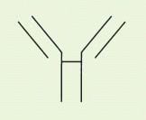
Heavy (H) and light (L) chains are normally paired 1:1 with each other, but the smallest stable unit is a four-chain (HL)2 structure (Fig. 25-1), for a nominal total mass of 150 to 160 kDa for IgG and higher for other isotypes (Table 25-1). IgE and IgG are composed of a single (HL)2 unit, whereas IgM exists as a pentamer of (HL)2 units joined by disulfide bonding with a third J-chain component. IgA exists mainly as a monomer in serum, but in secretions, it exists primarily as a dimer plus trimer and higher forms in which the oligomers are linked by J chain as well as the fragment of secretory chain (secretory piece) that is involved in the mucosal transport.


FIGURE 25-1 Antibody structure. The structural relationships and functions of domains of immunoglobulin G. (Reproduced from Wasserman RL, Capra JD. Immunoglobulins. In: Horowitz MI, Pigman W, eds. The Glycoconjugates. New York: Academic Press, 1977:323, with permission.)


FIGURE 25-2 Space-filling model of human immunoglobulin G1 antibody with CDRs in color representing anti-Tac-H; human myeloma protein Eu with CDRs grafted from murine anti-Tac. (Photo provided courtesy of Dr. C. Queen.) (Please see Color Insert.)

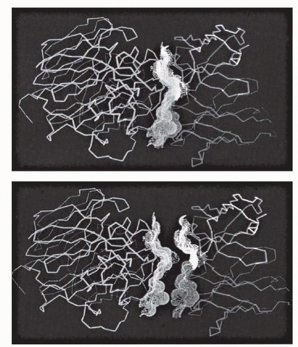
The V region itself is composed of subdomains—relatively conserved framework regions interdigitated with the complementarity-determining regions (CDRs; also termed “hypervariable segments” [HVSs]) that make primary contact with antigen (Fig. 25-1).6 Three CDRs are found in each heavy and light chain that may participate in antigen binding. The V regions should be seen as juxtaposed three-fingered gloves, with the CDRs covering the tips (Fig. 25-2), arrayed in a broad contact surface with antigen (Fig. 25-3).

Antibodies are glycoproteins. Glycosylation of proteins plays various roles related to solubility, transport, conformation, function, and stability. Carbohydrate is located mainly in C domains, with a lower frequency in V regions (see data on M195 later).7 IgG contains a major conserved glycosylation site in CH 2 that contributes to the conformation of this domain, which is crucial to the functional ability to bind to complement and to Fcγ receptors.

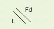
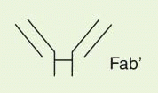
The IgG antibody “unit” has been defined in terms of susceptibility to proteases that cleave in the exposed, nonfolded regions of the antibody (Fig. 25-1). A summary of antibody fragments and engineered or synthetic products is presented in Table 25-2. Fab contains the V region and first C domain of the heavy chain (VH + C
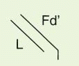
H 1 = Fd) and the entire light chain (L). Fab′ includes, in addition, a portion of the H chain hinge region and one or more free cysteines (Fd′). Fab′2 is a dimer of Fab′ linked through hinge disulfide(s). Fv is a semistable antibody fragment that includes only VH + VL, the smallest antigen-binding unit. Fc is the C-terminal crystallizable fragment that includes the complement and Fc receptor-binding domains (see below). Genetically engineered products include the δ constructs; these lack the second C domain of heavy chain and behave like Fab′2, with bivalence, abbreviated survival and lack of interaction with host effector systems, but they do not require enzymic processing.8 Another genetically engineered product, sFv (single-chain Fv), is Fv with a peptide linkage engineered to join the C-terminus of one chain to the N-terminus of the other for improved stability. More advanced products have been designed that conceptually represent the antigen-binding domain in a single peptide product9; this is not related structurally to an antibody and is therefore considered an antibody mimic.


FIGURE 25-3 Antigen-antibody binding surface juxtaposition. The variable (V) region (Fv) of antibody (right) binds to influenza virus protein neuraminidase (left) in the top panel. The VH (red) and VL (blue) regions are separately colored to show their respective binding contributions. The bottom panel offsets the two molecules by 8 Å to show the complementarity of surfaces that promotes the binding interaction. The stippled surface of the neuraminidase defines the antigen “epitope.” (Photo provided courtesy of Drs. P.M. Colman and W.R. Tulip, CSIRO Australia.) (Please see Color Insert.)


Ontogeny

Antibodies are a striking example of adaptation to the environment. This system allows the host to make molecules that bind antigens that the host has never encountered. The most diverse representation of classes and functions is found in Mammalia. The power of antigen recognition begins with an inherited array of duplicated and diversified germ-line V genes, a random mutational process that creates novel CDRs, a combinatorial selection process that amplifies the germ-line capabilities, and a controlled and directed mutational process that hones the specificity and matures the antibody into a high-affinity, antigen-specific reagent.

The biologic expression of antibody begins with the B-cell progenitor, which undergoes a series of maturation steps that begin with V gene selection for heavy chain followed by light chain V selection that yields surface expression and secretion by the mature B cell. On interaction with antigen, the B cells are activated
to proliferate, secrete antibody, undergo CDR mutagenesis and affinity maturation, and finally undergo chain switch and plasma cell differentiation. Plasma cells remain in tissues, spleen, or lymph nodes and secrete large quantities of antibody, which is the main function of this terminally differentiated cell.
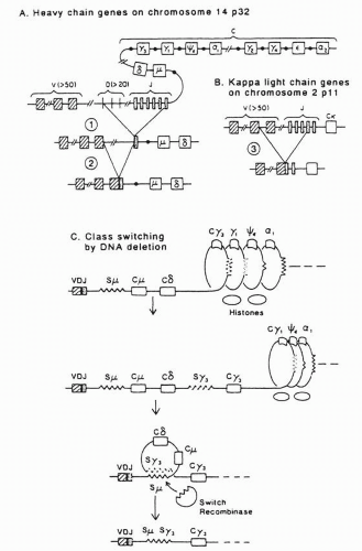
4


TABLE 25.2 Antibody fragment definitions


Designation


Representation


Description


Fabc


image


Complete immunoglobulin G


Enzyme-generated products


Fab


image


Papain digest; Fd + L


Fab′


image


Pepsin digest monomer; Fd′ + L


Fab′2


image


Pepsin digest dimer


Fv


image


V region digestion fragment; VH + VL


Fc (or Fc′)


image


C region digestion fragment; crystallizable fragment


pFc (or pFc′)


image


Smaller fragments of Fc


Genetically engineered products


δ CH 2


image


Deleted CH 2 domain; dimer of V – CH 1 – CH 3 + L


SFv


image


Single-chain Fv; VH and VL joined by peptide linker


Synthetic products


ABU


image


Antigen-binding unit; peptide mimic


C, constant region; CH, constant region of the heavy chain; L, light chain; V, variable; VH, variable region of the heavy chain; VL, variable region of the light chain.


The genes of heavy and light chains share important features of structure and maturation. Each gene locus contains widely separated V, C, and minigene domains that are placed into juxtaposition by DNA recombination mechanisms. The minigenes— diversity (D) and joining (J) regions for heavy chain and J regions for light chain—contribute to or constitute, with modifications, the CDR3.10 The κ and λ light chain loci are located on chromosomes 2 and 22, respectively, but all heavy chains are contained within a single massive locus on chromosome 14.

Germ-line diversity is essential to the generation of the antibody repertoire. On the heavy chain locus are an estimated 80 functional VH genes, 12 D regions, and 6 J regions for a potential of 6,000 combinations (Fig. 25-4).10, 11, 12 Roughly 80 Vκ light chain and 5 Jκ domains are found, which, randomly associated, can generate 400 combinations (the λ locus contains a smaller number of distinct V genes). A simple arithmetical calculation suggests that Vκ VH combinations alone could generate a diversity of approximately 2 × 10⁶. Yet even this number is conservative because this diversity is amplified in turn by errors in recombination and processes called N and P nucleotide addition in CDR3, which add enormously to the potential complexity. This, in theory, exceeds the total lifetime B-cell output by several orders of magnitude.10 Many authors, however, have cautioned that the mathematical diversity does not allow for the redundancy in configurations that could provide equivalent binding domains; in terms of antigen binding, the practical diversity is probably in range of 1 × 10⁷.

V gene selection is based on random expression followed by specific amplification. The argument has been made on physicochemical grounds that 105 different antibody molecules are sufficient to create a topologic set that recognizes any antigen surface with an
affinity of 105 to 106 M−1,13 a weak but biologically important number that corresponds to recognition affinities of naive antibody-antigen contacts that are often broadly polyreactive. B cells express antibody, principally IgM and IgD, on their membranes. On contacting antigen, these cells are stimulated to divide and undergo CDR mutations. Subsequent binding and stimulation are in proportion to the strength of the binding reaction; hence, an in vivo selection occurs for mutations that enhance the affinity of the antibody for the antigen, a process termed “affinity maturation.”14 Simultaneous with this increased affinity is a narrowing of the specificity, with the antibody shedding its early polyreactive phenotype. The cell then undergoes “class switch” to one of the mature antibodies (IgG, IgA, IgE) by deleting DNA between the VDJ region and the new C region of the heavy chain, which brings this new C domain in juxtaposition with the V region (Fig. 25-4). (The light chain is unchanged.) Some time after commitment to a mature antibody, the cell ceases its CDR mutagenesis, affinity maturation is completed, and the B cell undergoes morphogenesis to a tissue-resident plasma cell.4


FIGURE 25-4 Generation of diversity. VJ and VDJ joining occur in L chain and H chain by excision of intervening DNA in the genome. Class switch involves deletion of intervening constant (C) domains (Cμ, Cδ, etc.) and transcription through the new proximal C region. C is finally joined to the V gene by splicing of the messenger RNA. C, constant; J, joining; V, variable. (Reproduced from Cooper MD. Current concepts: B lymphocytes—normal development. N Engl J Med 1987;317:1452, with permission.)


Antigen-Antibody Interactions

Affinity is a quantitative measure of the strength of the interaction between an antibody and its cognate antigen, analogous to the equilibrium constant in the chemical mass action equation:


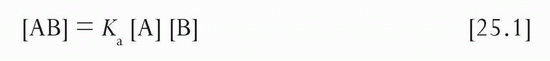
The equilibrium or affinity constant (Ka) is represented in units of M−1. In most instances studied by x-ray crystallography, contacts between antibody and protein antigen are dominated by noncovalent hydrogen bonds (O-H), with a lower frequency of salt bridges (COO− + H3N), for a total of 15 to 20 contacts. The effect of adding a new Hydrogen bond can be estimated from the free energy gain (0.5 to 1 kcal per mole) and from ΔG = −RT ln Ka to yield affinity increases of threefold to tenfold. Therefore, the affinity maturation that takes place (or affinity that may be lost in antibody engineering) changes quickly with a relatively small change in the number of bonds; that is, creating as few as three new hydrogen bonds may generate an affinity enhancement of more than 100-fold. This has been borne out by affinity changes that accompanied productive amino acid substitutions in V region engineering (see below). Of note, antibody affinities are generally much higher for protein antigens than for carbohydrate antigens, which may have less opportunity for hydrogen-bonding interactions (but are also “T-independent” antigens).

Although affinity and Ka directly express the binding potential of the antibody and are the most suitable measures for comparing affinities, the inverse of Ka, termed Kd or the dissociation constant, is expressed in molar units and indicates the concentration that is the middle of the range for the biologic action of the antibody:


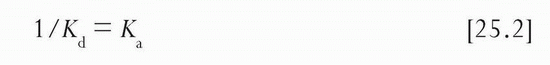
That is, Kd is the concentration of free antibody at which antigen is 50% saturated; if the antibody is in large excess, the input antibody concentration approximates the free concentration. Kd is a frequently used term, but its relationship to affinity must always be borne in mind; that is, low affinity equals high Kd, and high affinity equals low Kd. For example, a Ka of 2 × 10⁹ M−1 implies a Kd of 0.5 × 10⁻⁹ mol/L (0.5 nmol/L), or approximately 0.1 μg/mL antibody concentration for IgG. If antigen is in the picomolar (10−12 mol/L) range, this concentration of antibody will have half of the antigen saturated and half of the antigen will remain “free.” At 10-fold higher antibody concentration (1 μg/mL, 10 × Kd), antigen will be 90% saturated and 10% free, and at 10-fold higher concentration (10 μg/mL, 100 × Kd), antigen will be 99% saturated and only 1% unbound. A key point of understanding is that the ratio of antibody to antigen has very little impact on the degree of antigen saturation when antibody is in excess. If antibody concentration is 1 nmol/L with a Kd of 1 nmol/L, it does not matter whether antigen is 0.1 nmol/L at the Kd, 0.1 pmol/L, or 0.1 fmol/L; antigen in each case is 50% bound, although the ratio of antibody to antigen is 10, 104, and 107, respectively. Only the relation of free antibody to its Kd determines the degree of antigen saturation.

The affinity constant Ka is itself composed of two terms that describe the on-rate (forward; units of M−1 S−1) and off-rate (back; units of S−1) of the reaction:


To a first approximation, the forward rate is diffusion limited and is comparable for many antibodies reacting with macromolecules or cell-bound structures. Reactions of antibodies with haptens and other small molecules in solution are dominated by the faster linear and rotational diffusion rates of the smaller component.15 For example, when 0.1 nmol/L of dinitrophenyl-lysine (0.1 ng/mL) or 0.1 nmol/L of cell-bound HLA-A2 (50 ng/mL) is mixed with specific IgG antibody at 10 μg/mL (65 nmol/L), 0.1 second is required for the antibody to react with 50% of the antigen for the hapten but 4 minutes is required to react with the surface protein. Yet, they have virtually the same affinity constant.15 This similarity is due to the fact that the fast association rate is balanced by a fast dissociation rate for the hapten (clearance half-time [t1/2] = 0.7 seconds) whereas stability is longer for the protein antigen (t1/2 = 6 minutes).

Although exceptions exist, the on-rates of antibodies to protein and cell-bound antigens are primarily in this range and inversely proportional to antibody concentration for antibody in excess of antigen (i.e., at 1 μg/mL, the 50% on time would be on the order of 0.5 to 1 hour). Accordingly, differences in affinity between antibodies to the same cellular antigen are in many instances reflective of the off-rate (kb). For most purposes, an antibody is generally considered of “good” affinity if its Ka is equal to or greater than 109 M−1, where off-rate t1/2 values of an hour or more at 4°C are common. Association and dissociation times at 37°C are both accelerated relative to the times at 4°C, on the order of 5 or more, frequently with a net decrease in antibody affinity of 2- to 10-fold. Temperature effects on affinity must be explicitly tested, however, because in some instances, protein-ligand affinities are enhanced by higher temperature.16

The foregoing expresses basic principles of binding processes. A further important feature of antibodies is their multivalent structures. Although the on-rates for monovalent Fab and bivalent Fab′2 constructs are comparable, the bivalent off-times may be 10-fold longer than for the monovalent constructs, which yield affinities that are similarly enhanced.15 To discriminate the affinity that is intrinsic to the V region antigen interaction from the effective affinity in a bivalent or multivalent interaction, the latter is often referred to as avidity. For monovalent interactions, avidity equals affinity; for multivalent interactions, avidity is greater than or equal to affinity. Theory predicts avidity enhancements that vastly exceed observed numbers, but structural constraints undoubtedly restrain the energy advantage of multivalent binding.17,18 In the extreme, steric factors constrain some bivalent antibodies (e.g., anti-Tac)19 to bind only monovalently to antigens on cell surfaces although they will bind bivalently to antigen in solution. When antigens are presented multivalently on the surfaces of cells, viruses, or other pathogens, even the low-affinity IgM interactions can yield a high-avidity, stable binding to such targets in vivo.


Pharmacokinetics and Pharmacodynamics

The metabolism of immunoglobulins determines the duration of usefulness of antibodies in vivo. Under normal conditions, the serum levels of endogenous immunoglobulins are determined by a balance between synthetic and catabolic rates.20 When antibodies are administered as therapeutics, these catabolic rates effectively specify the dose and schedule necessary to maintain therapeutic blood levels when steady-state exposures are targeted. Table 25-1 lists the half-lives of human antibody survival in humans. IgG has the longest survival, 23 days (this value is for IgG1, IgG2, and IgG4; IgG3 survival is 7 days). A key element in the regulation of IgG catabolism is the Brambell receptor (FcRB), named after its discoverer, F.W.R. Brambell, who described this receptor more than 30 years ago (see Ref [21] for a review).21 This receptor is located in endosomes of endocytically active tissues, primarily vascular endothelium, which is mainly responsible for the catabolism of plasma proteins, including IgG. There, FcRB binds IgG, recycling it to the cell surface and diverting it from the pathway to lysosomes and the catabolism that is the fate of other, nonprotected proteins. In this role, FcRB is also termed the IgG protection receptor (FcRp). Yet FcRB is also responsible for transmission of IgG from mother to young, via yolk sac, placenta, or newborn intestine; in this manifestation, it is termed the IgG neonatal transport receptor (FcRn) for neonatal intestine, the tissue from which the receptor was initially cloned.

A substantial body of knowledge exists on the metabolism of immunoglobulins in various disease states. Conditions of protein wasting (enteropathies, vascular leak syndromes, burns), febrile states, hyperthyroidism, hypergammaglobulinemia, and inflammatory disorders are accompanied by significant acceleration of immunoglobulin catabolism.20 This information is of importance for understanding in vivo survival data in various clinical applications. In fact, the controlled conditions of testing immunoglobulin metabolism are rarely duplicated in practice, with antibody survivals typically shorter than suggested by the numbers above. Typically, murine antibody survival t1/2 values are in the range of less than 1 to 3 days, and antibodies with human gamma Fc domains (chimeric or humanized) have t1/2 values in the range of 1 to 15 days. Some of this acceleration in clearance is clearly due to disease-associated catabolic factors and to antigen binding in vivo, but subtle changes in the drug structure during product preparation may have a role in this acceleration as well. The influence of antigen expression on antibody clearance in vivo is considered later.

Antibody fragments have been studied because of their abbreviated survival and because their small size may translate into better tissue penetration. Fab and Fab′2 have survivals in vivo of 2 to 5 hours in mice, with comparable values in humans, and survival is dependent largely on kidney filtration mechanisms.20 Excretion is not based on size alone because the Fc fragment, which is comparable in size to Fab, is not filtered and has an in vivo survival of 10 days in humans. These rapidly catabolized fragments, like other filtered proteins, are largely absorbed in the proximal tubule and degraded to amino acids, which are returned to circulation. No intact immunoglobulin or fragments reenter circulation once filtered.22 In normal kidney, less than 5% of filtered light chain is excreted intact, whereas this fraction increases in the setting of renal tubular disease.23

The effect of circulating antigen on the efficacy of antitumor therapies has been evaluated. This was first encountered in antiidiotype therapies directed at the surface Ig on B-cell lymphomas, some of which secreted high levels of idiotype.24 Circulating idiotype prevented access of administered antibody to the idiotype on tumor cells, which effectively neutralized the drug, unless very high doses were given to overwhelm the secreted quantities of idiotype. Other targeted tumor antigens, including carcinoembryonic antigen (CEA),25 gangliosides GD2 and GD3,26,27 and Tac,28 also circulate at significant levels that might require adjustments to therapy.


Key observations include that there is (a) a direct relationship between the soluble antigen levels and the dose necessary to attain 50% (or 90% or 99%) bindability of administered antibody and (b) a predictable relationship between the rate of antigen synthesis and the time to antibody saturation.28 The actual partitioning of antibody between soluble and cellular antigen depends on several features, including the effective avidity of antibody for antigen on cells (which may be higher than for soluble antigen) and other factors influencing tumor penetration.

A special concern in this setting is that many soluble forms of antigen have short t1/2 values once shed from tumor cell surfaces, and the interaction with antibody may prolong their in vivo survival and increase their level in the whole body. When the target itself is a cytokine or cytokine receptor, adverse consequences conceivably could derive from the antibody treatment if the antigen retains activity in the antibody complex, as shown for interleukin-3, interleukin-4, and interleukin-7 complexes in vivo.29 If the antigen does not retain activity in complex, then this problem causes no concern because the free concentration of antigen cannot be increased by the presence of antibody, even after antibody is fully saturated with excess antigen. A different potential consequence of antigen load is that it may reduce transport of radioantibody to tumor for imaging or therapy. Studies have shown, however, that antibody can partition sufficiently between soluble and cellular antigen to yield targeting adequate for tumor-imaging purposes (CEA,25 Tac28).


Biologic Functions

Antibodies mediate several actions of potential therapeutic interest, some of which are part of the normal biologic function of antibodies and some of which are adapted in novel ways to the needs of specific settings.


Complement-Dependent Cytotoxicity

The complement (C′) system is at least as primitive evolutionarily as antibodies. One view is that the alternate (antibody-independent) pathway is the primordial system, which diversified to create the classic pathway to collaborate with antibody to direct the attack complex (C5-9) against antibody-coated targets. The most effective mediator of complement-dependent cytotoxicity (CDC) is IgM. Single IgM molecules can fix and activate complement on cell surfaces. In contrast, IgG-mediated CDC depends on the juxtaposition of pairs of IgG molecules to bind complement to cells,30 with substantially lower complement fixation and reduced killing efficiency relative to IgM. Human IgG1 and IgG3; mouse IgG2a, IgG2b, and IgG3; rat IgG2a; and rabbit IgG all fix and activate complement, whereas human IgG2 fixes C′ poorly, and human IgG4 and murine IgG1 normally do not fix C′.31 (Complement fixation depends on conserved residues in the CH 2 domain; short hinge regions are thought to hinder C1q access and binding with these latter isotypes.) Although human IgG3 fixes human C′ better than IgG1, actual target lysis may be better with IgG1 due to more efficient activation of C4.32 For the most part, human and murine antibodies function comparably well in directing CDC with rabbit C′ in in vitro tests and in fixing human or rabbit C′. Some cases may exist, however, in which murine IgG3 is more potent owing to the unique feature of Fc polymerization on cell surfaces apparently not being similarly available to human IgG.33

Despite these considerations, the impact of C′ fixation and CDC in therapeutic applications against cancer is uncertain. Two considerations may be relevant: first, the complete first component of human complement is very large (˜800 kDa) and, like IgM,20 probably has limited capacity to escape the blood stream and penetrate tissues. Second, the cells to which complement has ready access (e.g., cells of the hematopoietic system and vascular endothelium) are endowed with potent phosphoinositol-linked membrane protease activities, decay-accelerating factor (DAF, CD55), and homologous restriction factor (HRF, CD59), which act at steps subsequent to C1 fixation to inactivate the human complement cascade.34,35 Rabbit, guinea pig, or other heterologous sera can kill cells in vitro that are resistant to human C′ because they bypass the human species restriction of these protease activities.


Antibody-Dependent Cellular Cytotoxicity

In antibody-dependent cellular cytotoxicity (ADCC), target cells are coated with antibody and engage effector cells equipped with Fc receptors (FcRs) that bind to the Fc region of IgG, release cytolysins, and lead to cell killing. The only classes demonstrated to mediate ADCC are IgE and IgG. Because of the dangers of anaphylaxis that would be associated with IgE use, antitumor IgE antibodies are not likely to be developed, and further discussion therefore focuses on IgG.

Classically, cells that mediated ADCC were called “K (killer) cells,” and all bear FcRs on their surfaces. Five types of FcRs are defined; at least three IgG FcRs and two IgE FcRs. Table 25-3 lists the FcRs for IgG and their properties.36, 37, 38 All FcRs are capable of directing the ADCC of effectors against appropriate antibody-coated targets. Cytolytic mechanisms include perforins, a system closely related to the C9 protein of the complement attack complex (C5-9), serine proteases (granzymes) in large granular lymphocytes (LGLs); superoxides, free radicals, proteases, and lysozymes in monocytes-macrophages and granulocytes; tumor necrosis factor; and other components. As far as is known, the lytic mechanisms of LGLs and cytotoxic lymphocytes are similar or identical, and the monocyte-macrophage and granulocyte mechanisms also share numerous features. The monocyte-granulocyte mechanisms are probably adapted to killing and engulfment of microorganisms, whereas T cells, the closest lineage relative of LGL-NK cells, have the role of killing cells of self-origin that express foreign or neoantigens. In general, the most potent of the mediators of cellular killing in circulation have been the LGLs. These cells also perform natural killing (NK cells), which is an antibody-independent lectin-like ligand interaction system.39 Other effectors, for example, monocytes and granulocytes, have been shown to mediate ADCC against nucleated targets,40 but in most direct comparisons with LGLs, the LGLs were more potent than these numerically more dominant cells.41 Nevertheless, the most effective approaches may include a multipronged attack that enlists the collaboration of several cellular systems to kill antibody-coated tumor targets.

Among the features that influence the amount of killing in ADCC are (1) the species origin of the antibody, (2) the IgG subclass, (3) the number of antibody molecules bound per target cell, (4) the ratio of effector cells to targets, (5) the activation state of effectors, (6) the presence of irrelevant IgG, and, perhaps, (7) the presence of tumor cell protective factors, and (8) different classes of Fc receptors that enhance or inhibit effective activity.42, 43, 44 These are discussed in turn.


TABLE 25.3 FCγ receptors: properties and binding characteristics


Receptor


Size (kDa)


Ka


Cells


Mouse IgG


Human IgG


FcRI (CD64)


72


108


Mono, mac, act, PMN, eos


2a, 3


3 > 1 > 4


FcRII (CD32)


40


106


Mono, mac, gran, B cell, plt


2a, 2b, 1 (immune complexes or aggregates)


3, 1


FcRIII


50-70


5 × 10⁵


3, 2a > 1


3, 1 > 2, 4


(CD16A) transmembrane


LGL/NK, mac, few T, act, mono


(CD16B) PI-linked


PMN, act, eos


act, activated; eos, eosinophil; gran, granulocyte; LGL, large granular lymphocytes; mac, macrocytes; mono, monocytes; NK, natural killer cells; plt, platelet; PMN, polymorphonuclear leukocyte. Adapted from Van de Winkel GJ, Capel JA. Human IgG Fc receptor heterogeneity: molecular aspect and clinical implications. Immunol Today 1993;14:215; Simmons D, Seed B. The Fcγ receptor of natural killer cells is a phosphoinositol-linked membrane protein [later shown not PI-linked]. Nature 1988;333:568; Walker MR, Woof JM, Bruggemann M, et al. Interaction of human IgG chimeric antibodies with the human FcR1 and FcR11 receptors: requirements for antibody-mediated host cell-target cell interaction. Mol Immunol 1989;26:403.


  • The species origin appears to have a significant influence on the ability of an antibody to recruit human effectors to kill human tumors. Although human and rat antibodies mediate ADCC with human effectors, murine antibodies are often less potent in this role. Long ago, isologous antiserum was determined to be more effective with any species’ effector cells,45 which suggests that the match of antibody to effector cell Fc receptor is a significant feature of ADCC.


  • In principle, all IgG subclasses are capable of ADCC. However, the IgG1 subclass of humans, the IgG2a and IgG3 subclasses of the mouse, and the IgG2b and IgG3 subclasses of the rat have been inferred to be the most active with human effector cells. This does not always parallel the order of FcR binding affinity, and other factors of Fc-FcR binding must be postulated that influence the induction of cytolysis.46


  • The selection of highly expressed target antigens has a direct impact on the likelihood that the cell can be killed with ADCC. The control of the number of antibody molecules bound reveals that a nearly linear relationship exists with cytolysis.47 A corollary of this phenomenon is that the modulation of antigen by antibody binding (antigen-antibody complex internalization or shedding) reduces target susceptibility even when baseline antigen is highly expressed.


  • Higher effector-to-target ratios yield increased killing, although a plateau in efficacy typically is achieved at higher ratios.45,48 In vivo, the ratio of effector cells to targets is not so readily controlled, except by stimulating proliferation or supplementing effectors, but this effect may provide a stronger rationale for treatment in adjuvant settings when the tumor burden is small, or after debulking surgery or induction chemotherapy.


  • The activation state of effectors has been shown in several systems, both in vitro and in vivo, to play an important role in the lysis of targets. This activation is achieved with any of several agents and by the expression of different classes of activating or inhibiting Fc receptors.42, 43, 44 Evaluation of the application of cytokines specific to the range of potential effectors is beyond the scope of this review, but in each instance, cytolytic capacity has been strongly correlated with the degree of effector cell activation. Only LGLs (NK cells) appear to have significant antitumor potency in ADCC in the absence of cytokine activation, but here, too, activation with cytokines also increases ADCC killing (Fig. 25-5).48 Interleukin-2 (IL-2) activation of LGLs has been the most widely applied in clinical trials to date (see Chapter 36).


  • The presence of circulating IgG is probably the most problematic for exploiting ADCC in vivo due to its FcR blocking activity. The very existence of FcRs and the presence of cytolytic granules are indications of the relevance of this capability to biology. Although one might argue that monocytes-macrophages and granulocytes are adapted to combat microorganisms, the sole role of T cells is to kill nucleated cells of self that present viral or other abnormal peptides, for which they use distinct cytolytic mechanisms. As stated
    above, LGLs apparently duplicate the cytolytic mechanisms of T cells but, in addition, possess FcRs to enable them to interact with antibodies that will direct them to these targets through non-major histocompatibility complex mechanisms. The problem with this interaction is that monomeric Fc of IgG has an affinity of approximately 5 × 10⁵ M−1 (Kd = 2 mmol/L) for the dominant FcR on LGLs (Table 25-3). At a 1 g/dL in vivo concentration, IgG is 65 mmol/L and 30-fold above this Kd, which implies that more than 95% of the FcR on LGLs is occupied with IgG Fc. (The occupancy fraction on monocytes-macrophages with higher-affinity FcR type I is still higher.) Countering this in the biologic interaction is that the affinity of specific IgG for antigen is typically much higher than this, which yields a stable multivalent surface presentation of IgG Fc on the target that in turn may interact in a multivalent manner with the effector cell FcR. In practice, however, most ADCC assays are markedly inhibited by added human serum. (Assays using fetal calf serum are not thusly affected because IgG is absent due to lack of placental transport in ungulates.)20 Whether the longer-term in vivo incubations of days versus the brief duration (˜3 hours) of in vitro assays allow a therapeutic effect in a treatment program requires further study. The relevance of other immunoglobulins to tissuebased ADCC (as opposed to serum-based ADCC) is unclear because the concentration of antibody in the tissues is not well defined. However, observed clinical responses to antibody therapies (see below) suggest that ADCC may be operative in vivo.


  • Tumor-based factors may mediate resistance to ADCC. One such factor is the complement regulatory protein HRF. HRF was originally defined as acting at C2 and C9 of the complement cascade. The cytolytic protein perforin I that is released by LGL-NK cells and cytotoxic lymphocytes is also referred to as C9-related protein and is likewise subject to proteolysis by HRF, which thereby neutralizes the lytic power of the killer cell.49 (The observation has been made, however, that HRFrelated protein Ly6 [CD59] does not protect against perforin lysis.) HRF is present at high levels on activated T cells and NK cells and is thought to play a role in protecting these cells from autolysis during lysis of intended targets. Cells that are resistant to ADCC could be induced to become sensitive by blocking with anti-HRF antibodies.34 Another factor that may contribute to cellular resistance is the secretion of mucins, which inhibit the penetration of antibodies and other macromolecules to the cell surfaces. Other issues of tumor penetration are discussed later.


  • Finally, the relevance of the in vitro ADCC assay to in vivo function is much discussed. Only in a few instances has this been examined by comparing ADCC-competent and ADCCincompetent antibodies.44,50 In a complement-deficient leukemic AKR mouse model, a leukemia-specific IgG monoclonal antibody (mAb) suppressed tumor, whereas an IgM antibody of the same specificity was ineffective.51 This suggested that binding to antigen was not sufficient and that C′ played no role. Other reports of studies in mice showed that the only antibody to induce an in vivo response was that which showed ADCC activity in vitro.52 Several studies have shown that antibody plus IL-2 activation of effectors was much more effective than either alone, which implicates cooperation between the cellular and humoral immune systems that is exemplified by of ADCC. Human trials in which a leukemic patient received human IgG Fc coupled to a murine antibody showed a more effective response than when the antibody was without human Fc.53 A further human trial with class/isotype-switched Alemtuzumab antilymphocyte antibody showed a dramatic response in patients with B-cell chronic lymphocytic leukemia (CLL) only for the one isotype that mediates ADCC in vitro.54


FIGURE 25-5 Impact of interleukin 2 (IL-2) on ADCC after 16 hours of activation of peripheral blood lymphocytes. NK, natural killer cells. (Reproduced from Junghans RP. A strategy for evaluating lymphokine activation and novel monoclonal antibodies in antibody-dependent cell-mediated cytotoxicity and effector cell retargeting assays. Cancer Immunol Immunother 1990;31:207, with permission.)


Phagocytosis

Antibody-dependent phagocytosis may be mediated by cells of the granulocytic and monocyte-macrophage lineages. Furthermore, these cells have receptors for C3 fragments, which enhance binding of antibody-coated targets that also activate complement, leading to C3 fixation. Only activated macrophages, however, are capable of engulfing antibody-coated erythrocytes. The ability to engulf larger tumor cells has been uncertain, but one in vitro evaluation of activated monocytes demonstrated phagocytosis of melanoma and neuroblastoma targets when assays were appropriately monitored.55 Nevertheless, phagocytic cells in the liver (i.e., Kupffer cells) and spleen probably are the primary mediators of circulatory clearance of antibody-coated platelets in alloimmune and autoimmune settings56 and in the instances in which rapid clearance of leukemic cells was observed during antibody therapies. Whether these cells are trapped and then lysed by ADCC mechanisms rather than phagocytosis is uncertain.


Receptor Blockade

Antibody binding occurs without cooperation of other elements of the immune system. Therefore, just as antitoxin can prevent a toxin from acting at its target site in the body, antibody can also deny access of growth factors to tumors whose proliferation is factor dependent. This approach has been applied more widely in nonmalignant settings for the suppression of immune responses in autoimmune and alloimmune settings.57 This approach is limited in malignancy because most tumors appear to be autonomous. In principle, an antibody directed against a cytokine should have the same result. However, the short half-lives of most cytokines and the locally high antibody concentrations required may make this approach more difficult. Such autocrine or paracrine loops may be better interrupted by an antireceptor than by an anticytokine antibody. One report has documented a marked enhancement of cytokine activity by antibody to cytokine via t1/2 prolongation, which runs counter to the goal of suppressing cytokine activity.29

Design of such applications also must consider the receptor occupancy that is necessary for cell survival and proliferation. Only 10% occupancy of the receptor for granulocyte-macrophage colony-stimulating factor is sufficient to induce maximal activation of granulocytes.58 Similarly, one must block more than 90% of the α chain of IL-2 receptor with antibody to have a significant impact on IL-2-dependent, antigen-induced T-cell proliferation.
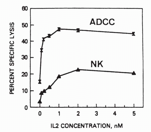
47

When the action of antibody is to attract effector cells, as with ADCC, then antigen expression correlates with target susceptibility. However, when the therapeutic action is to deprive a ligand access to its receptor, the level of expression of receptor antigen may have no bearing on the susceptibility of the target to antibody therapy. For example, the interleukin 2 receptor alpha chain (CD25)
confers high affinity binding of IL2 in conjunction the moderate affinity beta-gamma chain complex. On eosinophils, CD25 is present at levels that are below detection by flow cytometry or immunohistochemistry, yet antibody to the alpha chain is sufficient to ablate responsiveness of the eosinophils to interleukin-2 at physiologic concentrations.59,60 Similarly, much is made of the failure of the levels of epidermal growth factor receptor (EGFR) to predict responsiveness to anti-EGFR therapy, with “negative” tumors by immunohistochemistry being as responsive as positive tumor.61,62 This contrasts with another target of this family, Her2/neu, in which the responsiveness is correlated with increased receptor expression.63 The lesson here is that the receptor level that is detectable by standard methods is not necessarily reflective of the physiologic needs of the cell to respond to a receptor-ligand interaction, or predictive of the benefit to disrupting that interaction. Clinical testing of new antibodies should, therefore, not be restricted to patients whose tumors have been shown to express the target antigen.


Apoptosis

Apoptosis is a process by which signals are transmitted through cell surface receptors to induce autoenzyme-mediated cell death or by which downstream events are accessed to achieve the same result. This has been demonstrated most persuasively during development and in the programming of T-cell precursors in the thymus. The Fas antigen is probably the natural membrane receptor for this process and is expressed in liver, heart, thymus, lung, and ovary, although other antigens may exert similar effects. Anti-Fas antibody administration resulted in an extraordinarily complete and rapid tissue destruction in animal studies.64 One report suggests that part of the killing mechanism of T cells is to engage this receptor on target cells.65 Some hematologic malignant cells, like their normal counterparts, are Fas-positive and are potential targets of antibody therapy (a) if these antibodies are not cross-reactive for normal tissue, (b) if ways of engaging the receptor can be selectively achieved (i.e., bifunctional anti-Fas antitumor antibody), or (c) if other antigens unique to tumors can be found that also access this cellular process. To date, such tumor-specific, apoptosis-inducing antigens have not been described, but lineage-associated, apoptosis-inducing antigens have been targeted in the treatment of lymphoma (see below).


Ab2 Vaccines

Antibody recognition of antigen entails the presentation of a molecular surface that is the complement in space of the antigen (Fig. 25-3), termed a “mirror image.” In the Jerne network nomenclature, the designation of antigen and antibody becomes arbitrary. The antigen is Ab0, the antibody is Ab1, the antibody to the antibody idiotype is Ab2, and so forth. Although antibody can react with idiotype in many ways, a subset of Ab2 is still considered to exist that mimics Ab0 (antigen), and a fraction of Ab3 raised against Ab2 mimics Ab1 and reacts with Ab0 (antigen).66 Therefore, a tumor antigen may not be immunogenic in the human host that carries it, but a murine antibody (Ab1) can be raised to this antigen, and a goat antibody (Ab2) can be raised to this. This Ab2 antibody includes epitopes that mimic antigen but presents them in a novel context in which they may be immunogenic in the original host. Such Ab2s have been used as vaccines to induce Ab3 antibody responses in the host that can cross-react with antigen (Ab0) on tumor. Antibody therapy in this sense is applied to induce an endogenous antibody and occasionally a T-cell response against tumor.67,68


Immune Activation

The concept of an autogenous but weak immune response in some cancers, notably melanoma and renal cell carcinoma, underlies the application of antibodies as immune stimulators. This has been most prominently developed with blocking the T cell inhibitory receptor CTLA4 that competes with its homologous but activating partner CD28 on T cells in its interaction with B7 on antigen-presenting cells (APCs). By reversing CTLA4’s inhibitory function, tumor responses can be observed but also autoimmune responses.69,70 On the other side of this coin, superagonistic CD28 antibody TGN1412 was originally conceived to expand regulatory T cells to suppress rheumatologic disorders, but it was found in human tests to have potent activating effects on T cells generally.71 When the tolerable dose ranges are worked out for this agent, it may similarly find its way into an immune-stimulating role in cancer therapies.


Antibody Modifications for Therapy

As discussed later, despite all the functions that antibodies perform in vivo, not all antibodies are therapeutically successful. Two major factors have been studied for their role in resistance to antibody therapy: (a) immunogenicity and (b) lack of therapeutic and cytolytic potency. The approaches to improve potency have themselves been twofold: (a) to improve the collaboration of antibodies with the other components of the immune system and (b) to use the antibody as a vector to deliver toxic agents (toxins or radioactivity) to tumor cells. While modified antibodies may still activate intrinsic immune effectors, this latter approach does not depend on the immunologic collaboration of antibodies with the remainder of the immune system.


Reduction of Immunogenicity

Immunogenicity derives from the fact that most antibodies are of nonhuman origin and as such are foreign proteins in the human host. The human antiglobulin response to mouse antibodies is directed mainly against the C domains of the murine antibody, with typically lower titers against the V domains. To address this problem, three versions of the foreign protein have been prepared. First, there is a chimeric version, in which the mouse C domains are replaced by human C domains. Second, there is a “humanized” or hyperchimeric version, in which the murine framework regions are replaced by human framework sequences (Fig. 25-4). Third, entirely “human” IgG has been produced in vivo (in transgenic mice).72,73

The first humanized antibody for cancer therapy was the panlymphocyte antibody Alemtuzumab.74 Since that time, many others have been prepared and tested in humans. The hyperchimeric and humanized antibodies have been much more successful in avoiding antiglobulin75 responses, with an incidence of 4% with Alemtuzumab and comparably low antiglobulin response rates with
anti-Tac-H76 and HuM195.77,78 The less extensively substituted chimeric antibodies have been more widely applied, with anti-Vregion responses observed.66,79,80 Further experience is required to ascertain the rules governing these responses. The innovation of human combinatorial phage display libraries78,81 is also being applied for deriving antiself and antitumor human antibodies. Efforts to suppress human antimurine antibody with immunosuppressive drugs have not been successful to date. As more is learned about the induction of immune tolerance, additional strategies will be tested to prevent the host response to the therapeutic antibody. Finally, the lowest levels of immunogenicity were noted in the fully human anti-EGFR antibody, panitumumab, in the range of 0.5% by equivalent assays.82 Experience with more such antibodies will be necessary to generalize that human antibodies are better than humanized antibodies in this regard.


Improved In Vivo Survival

The importance of Fc to prolonged survival of IgG was revealed 60 years ago by Brambell and shown to be mediated by an MHC class-I related receptor on vascular endothelium that rescues endocytosed IgG and recycles it to the endothelial cell surface.82 Human IgG C domains (specifically CH2) can confer a longer in vivo t1/2, on the order of 23 days, for human IgG in humans versus the 1- to 3-day t1/2 of murine antibody in humans.20 One study compared a chimeric anti-idiotype antibody with the parental murine antibody, demonstrating a prolonged in vivo survival (with t1/2 longer than 10 days for a nonreactive chimeric construct).47 Other studies involving chimeric antibodies against colon carcinoma yielded survival t1/2 values of 3 to 6 days and 4 to 12 days.79,80 The humanized Alemtuzumab had survival t1/2 values of 1 to 6 days,75 and anti-Tac-H had values of 2 to 15 days.76 Thus, observed survivals fall short of those expected from controlled studies in normal volunteers. Some of this acceleration in clearance is clearly due to disease-associated catabolic factors,20 but in vivo antigen binding and product preparation may also play a role in rapid clearance of the antibody. Further studies are necessary to better understand the various factors that determine the t1/2 of the antibody in vivo.


Binding Affinity of Engineered Antibodies

The affinity for substrate (antigen) is an intrinsic characteristic of an antibody conferred by the particular amino acid sequence and spatial presentation of the CDRs. In the past, the affinity that was retrieved from a given hybridoma was an immutable feature of the antibody. Affinity could be altered only by reducing the valence (i.e., Fab versus Fab′2, IgG versus IgM), which invariably reduced affinity. IgG dimers, however, have shown a marked increase in affinity of up to 1,000-fold,83 which may improve Fc-dependent functions. With CDR manipulation in the humanization of antibodies, a major disturbance sometimes occurred in the affinity for antigen. Search for causes of this affinity loss revealed the importance of single, critical residues or carbohydrates to total affinity.84, 85, 86

Phage display technology offers a powerful tool to improve affinities by mutating the CDR’s. Fab molecules are expressed on the surface of phage and are selected against immobilized antigen and enriched in proportion to their affinity. This allowed a random CDR mutagenesis-selection procedure that recapitulates in vitro the in vivo process of affinity selection and maturation.87 By this procedure, the affinity for antigen of any low-affinity antibody can be enhanced 1,000-fold or more in a simple selection procedure. Finally, the issue of valence has been addressed to reduce the likelihood of antigen modulation during therapy by preparing univalent IgG that still retains the Fc effector domains and Fc-dependent functions.88,89


Complement-Dependent Cytotoxicity

The opportunities and problems of CDC as a means of killing tumors were outlined earlier. The humanization of antibodies has in some instances shown an improvement in cellular killing with heterologous complement, but for the most part, the effect has not been dramatic and is not sufficiently potent to kill human tumor cells with human complement when murine antibodies failed. On the other hand, the principle of the relative advantage of human over mouse antibodies has been clearly violated by occasional observations of failure to fix C′ with chimeric human IgG1 and IgG3 molecules when the murine antibodies have fixed C′.47,86,90 The humanization of IgG antibodies is not expected to render them as effective as IgM antibodies for CDC killing. In addition, the possibility exists that the murine IgG3 may be more potent than any human IgG given its capacity for polymerization on cell membranes with enhanced C′ fixation and lysis.33 Finally, dimeric forms of human IgG1 antibodies have been engineered that are far more effective in fixing complement than monomeric versions,91,92 but none have yet been tested for these effects in humans.


Antibody-Dependent Cellular Cytotoxicity

In contrast to CDC, ADCC with human antibodies show a fairly consistent advantage over mouse antibodies by the improved efficacy of their interaction with human effector cells. In several tests, a marked increase is seen in the potency of cellular killing in the chimeric constructs with human Fc domains. Of the human isotypes, IgG1 has been consistently the most effective, and chimeric and humanized antibodies are equivalent when normalized to molecules bound per target cell.47

Only gold members can continue reading. Log In or Register to continue

Stay updated, free articles. Join our Telegram channel

May 27, 2016 | Posted by in ONCOLOGY | Comments Off on Antibodies

Full access? Get Clinical Tree

Get Clinical Tree app for offline access