Alkylating Agents


Alkylating Agents


PART A CLASSICAL ALKYLATING AGENTS

Stanton L. Gerson

Alina D. Bulgar

Lachelle D. Weeks

Bruce A. Chabner

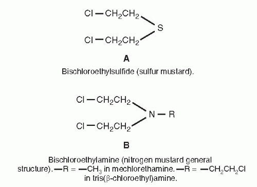
The alkylating agents are antitumor drugs that act through the covalent binding of alkyl groups to cellular molecules. This binding is mediated by reactive intermediates formed from a more parent alkylating compound. Historically, the alkylating agents have played an important role in the development of cancer chemotherapy. The nitrogen mustards mechlorethamine (HN2, “nitrogen mustard”) and tris(β-chloroethyl)amine (HN3) were the first nonhormonal agents to show significant antitumor activity in humans.1,2 The clinical trials of nitrogen mustards in patients with lymphomas evolved from the observation that lymphoid atrophy, in addition to lung and mucous membrane irritation, was produced by sulfur mustard during World War I. Antitumor evaluation3 showed that the related but less reactive nitrogen mustards, the bischloroethylamines (Fig. 14A-1), were less toxic and caused regression of lymphoid tumors in mice. The first clinical studies produced dramatic tumor regressions in some patients with lymphoma, and the antitumor effects were confirmed by an organized multi-institution study.1,2 This demonstration of efficacy encouraged further efforts to find chemical agents with antitumor activity, leading to the wide variety of antitumor agents in use today. Nonclassical alkylating agents include methylating agents such as procarbazine and temozolomide and are discussed later in this chapter. Alkylating agents, despite the enthusiastic development of targeted agents, continue to occupy a central position in cancer chemotherapy, both in conventional combination regimens and in high-dose protocols with hematopoietic cell transplantation (HCT). Because of their linear dose-response relationship in cell culture experiments,4 these drugs have become primary tools used in HCT for a variety of diseases. Better appreciation of resistance mechanisms and development of targeting agents to block these resistance pathways promise to improve the efficacy of alkylating agents.


FIGURE 14A-1 Structures of bischloroethylsulfide and bischloroethylamine. A. Bischloroethylsulfide (sulfur mustard). B. Bischloroethylamine (nitrogen mustard general structure).


Alkylating Reactions

An alkylation reaction can occur by two mechanisms: SN1 and SN2. In SN1 reactions, the rate-limiting step is the formation of a carbonium ion that can react rapidly with a nucleophile. This reaction follows first-order kinetics with a rate that depends solely on the concentration of the alkylating agent. In contrast, SN2 reactions follow second-order kinetics and depend on the concentrations of both the alkylating agent and the nucleophile. Such reactions involve a transition-state entity formed by both reactants that decompose to form the alkylated cellular constituent. Agents such as chloroethylnitrosoureas, through a SN1-type of mechanism, can form covalent adducts with oxygen and nitrogen atoms in DNA. Compounds with SN2 predominant mechanisms, such as busulfan, tend to react more slowly, with little alkylation of oxygen sites. Because alkylating agents are designed to produce reactive intermediates, the parent compounds typically have short elimination half-lives of less than 5 hours.

As a class, the alkylating agents share a common target (DNA) and are cytotoxic, mutagenic, and carcinogenic. The activity of most alkylating agents is enhanced by radiation, hyperthermia, nitroimidazoles, glutathione depletion, and inhibition of DNA repair. They differ greatly, however, in their toxicity profiles and antitumor activity. These differences are undoubtedly the result of differences in pharmacokinetic features, lipid solubility, ability to penetrate the central nervous system (CNS), membrane transport properties, detoxification reactions, and specific enzymatic reactions capable of repairing alkylation sites on DNA.5, 6, 7 Application of techniques such as magnetic resonance imaging and mass spectrometry to the study of the alkylation mechanism and the chemical nature of the intermediates involved have led to a detailed understanding of these reactions.8,9 Such approaches, coupled with improved techniques for studying cellular damage10,11 and for determining mechanisms of detoxification,12 make it possible to predict sites of alkylation of an agent and allow scientists to understand and modify the biologic consequences of such alkylations.


TABLE 14A.1 Key features of selected alkylating agents


Cyclophosphamide


Ifosfamide


Melphalan


BCNU


Busulfan


Bendamustine


Mechanism of action


All agents produce alkylation of DNA through the formation of reactive intermediates that attack nucleophilic sites.


Mechanisms of resistance


Increased capacity to repair alkylated lesions, for example, guanine O6-alkyl transferase (nitrosoureas, busulfan)
Increased expression of glutathione-associated enzymes, including γ-glutamyl cysteine synthetase, γ-glutamyl transpeptidase, and glutathione-S-transferases
Increased ALDH (cyclophosphamide)
Decreased expression or mutation of p53


Dose/schedule (mg/m2)


400-2,000 IV


1,000-4,000 IV


8 PO qd × 5d


200 IV


2-4 mg qd


70-100 mg daily, on day 1 and 2 of a 28-day cycle


100 PO qd


Oral bioavailability


100%


Unavailable


30%


Not known


50% or greater


?


Pharmacokinetics
Primary elimination t1/2 (h)


3-10 (parent)
1.6 (aldophosphamide)
8.7 (phosphoramide mustard)


7-15 (parent)


1 (parent)


0.25-0.75a (nonlinear increase with dose from 170 to 720 mg/m2)


2-3 h


0.5 (parent)


Metabolism and excretion


Microsomal hydroxylation activates, then chemical decomposition


Microsomal hydroxylation activates, then chemical decomposition


Spontaneously decomposes


Decomposes to active and inert products; also P450-mediated inactivation


Enzymatic conjugation with glutathione


Chemical decomposition


Hydrolysis to phosphoramide mustard (active) and acrolein


Hydrolysis to iphosphoramide mustard and acrolein


20%-35% excreted unchanged in urine


Excretion as inactive oxidation products


Excretion as inactive oxidation and dechloroethylated products


Excretion primarily in feces


Toxicity


Bone Marrow


Acute, platelets spared


Acute but mild


Delayed, nadir at 4 wk


Delayed, nadir 4-6 wk


Acute and delayed marrow aplasia


Acute but mild


Other


Hemorrhagic cystitis, cardiac toxicity, IADH


Hemorrhagic cystitis, encephalopathy


Pulmonary fibrosis, renal failure, hypotension


Addisonian syndrome, seizures, pulmonary fibrosis, venoocclusive disease


Mucositis, infections, tumor lysis syndrome


Precautions


Use MESNA with high-dose therapy


Always coadminister MESNA


Decomposes if administered over < 1 h


Monitor AUC with high-dose therapy Induces phenytoin metabolism


a See reference 296.


AUC, area under the concentration time curve; BCNU, bischloroethylnitrosourea; IADH, inappropriate antidiuretic hormone syndrome; IV, intravenously; MESNA,
2-mercaptoethane sulfonate; PO, per os; t1/2, plasma half-life.


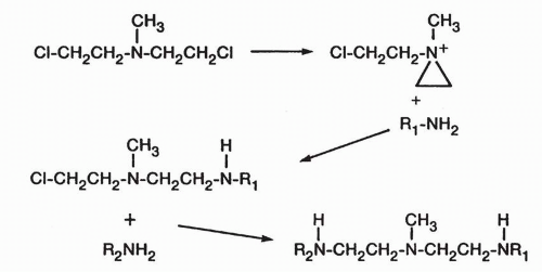
FIGURE 14A-2 Alkylation mechanism of nitrogen mustards. (From Colvin M. Molecular pharmacology of alkylating agents. In: Cooke ST, Prestayko AW. Cancer and Chemotherapy, vol 3. New York: Academic Press, 1981:291.)


Alkylating Agents Used Clinically

The important pharmacologic properties of the clinically useful alkylating agents are summarized in Table 14A-1.


Nitrogen Mustards

The prototypic alkylating agents have been the bischloroethylamines or nitrogen mustards. The first nitrogen mustard to be used extensively in the clinic was mechlorethamine (mustine) (Fig. 14A-1), sometimes referred to by its original code name HN2 or by the term nitrogen mustard. The mechanism of alkylation by the nitrogen mustards is shown in Figure 14A-2. In the initial step, chlorine is lost and the β-carbon reacts with the nucleophilic nitrogen atom to form the cyclic, positively charged, and very reactive aziridinium moiety. Reaction of the aziridinium ring with a nucleophile (electron-rich atom) yields the initial alkylated product. Formation of a second aziridinium by the remaining chloroethyl group allows for a second alkylation, which produces a cross-link between the two alkylated nucleophiles.


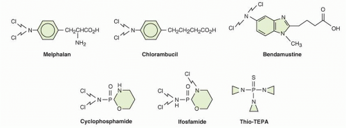
FIGURE 14A-3 Alkylating agent structures.

Numerous analogs of mechlorethamine were synthesized in which the methyl group was replaced by a variety of chemical groups that stabilized the molecule. Most of these compounds proved to have less antitumor activity than mechlorethamine, but many other derivatives have a higher therapeutic index, a broader range of clinical activity, and can be administered both orally and intravenously. These drugs, which for the most part have replaced mechlorethamine in clinical use, are melphalan (L-phenylalanine mustard), chlorambucil, bendamustine, cyclophosphamide, and ifosfamide (Fig. 14A-3). The latter two agents are unique in that they require metabolic activation and undergo a complex series of activation and degradation reactions (to be described in detail later in this chapter).

These derivatives have electron-rich groups substituted on the nitrogen atom. This alteration reduces the electrophilicity of the nitrogen and renders the molecules less reactive. Melphalan and chlorambucil retain alkylating activities and seem to be more tumor selective than nitrogen mustard. Cyclophosphamide and ifosfamide, on the other hand, possess no intrinsic alkylating activity and must be metabolized to produce alkylating compounds.

Cyclophosphamide remains the most widely used alkylating agent.13 It is an essential component of drug regimens for non-Hodgkin’s lymphoma (NHL) (CHOP—cyclophosphamide, doxorubicin, vincristine (oncovin), prednisone), other lymphoid malignancies, and solid tumors in children. Additionally, it is used in combination treatments for breast cancer, and in high-dose chemotherapy with bone marrow restoration.

Ifosfamide, an isomeric analog of cyclophosphamide, was introduced into clinical use in 1972. It is currently approved for the treatment of relapsed testicular germ cell tumors14 and for the treatment of both pediatric and adult soft tissue sarcomas.15,16 It is used in combination with etoposide and carboplatin for relapsed lymphomas.

Melphalan is primarily employed in multiple myeloma,17 occasionally in malignant melanoma and in high-dose chemotherapy with marrow transplantation.


Chlorambucil, an oral medication, has single agent activity against chronic lymphocytic leukemia (CLL) and small B-cell lymphomas.18

Originally described in 1963, bendamustine (Fig. 14A-3) has emerged as an effective treatment for patients with CLL and indolent NHL.19, 20, 21, 22, 23, 24, 25, 26, 27


Aziridines

The stable aziridines are analogs of the reactive ring-closed intermediates of the nitrogen mustards. Compounds bearing two or more aziridine groups, such as thiotepa (Fig. 14A-3; [thiotepa, triethylenethiophosphoramide]), have clinical activity against breast and ovarian cancer,28 but thiotepa is currently used as an occasional component of high-dose regimens.29 It was originally tested for antitumor activity because the nitrogen mustards alkylate through an aziridine intermediate. Both thiotepa and its primary desulfurated metabolite TEPA (triethylenephosphoramide) have cytotoxic activity in vitro.

Altretamine, with hydroxymethylmelamine as the active metabolite, is only rarely used as salvage therapy in recurrent ovarian cancer.30 It is less toxic than other alkylating drugs but has a low level of antitumor activity for this disease. Although the mechanism of action of these compounds has not been explored thoroughly, they presumably alkylate through opening of the aziridine rings, as shown for the nitrogen mustards. The reactivity of the aziridine groups is increased by protonation and thus is enhanced at the low pH more characteristic of tumors than normal tissues.


Alkyl Alkane Sulfonates

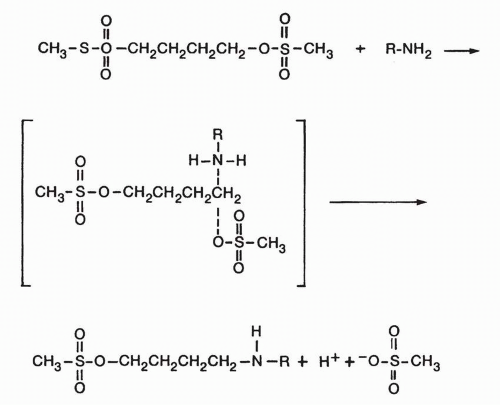
The major clinical representative of the alkyl alkane sulfonates is busulfan, which is widely used in high-dose regimens for the treatment of acute myelogenous leukemia.31 Of the alkyl alkane sulfonates, compounds with one to eight methylene units between the sulfonate groups have antitumor activity, but maximal cross-linking and activity are achieved by compounds with four units.32 The mechanism of action of the alkyl alkane sulfonates is shown in Figure 14A-4.

Busulfan exhibits second-order alkylation kinetics. The compound reacts more extensively with thiol groups of amino acids and proteins33 than do the nitrogen mustards, and these findings have prompted the suggestion that the alkyl alkane sulfonates may exert their cytotoxic activities through such thiol reactions along with interactions with DNA.33,34 Brookes and Lawley35,36 were able to demonstrate the reaction of busulfan with the N-7 position of guanine. The cytotoxic potential of busulfan correlates with adenine-to-guanine cross-linking.37 Busulfan is markedly cytotoxic to hematopoietic stem cells. This effect is seen clinically in the prolonged aplasia that may follow busulfan administration and can be shown experimentally in stem cell cloning systems.38 The pharmacologic basis for this property of busulfan is not well understood but may involve damage to the mesenchymal stem cells in the microenvironment. In recent years, an intravenous formulation has simplified the dose appropriate administration of busulfan to achieve optimal blood levels during high-dose myeloablative treatments.


FIGURE 14A-4 Structure and alkylating mechanism of busulfan, an alkane sulfonate. (From Colvin M. Molecular pharmacology of alkylating agents. In: Cooke ST, Prestayko AW. Cancer and Chemotherapy, vol 3. New York: Academic Press, 1981:291.)


Nitrosoureas

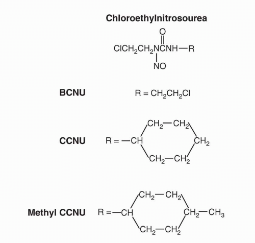
The nitrosourea antitumor agents were discovered in a drug screening effort that focused on analogues of methylnitrosoguanidine and methylnitrosourea.39 Chloroethyl derivatives such as chloroethylnitrosourea and BCNU (carmustine) (Fig. 14A-5) possess marked antitumor activity and had activity against tumor in the CNS.39,40 In addition to chloroethyl alkylating activity, the available nitrosoureas can also carbamoylate nucleophiles.41 Closely related methylating agents, procarbazine, temozolomide, and dacarbazine (DTIC), are lipophilic and penetrate the CNS (see nonclassical alkylating agents, this chapter).

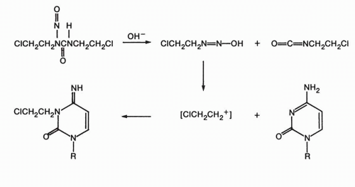
The nitrosoureas exhibit only partial cross-resistance with other alkylating agents,40 and a number of studies established unique aspects of the mechanism of the alkylation reaction for these compounds (Fig. 14A-6). BCNU cross-links DNA after the formation of initial monoadducts, particularly at the N-7 position of guanine. As shown
in Figure 14A-6, the diazonium hydroxide intermediate formed during BCNU hydrolysis decomposes to form a 2-chloroethyl carbonium ion (or equivalent), a strong electrophile, capable of alkylation of guanine, cytidine, and adenine bases.42 In a subsequent step occurring over hours, the chloride is displaced by electron-rich nitrogen on the complementary DNA strand base to form a cross-link. DNA-protein cross-links are also possible by initiating chloroethylation at the amino or sulfhydryl group of protein.43


FIGURE 14A-5 Structures of nitrosoureas. BCNU, bischloroethylnitrosourea; CCNU, cyclohexylchloroethylnitrosourea.


FIGURE 14A-6 Alkylation of nucleoside by bischloroethylnitrosourea (BCNU).

Isocyanates resulting from the spontaneous breakdown of many of the methyl- and chloroethylnitrosoureas are also shown in Figure 14A-6. The role of isocyanate-mediated carbamoylation in antitumor effects is incompletely understood, but this activity may be responsible for some toxicities associated with nitrosourea therapy.44

High-dose BCNU, etoposide, and cisplatin comprise the BEP regimen used for autologous stem-cell transplantation in patients with refractory or relapsed lymphoma.45 Another high-dose BCNU-containing regimen, BEAM (BCNU, etoposide, cytarabine, melphalan), has also been used with success with autologous hematopoietic stem-cell transplant in patients with NHL.46 In the 1980s, BCNU attracted interest as an adjuvant to radiation therapy in the treatment of patients with grade III and IV astrocytoma47 but has been replaced by temozolomide.48 BCNU-impregnated polymer wafers implanted in the tumor bed at the time of surgical resection provide a controlled release form of local chemotherapy.49

Streptozotocin is a unique methylnitrosourea with methylating activity that lacks carbamoylating activity. It is used exclusively in the treatment of metastatic islet cell carcinoma of the pancreas and malignant carcinoid tumors.50 The dose-limiting toxicities in humans have been gastrointestinal and renal, but not hematopoietic.


Alkylating Agent-Steroid Conjugates

Steroid receptors may serve to localize and concentrate appended drug species in hormone-responsive cancers. A number of synthetic conjugates of mustards and steroids have been developed. Of these drugs, two have made the transition into clinical application. Prednimustine, an ester-linked conjugate and slow release form of chlorambucil and prednisolone, is no longer available for clinical use.51 Estramustine is a carbamate ester-linked conjugate of nornitrogen mustard and estradiol but functions as an inhibitor of tubulin polymerization (see Chapter 13).


Prodrugs of Alkylating Agents

Therapy with alkylating agents is compromised by a high level of toxicity to normal tissues and a lack of tumor selectivity. Cyclophosphamide and ifosfamide were prodrugs synthesized in the hope that high levels of phosphamidases in epithelial tumors would selectively activate the drugs.52 Strategies for more selective delivery of alkylating agent to tumor have been explored including cleavable tumor-directed antibodyalkylating agent conjugates,53 alkylating agent-glutathione conjugates (which might be selectively cleaved by glutathione transferase (GST) P1 expressed in high levels in tumor cells)54 or viral vectors delivering activating enzymes to tumor cells.55


Cellular Pharmacology


Cellular Uptake

The uptake of alkylating agents into cells is an important determinant of cellular specificity. Many are highly lipid soluble (including the active metabolites of the methylating agents, cyclophosphamide, and ifosfamide, as well as chlorambucil) and readily enter cells by passive diffusion. Mechlorethamine uptake depends upon the choline transport system.56 Melphalan is transported into several cell types by at least two active transport systems, which also carry leucine and other neutral amino acids across the cell membrane. 57,58 High levels of leucine in the medium protect cells from the cytotoxic effects of melphalan by competing with melphalan for transport.59 In contrast to mechlorethamine and melphalan, the highly lipid-soluble nitrosoureas BCNU and CCNU enter cells by passive diffusion.60 Chlorambucil uptake also occurs through simple passive diffusion.

Studies of cellular uptake of alkylating agents that require metabolic activation (such as cyclophosphamide or ifosfamide) are hampered by uncertainty about which metabolite, or even parent drug, is the most critical moiety for transport.


Sites of Alkylation

Any alkylating agent producing reactive intermediates binds to a variety of cellular constituents61 including nucleic acids, proteins, amino acids, and nucleotides. As an example, the active alkylating
species from a nitrogen mustard demonstrates selectivity for nucleophiles in the following order: (a) oxygens of phosphates, (b) oxygens of bases, (c) amino groups of purines, (d) amino groups of proteins, (e) sulfur atoms of methionine, and (f) thiol groups of cysteinyl residues of glutathione.62 This ranking, however, assumes there are no steric or hydrophilic/hydrophobic barriers to the tissue nucleophile, and this is seldom the case. In addition, glutathione conjugation is often favored in the presence of GSTs, which offer catalysis. Thus, generalizations about alkylating agent targets are fraught with difficulty. In addition, it seems likely that a matrix of biochemical targets of alkylating agents may contribute to cytotoxicity, though DNA is generally favored as the primary target. Proof of this hypothesis may be emerging from three areas of research where cytotoxicity correlates with (a) activity of DNA repair enzymes, perhaps best shown for BCNU and repair by alkyl guanine alkyltransferase (AGT),63 (b) changes in a matrix of genetic and epigenetic events measured and analyzed by gene expression arrays,64 and (c) specific DNA adducts shown by mass spectrometric analysis.65 The stringency of such analyses requires that alternative toxic pathways not involving DNA must be excluded, a difficult requirement to meet. For this reason, mechanistic understanding of alkylating agent activity must be considered incomplete.

In the DNA molecule, the phosphoryl oxygens of the sugar phosphate backbone are obvious electron-rich targets for alkylation. Alkylation of the phosphate groups occurs66,67 and can result in strand breakage from hydrolysis of the resulting phosphotriesters. Although the biologic significance of the strand breakage caused by phosphate alkylation remains uncertain, the process is so slow that it seems unlikely to be a major determinant of cytotoxicity, even for monofunctional agents.68

Extensive studies with carcinogenic alkylating agents such as methyl methane sulfonate have shown that virtually all the oxygen and nitrogen atoms of the purine and pyrimidine bases of DNA can be alkylated to varying degrees. The relative significance of these sites of alkylation of specific bases and of specific sites on DNA in determining cytotoxicity, specific organ toxicities, or carcinogenesis remains uncertain. Alkylation of the O-6 atom and of the extracyclic nitrogen of guanine appears to be particularly important for carcinogenesis.69, 70, 71

Studies of the base specificity of alkylation by the chemotherapeutic alkylating agents have been much less extensive. Busulfan and mechlorethamine alkylate the N-7 position of guanine. Guanine cross-links (two guanine molecules abridged at the N-7 position by an alkylating agent) have been isolated from acid hydrolysates of the reaction mixtures.35,36

Reaction of the nitrogen mustard with native DNA, however, produces alkylation of the N-1 position of adenine in addition to N-7-alkylated guanine. The enhanced alkylation of the N-7 position of guanine may result from base stacking and charge transfer that enhance the nucleophilic character of the N-7 position.72 Melphalan preferentially alkylates guanine N-7 or adenine N-3.65

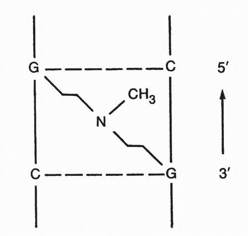
Base sequence influences the alkylating reaction. The N-7 position of guanine is most electronegative and, therefore, most vulnerable to attack by the aziridinium cation intermediate of the nitrogen mustards when the base is flanked by guanines on its 3′ and 5′ sides. The key site of DNA attack for the nitrosoureas as well as nonclassic methylating agents such as procarbazine and dacarbazine seems to be the O-6 methyl group of guanine.7 Enhanced repair of this site is associated with drug resistance.73 Thus, the preferred sites for alkylation vary by alkylating agent and chemical environment around the DNA base in question.


DNA Cross-Linking

On the basis of their isolation of the guanines linked at N-7 by alkylating agents, Brookes and Lawley72,74 postulated that the bifunctional alkylating agents such as the nitrogen mustards produced interstrand and intrastrand DNA-DNA cross-links and that these cross-links were responsible for the inactivation of the DNA and cytotoxicity. On the basis of the Watson-Crick DNA model, these authors suggested that appropriate spatial relationships for cross-linking by nitrogen mustards or sulfur mustard occurred between the N-7 positions of guanine residues in complementary DNA strands (Fig. 14A-7).

The importance of cross-linking is supported by the fact that the bifunctional alkylating agents, with few exceptions, are much more effective antitumor agents than the analogous monofunctional agents. Furthermore, increasing the number of alkylating units on the molecule beyond two does not usually increase the antitumor activity of the compound.

Direct evidence that DNA cross-linking occurs as the result of treatment of DNA or cells with bifunctional alkylating agents was provided initially by relatively insensitive physical techniques, including sedimentation velocity studies and denaturation-renaturation studies.75 These techniques, however, could not detect DNA interstrand cross-linking in mammalian cells exposed to therapeutic levels of alkylating agents in vitro or in tissues after in vivo drug administration. In 1976, a more sensitive assay for DNA interstrand cross-linking in cells, the alkaline elution method,76 was reported and had the necessary sensitivity to detect DNA cross-linking in cells and tumor-bearing animals exposed to minimal cytotoxic levels of alkylating agents.77,78 These studies and others using ethidium bromide fluorescence to detect cross-links have shown that DNA cross-linking by bifunctional alkylating agents correlates with cytotoxicity and that DNA in drug-resistant cells has lower levels of cross-linkage.79,80 The alkaline elution technique also detects DNA-protein as well as DNA-DNA cross-links.81 DNA-protein crosslinks likely do not play a major role in cytotoxicity and may be repaired by replication bypass mechanisms.81


FIGURE 14A-7 Cross-linking of DNA by nitrogen mustard. (Modified and reproduced from Brookes P, Lawley PD. The reaction of monofunctional and difunctional alkylating agents with nucleic acids. Biochem J 1961;80:486, with permission.)


In addition to these target effect-response studies, silencing of the AGT promoter in gliomas correlates with improved antitumor activity and survival in patients treated with BCNU.82 Because AGT repairs guanine alkylation products produced by BCNU, decreased enzyme activity would be expected to increase DNA alkylation, implying that DNA is a critical target for BCNU effects. Thus, evidence increasingly supports the hypothesis that DNA adduct formation is the major mechanism of alkylating agent cytotoxicity.

Chloroethylnitrosoureas cross-link via a unique mechanism.43 The spontaneous decomposition of the chloroethylnitrosoureas generates a chloroethyldiazonium hydroxide entity that can alkylate DNA bases to produce an alkylating chloroethylamine group on the nucleotide in the DNA strand. This group could then alkylate an adjacent nucleotide on the complementary DNA strand in a slower step, producing an interstrand cross-link. The mechanism of alkylation by thiophosphates such as thiotepa likely begins with protonation of the aziridine N, which leads to ring opening. Cross-linking can proceed by one of several mechanisms, either activation of the free chloroethyl carbon or activation of a second aziridine ring on the original molecule. Although interstrand cross-links are important mediators of the cytotoxic effects of alkylating agents, the monofunctional DNA alkylations exceed cross-links in number and are potentially cytotoxic. This hypothesis is supported by the fact that certain clinically effective agents, such as procarbazine and dacarbazine, are monofunctional alkylating compounds and do not produce cross-links in experimental systems. The basis of the cytotoxic effects of monofunctional alkylation appears to be through mismatch repair-mediated processes, since cells lacking mismatch repair are often methylating agent tolerant. The futile cycling of repair reactions through mismatch repair efficiently removes bases opposite O6-methylguanine and reinserts a thymine, with repeated attempt to repair the O6-mG:T mismatch and further futile repair. This produces single and double strand breaks that are cytotoxic, especially in the second round of DNA synthesis, after formation of the O6-mG:T mismatch.

Data suggest that alkylation is nonuniform along the DNA strand and may be concentrated in specific regions. One determinant of regional specificity of DNA alkylations may be chromatin structure11,83; areas of active transcription seem to be most vulnerable. Additionally, evidence shows that nitrogen mustards such as mechlorethamine preferentially cross-link at 5′-GNC sequence.84

In summary, the preponderance of evidence supports the hypothesis that the major factor in the cytotoxicity of most clinically effective alkylating agents is interstrand DNA cross-linking, which results in inactivation of the DNA template, cessation of DNA synthesis, and ultimately cell death. Cell-cycle checkpoint proteins, including most prominently p53, are responsible for the recognition of DNA alkylation and strand breaks. Recognition of DNA damage leads to a halt in cell-cycle progression and initiation of programmed cell death. Cells containing mutated p53 have greater resistance to alkylating agents.85

An increased knowledge of alkylation mechanisms and targets may make it possible to improve the therapeutic index of these agents. For example, the therapeutic index of alkylating agents should improve if the alkylation of tumor cells were increased without a simultaneous increase in normal tissue alkylation. This might be accomplished by maneuvers designed to inhibit drug activation by glutathione or GST P1,86 or by blocking the repair processes, as discussed below.


Tumor Resistance

The emergence of alkylating agent-resistant tumor cells is a major problem that limits the clinical effectiveness of these drugs. Cellular resistance mechanisms identified in preclinical experimental settings include drug uptake, enhanced anti-apoptosis pathways, activation of survival pathways, enhanced intratumoral drug inactivation, and changes in DNA repair.


Decreased Cellular Uptake of Selected Alkylating Agents

Several of the drugs (melphalan, nitrogen mustard) of this class require active transport into cells. One mechanism for drug resistance is decreased drug entry into the cell. This mechanism was best demonstrated in L5178Y lymphoblast cells resistant to mechlorethamine.56 The extracellular domain of the leucine-melphalan transporter expresses CD98. Reduced expression of CD98 on human myeloma cells is associated with melphalan resistance.87 The glutathione-dependent efflux transporters MRP1 and MRP2 can confer resistance to chlorambucil.89


Resistance due to Inactivation by Glutathione or GSTs

Intracellular inactivation of alkylating agents has been implicated in human tumor resistance. Early studies showed increased levels of sulfhydryls associated with resistance in experimental tumors, and increased nonprotein sulfhydryl content, particularly in the form of glutathione, in resistant tumor cell lines.88 While increased intracellular glutathione content may be found in resistant cells, elevated GST activity may also play a role90 and increased aldehyde dehydrogenase (ALDH) activity, which converts aldophosphamide to the inactive carboxyphosphamide, was present in cells resistant to cyclophosphamide.91, 92, 93

Alterations in the GSH/GST system found in alkylating agent resistance phenotypes include increased intracellular GSH levels, elevation of GST activity, and changes in the expressed levels of one or more GST isozymes. Currently, several GSH-related mechanisms may explain the observed tumor cell resistance to alkylating agents, including (a) enhanced inactivation of electrophilic alkylating agents, such as melphalan94 by direct conjugation to GSH; (b) GSHdependent denitrosation of nitrosoureas, a reaction that is preferentially catalyzed by one of the rat liver GST μ enzymes in the case of BCNU; (c) scavenging for reactive organic peroxidases, a process that is catalyzed by GSH peroxidase;95 and (d) quenching of chloroethylated-DNA monoaducts.96

Inherited polymorphisms of functional significance have been reported in genes that encode glutathione-S-transferases (GSTs) and may contribute to resistance (see Chapter 6). There are four cytosolic families of GSTs, including GST α, GST μ, GST θ, and GST π.97 Gene clusters of GST μ (GSTM1, M2, M3, M4, and M5) and GST θ (GSTT1 and T2) are located on chromosomes 1 and 22, respectively.98 Independent gene deletions at GSTM1 and GSTT1 loci result in a lack of active protein in ≈50% and 20% of Caucasians, respectively.99 GST π or GSTP1, encoded by a single locus (GSTP1) on chromosome 11, is also subject to polymorphic variation.100 Codon 105 residue forms part of the GSTP1 active site for binding of hydrophobic electrophiles,101 and the Ile-Val substitution affects substrate-specific catalytic activity and thermal stability of the encoded protein.102 Reactive metabolites of ifosfamide, busulfan, and chlorambucil are substrates for GSTP1-mediated
glutathione conjugation in vitro. Allelic variants of GSTP1 differ significantly in their efficacy in catalyzing the GSH conjugation and hence their ability to detoxify alkylating agents.103 The effect of these polymorphisms on clinical conjugation, toxicity, and antitumor response is uncertain, and the studies to date are summarized in Chapter 6.


Resistance to Cyclophosphamide due to Elevated Aldehyde Dehydrogenase Activity

Resistance to cyclophosphamide may also be determined by the activity of cellular ALDH.93,104 ALDH is an enzyme responsible for the oxidation of intracellular aldehydes.105 The cytoplasmatic ALDH isozyme converts activated cyclophosphamide to the inactive excretory product, carboxyphosphamide, in both murine and human cell lines.106 There is no clear evidence that ALDH activity confers resistance in human tumors.

ALDH may have an important role in early differentiation of hematopoietic stem cells by oxidizing retinol to retinoic acid.107 It is hypothesized that cancer stem cells survive cyclophosphamide treatment due to ALDH. Murine and human hematopoietic and neural stem and progenitor cells have high ALDH activity.108,109 Increased ALDH activity has also been found in malignant stem cell populations in multiple myeloma and acute myeloid leukemia.110


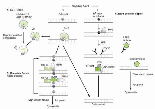
FIGURE 14A-8 DNA repair pathways that play an important role in resistance to alkylating agents. A. AGT. B. Mismatch repair. C. Base excision repair. Targeted therapies to improve tumor sensitivity including O6-benzylguanine, PARP inhibition, and methoxyamine are indicated. AGT, alkylguanine transferase; O6-meG, O6-methylguanine; N7-meG, N7-methylguanine; N3-meA, N3-methyladenine; O6-BG, O6-benzylguanine; Ub, ubiquitin; PARP, Poly-ADP-ribose polymerase; Polβ, DNA Polymerase β; MPG, methylpurine glycosylase. Not shown are components of the double-strand break repair complexes. (Adapted from Sarkaria JN, Kitange GJ, James CD, et al. Mechanisms of chemoresistance to alkylating agents in malignant glioma Clin Cancer Res 2008;14:2900-2908, with permission.)


DNA repair and Alkylating Agent Resistance

Enhanced repair of DNA lesions generated by alkylation plays a clearly established role in resistance of experimental and human tumor cells to alkylating agents. Because DNA appears to be the most critical target for the alkylating agents, its repair has been a major focus of study and several mechanisms involved in repairing alkylation and strand breaks are summarized in Figure 14A-8.

Enhanced excision of alkylated nucleotides from DNA as a mechanism of resistance to alkylating agents was first demonstrated in bacteria75 and later in mammalian cells. Bacterial, fungal, and mammalian cells are capable of excising and repairing sites of alkylation, as well as removing cross-links and repairing single- and double-strand breaks.


Alkylguanine DNA Alkyltransferase Mediated repair

Repair of DNA alkylation products and cross-links involves multiple systems, each composed of one or several distinct enzymes
(see Fig. 14A-8). The simplest of these catalyzes the transfer of alkyl substituents (methyl-, ethyl-, benzyl-, 2-chloroethyl-, and pyridyloxobutyl-) from the O6-position of guanine to an active cysteine acceptor site within the protein in a single enzyme repair process. This enzyme, alkylguanine-O6-alkyl transferase (AGT), is encoded by the MGMT (O6-methylguanine methyltransferase) gene in humans and is homologous to the bacterial alkyltransferase gene, ada.

Several clinical trials have established an inverse correlation between AGT content in brain tumors and the response to treatment of brain tumor patients receiving BCNU, temozolomide, and other alkylating agents.111, 112, 113 Lower levels of AGT activity result from epigenetic silencing due to MGMT promoter methylation. Tumors with methylated MGMT are highly responsive to these agents, while those with fully expressed AGT tend to be resistant. The availability of 5′ cytosine methylation-specific PCR (MSP) provides a facile assessment of MGMT promoter methylation and has value as a predictive assay for response to alkylating agent-based chemotherapy. MGMT promoter methylation has been correlated with survival in patients with glioma treated with nitrosoureas and temozolomide82,114 and prolonged progression-free survival (PFS) in patients treated with temozolomide.115,116 Together, these data illustrate a role for MGMT gene function in glioma chemoresistance to alkylating agents. Clinical trials attempting to modulate AGT activity are discussed below.111,117,118


Mismatch Repair

A second DNA repair system, mismatch repair (MMR), recognizes the mismatch created by alkylation of DNA bases, and, after unsuccessful attempts at repair, triggers cell-cycle arrest and apoptosis. For example, O6 alkylation of guanine leads to mispairing of the damaged base with thymine, creating a distortion in the DNA double helix, which is recognized by components of the MMR complex. MMMR deficiency has been associated with resistance to alkylating agents owing to the inability to recognize the mismatch and initiate the cycle of futile repair attempts, cell-cycle arrest, and apoptosis.119,120 Several proteins comprise the MMR pathway (hMLH1, hPMS2, hMSH2, hMSH3, and hMSH6), which is programmed to correct erroneous DNA base pairing.

In MMR competent cells during DNA replication, DNA polymerase mispairs unresolved O6-methylguanine with thymine. The mismatch triggers attempts to remove the mispaired thymine. If repair is successful, subsequent rounds of replication continue to mispair O6-methylguanine with thymine, resulting in repetitive futile cycles of MMR. This futile cycle may induce double-strand breaks, which in turn, trigger p53-dependent cell-cycle arrest and apoptosis.121 Loss of MMR competence creates tolerance to the mispaired bases and allows cell replication and survival, provided the DNA lesions are compatible with viability. The loss of MLH6 is a frequent event in glioma cells selected by resistance to temozolomide.122

Specific interactions of MMR proteins with cell-cycle checkpoint proteins have been implicated in apoptosis. hMLH1 has been associated with signaling ATR-dependent G2 cell-cycle arrest in response to DNA methylation,123 and MMR proteins may initiate degradation of cyclin D1 following alkylation to promote cell-cycle arrest in response to alkylation.124 Functional MSH2 and hMLH1 are also believed to activate p73-dependent apoptosis pathways via c-Abl.125

Resistance to alkylating agents conferred by MMR deficiency is further illustrated by hereditary nonpolyposis colon cancer, which is caused by mutations in hMLH1 or hMSH2 genes. Colon cancer cells harboring these mutations are resistant to alkylating agents,126 as are other cell lines deficient in hMLH1.127


DNA Excision Repair

NA excision repair pathways provide a comprehensive mechanism for recognizing and removing damaged bases or nucleotide segments from a single DNA strand and then resynthesizing the new DNA segment, using the opposing undamaged strand as a template. Excision repair complexes includes base excision repair (BER) and nucleotide excision repair (NER).


Base Excision Repair

In response to DNA alkylation, BER is initiated by the action of damage-specific DNA glycosylases that recognize and excise single base lesions such as N3 methyladenine and N7-methylguanine. Release of the damaged base produces an apurinic/apyrimidinic (AP) site, which is excised by the APE endonuclease (Fig. 14A-9). The missing segment is then resynthesized by DNA polymerase and sealed by DNA ligase. Persistent AP sites are recognized by topoisomerase I and II and these may form cleavable complexes that induce apoptotic signals. The enzyme PARP plays a pivotal role in the recognition of strand breaks and in the formation of DNA strand break intermediates that attract repair complexes. The N3-methyladenine and N7-methylguanine are the most common adducts created by alkylation and account for greater than 80% of all methylation events. However, these lesions contribute modestly to the cytotoxicity of alkylating agents due to the efficiency of the BER pathway. Perturbation of BER capacity through alterations of glycosylase expression or through pathway inhibition greatly decreases the efficiency of N3 and N7 methyl adduct repair.128 Targeting of BER, as described below, is particularly effective in enhancing the sensitivity to platinating agents in various cell lines tumors, especially breast cancers deficient in BRCA 1 and BRCA 2, and to alkylating agents in MMR-deficient tumors.129


Nucleotide Excision Repair

NER is an additional mechanism for excising bulky alkylation products and DNA intrastrand cross-links. The pathway includes multiple proteins that recognize DNA adducts, such as those produced by alkyl lesions and incise 3′ and 5′ to the damaged base(s), causing release of the damaged nucleotides and surrounding segments of DNA. Excision is followed by resynthesis of the missing segment, using the opposing strand as a template. Components of the NER complex also have a role in repair of double-strand breaks. NER-deficient mammalian cells, such as those derived form patients with xeroderma pigmentosum (XP), are hypersensitive to alkylating and cross-linking agents.130,131 Studies of the effects of NER deficiency
on the toxicity of alkylating agents are most extensive in rodent models, but there is evidence that polymorphic variants of ERCC1 confer increased alkylating agent sensitivity for both normal and malignant tissues, influence response to treatment, and greater toxicity.132 The role of NER in alkylating agent sensitivity and resistance in clinical cancer treatment is under active investigation.


FIGURE 14A-9 Hydrolysis products of mechlorethamine.


Cross-Link Repair

Interstrand cross-links covalently tether strands of DNA, preventing unwinding of duplex DNA and prohibiting polymerase access. Both strands of DNA are involved in this lesion, precluding straightforward excision repair and gap-filling pathways. Consequently, the repair of interstrand DNA cross-links is complex, integrating elements of the NER pathway, a variety of less well-understood activities to form a double-strand break, insertion of new bases, and homologous recombination (HR). Though mechanisms are incompletely understood several, mammalian cell types have extreme sensitivity to cross-linking agents. Faconi anemia133 and Bloom’s (BLM) syndrome cells134 are both hypersensitive to alkylating agents that cause interstrand cross-links. The Fanconi anemia pathway and the BLM helicase are believed to be activated in response to replication stalling due to cross-linked DNA; their dysfunction in the inherited disorders accounts for alkylating agent sensitivity.135,136 Furthermore, mutations in excision repair genes ERCC1 and ERCC4 (XPF) also render cells sensitive to cross-linking by alkylating agents, suggesting these genes play a role in the repair of cross-links in addition to their role in NER.137


Akt

The Akt family in humans is comprised of three genes (Akt1, Akt2, and Akt3) encoding for serine/threonine protein kinases (PKB). Akt activation occurs downstream of various receptor tyrosine kinases and phosphatidylinositol 3-kinase (PI3K). The PI3K/Akt pathway is frequently activated in human cancer and has been implicated in tumor cell proliferation, cellular survival, and chemotherapy resistance (see Chapter 30). In response to alkylation by the agent temozolomide, Akt is induced in lymphoblastoid, colon, and breast cancer cells in a mismatch repair-dependent manner.138,139

Upon activation, PI3K/Akt signaling confers resistance to chemotherapy through its antiapoptotic effects as mediated by phosphorylation of several downstream targets. Activated Akt is thought to inhibit apoptosis by phosphorylating molecules upstream and downstream of the mitochondrial apoptotic pathway. Akt-dependent phosphorylation of proapoptotic BH3 family members such as Bad, Bax and Bim-EL decreases the ability of these proteins to hold mitochondria in an open configuration, resulting in a reduction in cytochrome c release. Akt can phosphorylate caspase-9 to inhibit its ability to activate executioner caspase 3.140,141 Akt reduces cytochrome c release through modification of the antiapoptotic Bcl-2 homologous Mcl-1.142,143 Akt exerts indirect inhibition of apoptosis through effects on p53, the most important regulator of apoptosis.144

Akt also drives chemoresistance by promoting cell growth. Akt is involved in the survival pathway of mammalian target of rapamycin (mTOR), a serine/threonine kinase that is implicated in protein synthesis control.145 Akt activates mTOR complex 1 (mTORC1; or mTOR-raptor complex) indirectly by inhibiting phosphorylation of tuberous sclerosis complex 2, thereby allowing Ras-related small G protein (Rheb)-GTP to activate mTORC1 signaling.146


Defects in Cell Cycle Arrest and Apoptosis

In addition to the mechanisms described above, mutation or silencing of genes that induces cell-cycle arrest and apoptosis may lead to alkylator resistance.

In cells that experience genotoxic stress during replication, activation of these factors triggers the signaling cascades, leading to delayed progression through S-phase in order to allow time for DNA repair.147 In the presence of DNA damage, an S-phase cell-cycle checkpoint is activated by the checkpoint kinases ataxia-telangectasia mutated (ATM) and ATM- and Rad3-related (ATR). Activation depends on the type of DNA damage: ATM is recruited to DNA double-strand breaks (DSBs) induced by agents such as ionizing radiation (IR), whereas ATR is recruited to sites of replication protein A (RPA)-coated single-stranded DNA (ssDNA). These sites accumulate at stalled replication forks or at sites of single-strand damage.148,149 The involvement of ATM and ATR in the response to carcinogen-induced DNA damage has been established,150 by showing an enhanced sensitivity of ATM- and ATR-defective cells to methylating agents.

Two parallel branches of the DNA damage-dependent S-phase checkpoint are thought to cooperate by inhibiting distinct steps of DNA replication. One branch is activated by the phosphorylation of structural maintenance of chromosomes 1 (SMC1), a cohesin that is activated by ATM or ATR.151 The second branch, consisting of the ATR-Chk1- or ATM-Chk2-complexes, regulates turnover of CDC25A, a phosphatase that regulates Cdk2 and consequently blocks the initiation of replication.152

Defects in damage recognition or apoptotic signaling may lead to relative resistance.153 For example, loss of normal p53 function, up-regulation of the antiapoptotic proteins, Bcl-2 or Bcl-XL, or overexpression of the epidermal-growth factor receptor (EGFR) can disrupt the normal apoptotic response to DNA damage caused by alkylating agents.154,155 As mentioned previously, apoptotic cell death after DNA damage is mediated through p53, which blocks cell-cycle progression, initiates attempts to repair damage, and ultimately activates apoptotic pathways.

Clinical Pharmacology

The primary pharmacokinetics properties of standard alkylating agents are given in Table 14A-1. Although some agents are too reactive chemically to provide more than momentary exposure of tumor cells to parent drug (the best examples are mechlorethamine and BCNU), others are stable in their parent form and even others require metabolic activation, as in the case of cyclophosphamide and ifosfamide. Doses of some alkylating agents may need to be adjusted for organ dysfunction and may require pharmacokinetic monitoring in individual patients, as with high-dose busulfan, to execute rational treatment regimens.


Activation, Decomposition, and Metabolism


Decomposition Versus Metabolism

A principal route of degradation of most of the reactive alkylating agents is spontaneous hydrolysis of the alkylating entity (i.e., alkylation by water). For example, mechlorethamine rapidly undergoes reaction to produce 2-hydroxyethyl-2-chloroethylmethylamine and bis-2-hydroxyethylmethylamine (Fig. 14A-9). Likewise, both melphalan and chlorambucil undergo similar hydrolysis to form the monohydroxyethyl and bishydroxyethyl products, although less rapidly than the aliphatic nitrogen mustards.187,188 The mono hydroxylated products are less active alkylators than their chloroalkyl precursors.

Most alkylating agents also undergo some degree of enzymatic metabolism. For example, if mechlorethamine radiolabeled in the methyl group is administered to mice, approximately 15% of the radioactivity can be recovered as exhaled carbon dioxide, which indicates that enzymatic demethylation is occurring. For the phosphoramide mustards and nitrosoureas, enzymatic metabolism plays a significant role in determining their pharmacokinetic profile.


Cyclophosphamide and Ifosfamide

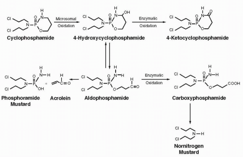
Cyclophosphamide is activated to alkylating and cytotoxic metabolites by cytochrome P450 isozymes 2B6, 2C9, and 3A4.189 The complex metabolic transformations are illustrated in Figure 14A-10. The initial metabolic step is the oxidation of the ring carbon adjacent to the nitrogen to produce 4-hydroxycyclophosphamide, which establishes equilibrium with aldophosphamide. Aldophosphamide undergoes a spontaneous (nonenzymatic) elimination reaction to form phosphoramide mustard and acrolein. Phosphoramide mustard, which is generally believed to be the DNA cross-linking agent of clinical significance, is a circulating metabolite that does not enter cells easily due to its anionic form. Thus, the intracellular generation of phosphoramide mustard from aldophosphamide is believed to be important to a therapeutic result. A major detoxification route is the oxidation of aldophosphamide to the inactive carboxyphosphamide by ALDH1A1 and, to a much lesser extent, by ALDH3A1 and ALDH5A1 in liver and in red blood cells. The concentration of ALDH in a variety of cell types appears inversely proportional to cytotoxicity, supporting its crucial role in determining cytotoxicity.93 The high enzyme concentration in hematopoietic progenitor cells may explain the ability of cyclophosphamide to produce major myelosuppression without myeloablation in patients receiving high doses without transplantation.190 Likewise, cancer stem cells expressing ALDH may also be drug resistant.

Multiple metabolites can react with glutathione (GSH). Some of these reactions with GSH may be reversible while others are irreversible; the latter are associated with detoxification pathways. Several-fold differences in the extent of metabolite formation have been observed among patients and these interindividual differences may be due to polymorphisms in cytochrome P450 enzymes (see Chapter 4). CYP3A4 and 3A5 genotypes may influence response or survival in patients treated with cyclophosphamide.

A minor (˜10%) alternative oxidative pathway leads to N-dechloroethylation and the formation of the neurotoxic chloroacetaldehyde. Cytochrome P450 3A4 is the main enzyme responsible for this undesirable secondary oxidation with a minor contribution from cytochrome P450 2B6.

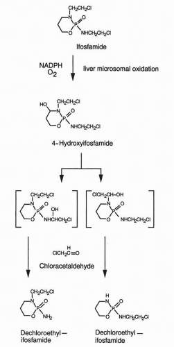
The metabolism of ifosfamide (Fig. 14A-11) parallels that of cyclophosphamide but with some differences in isozyme specificities and reaction kinetics. Activation of ifosfamide to 4-hydroxyifosfamide is catalyzed by the hepatic cytochrome P450 isoform 3A4. Aldo-ifosfamide partitions between ALDH1A1-mediated detoxification to carboxyifosfamide and a spontaneous (nonenzymatic) elimination reaction to yield isophosphoramide mustard and acrolein. Isophosphoramide is the DNA cross-linking agent of clinical significance. Hydroxylation proceeds at a slower rate for ifosfamide than for cyclophosphamide, which results in a longer plasma half-life for the parent compound. Dechloroethylation of ifosfamide produces inactive metabolites (primarily mediated by cytochrome
P450 isozyme 2B6 and 3A4) and competes with the activation step as a major pathway of elimination.191, 192, 193, 194


FIGURE 14A-10 Metabolism of cyclophosphamide.

Both cyclophosphamide (above doses of 4 g/m2) and ifosfamide (above doses of 5 g/m2) exhibit dose-dependent nonlinear pharmacokinetics, with significant delays in elimination at higher doses.195 Interestingly, both drugs also induce their own metabolism, resulting in significant shortening of the elimination half-life for the parent compound when the drugs are administered on multiple consecutive days.196

Both agents can undergo further chemical reaction to form acrolein, which is toxic to bladder. This compound may also form O6G adducts, and these may be recognized and removed by AGT.


Nitrosoureas

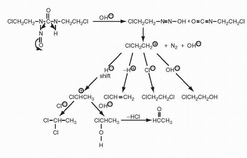
The decomposition of nitrosoureas to generate the alkylating chloroethyldiazonium hydroxide entity has been mentioned, and the products generated by this decomposition in aqueous solution are illustrated in Figure 14A-12.


FIGURE 14A-11 Metabolic activation of ifosfamide to its active form, 4-hydroxyifosfamide, and further metabolic transformation to chloracetaldehyde and other end products. NADPH, reduced form of nicotinamide adenine dinucleotide phosphate.

The nitrosoureas also undergo metabolic transformation. BCNU can be inactivated through denitrosation reactions catalyzed by both cytosolic and microsomal enzymes. Class μ glutathione S-transferase is a major catalyst of the cytosolic denitrosation reaction. Enhancement of P450 activity in vivo by phenobarbital abolished the therapeutic effect of BCNU against the 9L intracerebral rat tumor and decreased the therapeutic activity of CCNU and BCNU against this tumor.197 The phenobarbital-treated rats had increased plasma clearance of BCNU. The plasma clearance of parent BCNU decreases and the plasma half-life increases as doses escalate from standard-dose (150 to 200 mg/m2) to high-dose regimens (600 mg/m2) (Table 14A-1).

CCNU and methyl-CCNU undergo hydroxylation of their cyclohexyl ring to produce a series of metabolites that represent
the major circulating species after treatment with these drugs.198 These metabolites have increased alkylating activity but diminished carbamoylating effects.198


FIGURE 14A-12 Decomposition of bischloroethylnitrosourea (BCNU) in buffered aqueous solution.


Clinical Pharmacokinetics

Gas chromatography-mass spectrometry and high-pressure liquid chromatography (HPLC) have generated pharmacokinetic information (Table 14A-1) for alkylating agents and their metabolites.


Melphalan

In patients who received 0.6 mg/kg of the drug intravenously, the peak levels of melphalan, as measured by HPLC, were 4.5 to 13 μmol/L (1.4 to 4.1 μg/mL), and the mean terminal-phase half-life (t1/2β) of the drug in the plasma was 1 hour. Dose adjustment is not indicated at conventional doses, but in high-dose regimens, there are conflicting data regarding adjustment for renal function. 199 The 24-hour urinary excretion of the parent drug averaged 13% of the administered dose. Inactive monohydroxy and dihydroxy metabolites appear in plasma within minutes of drug administration.170

Other studies have demonstrated low and variable systemic availability of the drug after oral dosing.187,200 Food slows its absorption. After oral administration of melphalan, 0.6 mg/kg, much lower peak levels of drug of approximately 1 μmol/L (0.3 μg/mL) were seen. The time to achieve peak plasma levels varied considerably and occurred as late as 6 hours after dosing. The low bioavailability was caused by incomplete absorption of the drug from the gastrointestinal tract because 20% to 50% of an oral dose could be recovered in the feces.200 Regional administration of melphalan is possible by both intracavitary201 and limb perfusion methods.202


Chlorambucil

After the oral administration of 0.6 mg/kg of chlorambucil,187,188 peak levels of 2.0 to 6.3 μmol/L (0.6 to 1.9 μg/mL) occur within 1 hour. Peak plasma levels of phenylacetic acid mustard, an oxidation product of chlorambucil with alkylating activity, range from 1.8 to 4.3 μmol/L (0.5 to 1.18 μg/mL), and the peak levels of this metabolite are achieved 2 to 4 hours after dosing. The terminal-phase half-lives for chlorambucil and phenylacetic acid mustard are 92 and 145 minutes, respectively. Less than 1% of the administered dose of chlorambucil is excreted in the urine as either chlorambucil (0.54%) or phenylacetic acid mustard (0.25%). Approximately 50% of the radioactivity from carbon-14-labeled chlorambucil administered orally is excreted in the urine in 24 hours. Of this material, over 90% appears to be the monohydroxy and dihydroxy hydrolysis products of chlorambucil and phenylacetic acid mustard.


Cyclophosphamide

Cyclophosphamide is well absorbed after oral administration to humans196 The systemic availability of the unchanged drug after oral administration of 100-mg doses (1 to 2 mg/kg) was 97% of that after intravenous injection of the same dose.196 Juma et al.203 found the systemic availability of the drug to be somewhat less and more variable (mean, 74%; range, 34% to 90%) after oral administration of larger doses of 300 mg (3 to 6 mg/kg). A comparison of oral versus intravenous cyclophosphamide in the same patient revealed no difference in the AUC for the primary cytotoxic metabolites, hydroxycyclophosphamide and phosphoramide mustard.204 After intravenous administration, the peak plasma levels of the parent compound are dose dependent. Peak levels are 4, 50, and 500 nmol/mL (Table 14A-2) after the administration of 1 to 2,196 6 to 15,203 and 60 mg/kg,205 respectively. The terminal-phase half-life of cyclophosphamide varies considerably among patients (3 to 10 hours). In patients less than 19 years of age, the plasma half-life of parent drug is 1.5 hours.206 Less than 15% of the parent drug is eliminated in the urine; the major site of clearance is the liver. Peak alkylating levels are achieved 2 to 3 hours after drug administration and the terminal half-life of plasma alkylating activity is 7.7 hours with a plateau-like level of plasma alkylating activity maintained for at least 6 hours.


TABLE 14A.2 Pharmacokinetics of cyclophosphamide and metabolites in humans


Parent or metabolite


Cyclophosphamide dose (mg/kg)


Cpeak/plasma (μM)


Plasma t1/2 (h)


References


Parent


1-2


4


196


Parent


6-15


50


3-10


203


Parent


60


500


205


Total alkylating activity


40-60


10-80


7.7


203,210,306


Phosphoramide mustard


4-12


3-18


8.7


307


Phosphoramide mustard


60-75


50-100


205


Nor-nitrogen mustard


4-9


4-15


3.3


Aldophosphamide/hydroxycyclophosphamide


10


1.4


1-1.5


204,206,207


Aldophosphamide/hydoxycyclophosphamide


20


2.6


308,309


The predominant metabolites found in plasma are nornitrogen mustard and phosphoramide mustard, with lesser concentrations of the putative transport forms aldophosphamide and 4-hydroxycyclophosphamide (Table 14A-2).

The peak plasma levels of the major metabolites of cyclophosphamide, 4-hydroxycyclophosphamide/aldophosphamide, were 1.4 and 2.6 nmol/mL after injection of doses of 10 and 20 mg of radiolabeled cyclophosphamide per kilogram, respectively. Subsequent studies have determined that 4-hydroxycyclophosphamide/aldophosphamide has a half-life of approximately 1.5 hours in children206 and 1 to 5 hours in adults receiving conventional204 or high-dose207 cyclophosphamide. The AUC for 4-hydroxycyclophosphamide and aldophosphamide at conventional doses of drug ranged from 3 to 19 nmol/mL × hours and seems independent of either peak plasma levels or the plasma half-life of the parent drug or hydroxycyclophosphamide.

Because the initial metabolism of cyclophosphamide is hepatic, modulation of the activity of P450 in vivo might be expected to alter the pharmacokinetics of the drug. Pretreatment with phenobarbital, a known P450 inducer, reduces the plasma half-life of the parent compound in both humans and experimental animals.208 Also, with repeated doses of cyclophosphamide, the plasma half-life of parent compound becomes progressively shorter,196,209 and that of 4-hydroxycyclophosphamide increases, which indicates that cyclophosphamide can induce the P450 enzymes responsible for its metabolism.

The pharmacokinetics of cyclophosphamide and its metabolites has been incompletely studied in patients with renal failure. Because both parent drug and active metabolites are excreted, albeit to a limited extent, by the kidneys, caution should be used when cyclophosphamide is administered to such patients.210


Ifosfamide

After single doses of 3.8 to 5.0 g/m2, the terminal half-life of ifosfamide was 15 hours, considerably longer than the previously cited values of 3 to 10 hours for cyclophosphamide. At ifosfamide doses of 1.6 to 2.4 g/m2, however, the half-life of the drug was similar to that of cyclophosphamide. Also, the alkylating activity excreted in the urine was similar for these doses of the two analogs and ranged from 6% to 15% for ifosfamide, although urinary excretion may approach 50% at high single doses.211 As with cyclophosphamide, ifosfamide clearance increases during continuous infusion or with multiple daily doses, reaching a steady state 2 to 3 days after drug administration is begun.212 Whereas less than 10% of an administered dose of cyclophosphamide is dechlorethylated, as much as 50% of a dose of ifosfamide may be excreted in the urine as dechlorethylated products.

Because the slower activation rate of ifosfamide results in more prolonged exposure to the bladder-toxic metabolite acrolein, the disulfide detoxifier MESNA is routinely administered in association with ifosfamide.


Thiotepa

Thiotepa is rapidly desulfurated to TEPA and other alkylating species.165,213, 214, 215, 216 The conversion of thiotepa to TEPA is mediated by P450 isoenzymes. Both thiotepa and TEPA have cytotoxic activity. Aside from individual variability, the plasma terminal half-life of intact thiotepa is a relatively consistent 1.2 to 2 hours. TEPA appears in plasma within 5 minutes of thiotepa administration. In 120 minutes, its plasma concentration reaches that of thiotepa, but it persists longer, with a half-life of 3 to 21 hours, so that after 24 hours TEPA concentration × time exceeds that of the parent drug. In 24 hours, only 1.5% of the administered thiotepa is excreted in the urine unchanged, together with 4.2% as TEPA and 23.5% as other alkylating species.213 The pharmacokinetics in children resembles that in adults.214,215


Bendamustine

Bendamustine has a similar mechanism of action to the other chloroethylating agents. It has three distinct structural elements: a mechlorethamine group, which acts as the chloroethylating agent; a benzimidazole ring, which may interfere with nucleotide metabolism; and a butyric acid side chain, which may react with proteins and membranes (Fig. 14A-4).19 Clinically bendamustine acts as an alkylating agent and not an antimetabolite. Bendamustine produces strand breaks at a number far greater than equimolar concentrations of cyclophosphamide or melphalan.20 Furthermore, bendamustine-induced strand breaks are repaired slowly compared to those produced by other alkylating agents perhaps because of the bulk of the adduct. Furthermore, bendamustine shows only partial cross-resistance with other alkylating agents.20 This improved activity in resistant populations has been attributed to both extensive strand break formation and failed repair.


Current use of bendamustine is in lymphoid malignancies. It is indicated for fludarabine-refractory CLL21,22 where response rates in excess of 50% are noted, including patients with complete responses. A recent phase III trial compared chlorambucil with bendamustine. One-hundred-and-sixty-two patients received bendamustine and one-hundred-and-fifty-seven chlorambucil. A complete or partial response was noted during the course of treatment in 68% of bendamustinetreated patients compared with a complete or partial response rate of only 31% in chlorambucil-treated patients (P < 0.0001).23 Bendamustine is also active against refractory indolent B-cell lymphomas with a more favorable therapeutic index than CHOP-rituximab (α-CD20) treatment.22 Additionally, a clinical trial evaluating the efficacy of bendamustine-rituximab combination therapy found that the combination is well tolerated and active against indolent lymphomas.24

Bendamustine is tightly bound to human serum plasma proteins (94% to 96%) and is mainly found in extracellular spaces. Bendamustine is primarily metabolized via spontaneous hydrolysis of either of the chloroethyl arms to hydroxyethyl metabolites, which are significantly less active than parent drug. Two active minor metabolites, M3 (a hydroxy metabolite) and M4 (an N-desmethyl product), are formed via cytochrome P450 1A2. However, concentrations of these metabolites in plasma are 1/10 and 1/100 that of the parent compound, respectively, suggesting that the cytotoxic activity is primarily due to bendamustine. Bendamustine clearance in humans is approximately 700 mL/min. After a single dose of 120 mg/m2 bendamustine IV over 1-hour, the intermediate t1/2‘s of the parent compound is approximately 40 minutes. The mean apparent terminal elimination t1/2 of M3 and M4 is approximately 3 hours and 30 minutes, respectively.


Nitrosoureas

In humans after short-term infusion (15 to 75 minutes) of 60 to 170 mg/m2, initial peak levels of up to 5 μmol/L of BCNU are achieved. The plasma concentration decay curves were biexponential, with a distribution-phase half-life of 6 minutes and a second-phase half-life of 68 minutes. With high-dose BCNU, longer elimination half-lives of 22 to 45 minutes have been reported.217


Busulfan

Busulfan is primary employed in high-dose regimens associated with bone marrow reconstitution. It is routinely administered over 3 to 4 consecutive days, with dosing every 6 hours or once daily. The daily dose is usually in the range of 3.2 mg/kg, or approximately 120 mg/m2. The drug bioavailability by the oral route is variable among patients.218 The drug exhibits circadian rhythmicity in its pharmacokinetics, particularly in children, with higher drug levels and slower elimination in the evening. The primary elimination half-life is approximately 2.5 hours in both children and adults, although interpatient variability is considerable at both low and high doses.

The relationship between intravenous dose and AUC within the same patient appears to be predictable over multiple days of administration during high-dose therapy.199 Because of variable bioavailability, there is less consistency with the oral formulation. A range of threefold AUC in plasma levels after oral dosing is found among individuals in various cohorts studied.219 Thus, intravenous administration is the preferred route. Clearance declines with age and with increases in body weight, which leads to potential underdosing of children in high-dose regimens.220 Busulfan clearance for patients older than 18 years averages 2.64 to 2.9 mL/min/kg, whereas for children aged 2 to 14 clearance averages 4.4 to 4.5 mL/min/kg, and for children aged 3 or younger, it is 6.8 to 8.4 mL/min/kg.221 Mean volume of distribution at steady state was larger in children less than 1 year of age (0.77 ± 0.24 versus 0.64 ± 0.11 L/kg; P = 0.040) and children less than 4 year of age (0.73 ± 0.18 versus 0.64 ± 0.11 L/kg; P = 0.001) than in older children.222 Thus, larger doses must be used in the younger age groups to achieve the desired cytotoxic exposure.

Because of its high lipid solubility and low level of protein binding, busulfan penetrates readily into the brain and cerebrospinal fluid. The ratio of drug concentration in cerebrospinal fluid to plasma approximates 1.223 Positron-labeled busulfan has been used to track uptake into the brain, revealing that approximately 20% of a standard dose rapidly enters the CNS.224 This access to the brain may enhance the activity of this drug against leukemia and lymphoma cells in the CNS, but it also may explain its propensity to cause seizures. Prophylaxis with anticonvulsants is required in patients receiving high-dose busulfan. Busulfan enhances the clearance of phenytoin (dilantin) and, in some patients, lowers the drug’s plasma concentration below the therapeutic range, which increases the risk of seizures.225 Phenytoin levels should be monitored in the setting of busulfan therapy or an alternative, non-P450 metabolized anticonvulsant should be used.

Monitoring of drug levels and adjustment of doses to reach desired levels of drug exposure (AUC) have become an accepted aspect of high-dose intravenous busulfan with stem-cell transplantation.

In an early study of patients receiving 16 to 32 mg/kg busulfan as part of a high-dose regimen preceding allogeneic bone marrow transplantation, a mean steady-state plasma busulfan concentration of 200 to 600 ng/mL (0.8 to 3.2 μM) was associated with an optimal outcome: a low rate of graft rejection or relapse, and a low incidence of serious or fatal toxicity (veno-occlusive disease and neurotoxicity).226 The clearance rate fell almost fourfold, from 20 mL/min/kg in the youngest patients (<age 2) to 5 mL/min/kg in patients over age 20. These findings argued for flexible dosing adjusted for patient age. Later studies established the efficacy of drug level monitoring in allowing dose adjustment in individual patients to achieve optimal results.227, 228, 229, 230 Current approaches utilize limited sampling techniques to define the AUC of the first dose or a test dose, and to adjust subsequent doses accordingly.

Exposures below 900 μM × min are associated with a high risk of relapse, while AUC levels above 1,500 μM × min increase the risk of venous occlusive disease of the liver and other potentially fatal adverse events.199,226 A target AUC of 1,200 μM × min., with dose adjustment based on pharmacokinetic monitoring after the first dose of busulfan, seems to be a reasonable and safe target. An alternative approach is to administer a small, test dose (0.8 mg/kg) of busulfan, from which an AUC parameter is calculated, and used to make an extrapolation to the desired AUC.229

Only gold members can continue reading. Log In or Register to continue

Stay updated, free articles. Join our Telegram channel

May 27, 2016 | Posted by in ONCOLOGY | Comments Off on Alkylating Agents

Full access? Get Clinical Tree

Get Clinical Tree app for offline access