Duane R. Hospenthal
Agents of Chromoblastomycosis
Chromoblastomycosis (chromomycosis) is a chronic, localized fungal infection of the skin and subcutaneous tissue that produces raised scaly lesions, usually of the lower extremities. The lesions of chromoblastomycosis are frequently warty or cauliflower like in appearance, with pathognomonic muriform cells (also called “copper penny” or sclerotic bodies) found on histologic examination. This disease of tropical and subtropical distribution is produced by inoculation of the infecting fungi in association with minor trauma. Alexandrino Pedroso, for whom the major etiologic agent is named, first noted the disease in 1911, although the first publication to describe what was likely chromoblastomycosis appeared in 1914, authored by Max Rudolph.1 The first reports to include identification of the fungal cause of this disease were published 1 year later by Medlar and Lane, who described a patient with disease acquired not in the tropics but in the United States in New England.
Etiologic Agents
Infection is caused by one of several dark-walled (dematiaceous) fungi found in the soil and in association with cacti, thorny plants, and other live or decaying vegetation. Fonsecaea pedrosoi is the most common cause of chromoblastomycosis, although disease caused by Cladophialophora carrionii, Fonsecaea monophora, Fonsecaea nubica, Phialophora verrucosa, and Rhinocladiella aquaspersa also occurs. In the largest reports from Brazil,2 Mexico,3 Sri Lanka,4 and Japan,5 F. pedrosoi has been responsible for 86% to 96% of all infections. With the more recent recognition of F. monophora and F. nubica as new species, identification of these organisms as the etiology of both new cases and old cases (with re-identification of isolates previously identified as F. pedrosoi) of chromoblastomycosis will likely continue to expand.6–9 Fonsecaea compacta is currently believed to be a variant of F. pedrosoi and not a distinct species. Rare or isolated reports of disease caused by Botryomyces caespitosus, Chaetomium funicola, Cladophialophora arxii, Cladophialophora boppii, Exophiala (Wangiella) dermatitidis, Exophiala jeanselmei, Exophiala spinifera, Phaeosclera dermatioides, and Rhytidhysteron spp. have been published.1,10–12
Epidemiology
Chromoblastomycosis has been described to occur throughout the world, although most cases arise in tropical and subtropical regions, especially those with high annual rainfall. Large numbers of cases have been described from Madagascar, Brazil, Mexico, Venezuela, and Costa Rica. Disease is more prevalent in males (4 : 1 ratio), in those aged 40 to 69,2 in association with outdoor activities such as farming and woodcutting, and in the absence of footwear. In Madagascar, a unique epidemiology has been described in what is probably the largest focus of endemic disease.13 Madagascar has two distinct foci of infection, with disease secondary to F. pedrosoi occurring in the humid, rainy, northern evergreen forest region and disease secondary to C. carrionii found in the arid southern desert region. In a study of 1343 cases of disease over 40 years in that country, the prevalence of 1 case/1920 inhabitants in the southern desert region has been described, with an incredible 1 in 910 prevalence in a single district of that region.
Pathology and Pathogenesis
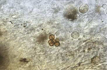
Traumatic inoculation of these fungi results in a mixed chronic suppurative and granulomatous host response.14 The epidermis typically becomes thickened in a process called pseudoepitheliomatous hyperplasia, a histologic morphology that may be misidentified as malignancy by more inexperienced microscopists. Foci of polymorphonuclear cells and microabscesses are seen in both the epidermis and dermis. In the dermis, granulomas that include multinucleated giant cells and epithelioid cells are present, along with varying amounts of fibrosis. Fibrosis is increased in older lesions and can extend into the subcutaneous tissue, although disease rarely extends deep into the subcutaneous tissue. The hallmark of chromoblastomycosis, the muriform cells—also called sclerotic, copper penny, or Medlar bodies—may be found intracellularly in macrophages or extracellularly in abscesses. These are darkly pigmented (brown-golden), thick-walled, rounded cells, 4 to 12 µm in diameter, with cross walls in one or two planes (Fig. 262-1). Hyphae may also occasionally be seen, usually in the epidermis. The host response to these structures results in a process termed transepithelial elimination, in which fungi and damaged tissue are expelled through the epidermis, a process similar to that seen in calcinosis cutis.15 The immunologic response of the host in this mycosis is still not well understood. Disease appears to be associated with an ineffective immune response to the organism, with chronic inflammation produced in response to persistence of the fungi in tissue. The cell-mediated immune response appears to play a central role. High interleukin-10 (IL-10) levels, low interferon-γ levels, and inefficient T-cell proliferation have been noted in patients with severe disease, with the opposite seen in those with milder chromoblastomycosis.16 Antibody responses have shown association with disease chronicity and extent but do not appear to provide any degree of protection in this infection.17
Clinical Manifestations
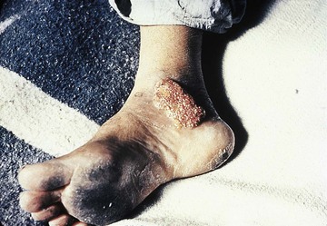
Weeks to months after inoculation of the causative organisms through minor trauma, subjects typically develop a small scaly papule on the lower extremity at the site of the trauma (Figs. 262-2 to 262-4). This lesion slowly develops into a superficial nodule, commonly with an irregular friable surface. Frequently, these nodules later spread out to become purplish, irregular, raised plaques. In descriptions by Carrion,18 lesions of chromoblastomycosis were categorized into five types: (1) early nodular lesions, described as soft and pink-violaceous, with smooth, verrucous, or scaly surfaces; (2) tumorous lesions, large, papillomatous, often lobulated masses with crusting, sometimes described as cauliflower like; (3) verrucous lesions with prominent hyperkeratosis; (4) plaque lesions; and (5) cicatricial lesions. Most lesions have black dots associated with their outer surface that are composed of fungi and necrotic debris, the products of transepithelial elimination. Although not typically painful, lesions may be associated with pruritus, are easily traumatized, and bleed readily. Ulceration is generally limited to those lesions with bacterial superinfection. Large lesions may become hyperkeratotic, and limb distortion, including elephantiasis, can occur as a result of blockage of normal lymphatic drainage.


