Acute Coronary Syndromes: Manifestations and Management


Acute Coronary Syndromes: Manifestations and Management


John Paul Vavalle

John H. Alexander

Matthew T. Roe


Coronary heart disease (CHD) is a global health epidemic that contributes to more than 7 million deaths per year and is the most frequent cause of death worldwide. Currently, more than half of the global burden of CHD is found in developing countries, and this percentage is growing rapidly.1 Within the United States, there has been a decrease in deaths from CHD over the last three decades due equally to improved therapeutics and risk factor modification. Nevertheless, the prevalence of CHD in the United States remains high with over 1 million hospitalizations for acute coronary syndrome (ACS) annually.2,3

ACS is a term that refers to the clinical syndrome of acute myocardial ischemia typically resulting from atherosclerotic plaque rupture that leads to occlusive or subocclusive intracoronary thrombus causing downstream myocardial ischemia or infarction. The term ACS encompasses three conditions: unstable angina (UA), non-ST-elevation myocardial infarction (NSTEMI), and ST-elevation myocardial infarction (STEMI).

The presence of myocardial necrosis, assessed by measurement of cardiac biomarkers such as troponin and creatine kinase MB (CK-MB), differentiates acute NSTEMI and STEMI from UA. STEMI is further distinguished from NSTEMI by the presence of ST-segment elevation on the electrocardiogram (ECG). Rapid identification, triage, and implementation of evidencebased therapies for ACS are essential to reducing the morbidity and mortality associated with this life-threatening condition.


PATHOPHYSIOLOGY OF ACS

The hallmark of ACS is myocardial ischemia that results from insufficient oxygen delivery to meet the oxygen demands of the myocardium. This phenomenon most commonly results from obstructed coronary blood flow due to intracoronary thrombus that has generated on the surface of a ruptured atherosclerotic plaque. However, other mechanisms of ACS have been described, including dynamic coronary obstruction from coronary spasm or vasoconstriction, progressive mechanical obstruction as would be observed with progressive atherosclerosis, coronary artery inflammation, coronary dissection, and secondary angina—where the increased oxygen demands of the myocardium outstrip a preserved supply (Table 90.1).3,4


Atherosclerosis

Coronary atherosclerosis develops over decades and is characterized by the progressive deposition of lipids, fibroblasts, and inflammatory cells beneath the endothelial layer within the arterial wall (see Chapter 89). In the coronary arteries, the majority of these atherosclerotic plaques never progress to cause an ACS event. However, the so-called vulnerable plaques that eventually rupture do so as a result of the disruption of the fibrous cap that exposes the thrombogenic lipid core to circulating blood and stimulates the formation of thrombi. A newly utilized term, atherothrombosis, has been developed to describe this process.


Plaque Inflammation and Rupture

Plaques prone to rupture have been termed vulnerable plaques and have been characterized histologically. Typically, these plaques have a lipid-rich or necrotic core with a thin fibrous cap.5 Activated macrophages and other inflammatory cells are generally present in higher concentrations in vulnerable plaques. Metalloproteinases and other enzymes secreted by these inflammatory cells can lead to thinning of the fibrous cap, making them prone to rupture. Autopsy studies have demonstrated dense infiltration of inflammatory cells into ruptured culprit plaques as compared to stable nonruptured plaques, supporting this notion of the inflamed vulnerable plaque.6

Identifying these plaques prone to rupture prior to an ACS event is an area of intense research, but virtual histology using intracoronary ultrasound, intracoronary optical coherence tomography, and molecular imaging with magnetic resonance imaging (MRI) or computed tomography (CT) has shown promise.7


Thrombosis

Intracoronary thrombosis is fundamental to the pathophysiology of ACS. At the moment of plaque rupture, platelet and plasma thrombosis transform a stable atherosclerotic plaque into an unstable coronary lesion. Intracoronary thrombi at the time of an ACS event have been demonstrated by angiography, atherectomy, and autopsy studies showing thrombus originating from the site of plaque rupture.8,9,10 Markers of coagulation activity, such as elevated plasma fibrinogen levels, prothrombin fragments, fibrinopeptide, and D-dimers, have all been detected in patients with ACS, but are nonspecific markers and cannot be used in isolation.11,12

At the core of the thrombotic process is platelet aggregation that begins when plaque rupture exposes collagen and tissue factor to circulating platelets. Platelet activation initiates with the binding of the glycoprotein Ib receptor with von Willebrand factor, resulting in conformational changes of the platelet, as well as release of α-granules, thromboxane A2 (TXA2), and other platelet chemoattractants.13 Mediators such as thrombin, adenosine diphosphate (ADP), and epinephrine amplify the platelet response by recruiting other platelets to the forming thrombus. The final pathway involves the activation of the glycoprotein IIb/IIIa (GP IIb/IIIa) receptor. Once activated, it is responsible for platelet aggregation and stabilization by binding fibrinogen, which cross-links the GP IIb/IIIa integrins between platelets.


Table 90.1 Causes of unstable angina and non-ST-elevation myocardial infarctiona


Thrombus or thromboembolism, usually arising on disrupted or eroded plaque


• Occlusive thrombus, usually with collateral vesselsb


• Subtotally occlusive thrombus on preexisting plaque


• Distal microvascular thromboembolism from plaque-associated thrombus


• Thromboembolism from plaque erosion


Non-plaque-associated coronary thromboembolism


Dynamic obstruction (coronary spasmc or vasoconstriction) of epicardial and/or microvascular vessels


Progressive mechanical obstruction to coronary flow


Coronary arterial inflammation


Secondary UA


Coronary artery dissectiond


a These causes are not mutually exclusive; some patients have two or more causes.


b DeWood MA, Stifter WF, Simpson CS, et al. Coronary arteriographic findings soon after non-Q-wave myocardial infarction.


N Engl J Med 1986;315:417-423.


c May occur on top of an atherosclerotic plaque, producing missed-etiology angina or UA/NSTEMI.


d Rare.


From Anderson JL, Adams CD, Antman EM, et al. ACC/AHA 2007 guidelines for the management of patients with unstable angina/non ST-elevation myocardial infarction: a report of the American College of Cardiology/American Heart Association Task Force on Practice Guidelines (Writing Committee to Revise the 2002 Guidelines for the Management of Patients With Unstable Angina/Non ST-Elevation Myocardial Infarction). Circulation 2007;116(7):e148-e304. Modified with permission from Braunwald E.


Unstable angina: an etiologic approach to management. Circulation 1998;98:2219-2222.


The activated platelet serves as the substrate upon which coagulation occurs. Tissue factor, released at the time of plaque rupture, leads to activation of factor X, converting it to factor Xa. Through multiple positive feedback loops involving both platelets and soluble coagulation factors, factor Xa leads to thrombin generation (factor IIa) that in turn converts fibrinogen to fibrin in the final step of thrombus formation.

As a result of the critical role that platelets and soluble coagulation factors play in ACSs, antiplatelet and anticoagulant therapies have become fundamental to the treatment of ACS. Current proven antiplatelet therapies include aspirin for the inhibition of TXA2 formation, P2Y12 receptor antagonists (clopidogrel, ticlopidine, prasugrel, and ticagrelor) for the inhibition of the ADP receptor pathway, and GP IIb/IIIa receptor antagonists that directly inhibit platelet aggregation. In addition, thrombin receptor antagonists that inhibit thrombin activation of platelets are currently under investigation. Currently proven anticoagulants include unfractionated heparin (UFH) and low molecular weight heparin (LMWH), indirect factor Xa inhibitors such as fondaparinux, and direct thrombin inhibitors (DTIs) such as bivalirudin. A number of additional anticoagulants, including the direct factor Xa inhibitor otamixaban and the RNA aptamer factor IXa inhibitor pegnivacogin, are currently under investigation.14,15


Other Mechanisms of ACS


Vasoconstriction

Dynamic coronary obstruction can occur as a result of intense focal vasoconstriction of a segment of an epicardial coronary artery. This is commonly labeled Prinzmetal (variant) angina. This focal spasm is the result of vascular smooth muscle hypercontractility and endothelial dysfunction.

Patients presenting with ACS from vasoconstriction usually have sudden onset of chest pain at rest without any antecedent increase in oxygen demand. Attacks can be precipitated by exposure to cold, emotional stress, adrenergic stimuli, exercise, and cocaine use.16,17,18 Variant angina episodes usually do not lead to acute myocardial infarction (MI), but prolonged episodes may result in acute myocardial necrosis, arrhythmias, and even sudden death.19,20 The diagnosis of variant angina is suspected by transient episodes of chest pain at rest that may be accompanied by dynamic ST-segment changes on ECG and may also be responsive to vasodilators such as nitroglycerin.


Progressive Mechanical Obstruction

Severe narrowing of the epicardial coronary arteries, in the absence of focal spasm or thrombus, may occur as a result of progressive atherosclerosis or may be due to intimal hyperplasia causing restenosis at the site of a previous percutaneous coronary intervention (PCI). In general, these patients present with angina symptoms that have been progressively worsening concurrent with the progression of the myocardial obstruction.


Coronary Inflammation and Dissection

Although much less common than other causes of ACS, it is important to recognize coronary artery inflammation and dissection as causes of coronary obstruction, as they may have devastating consequences. Coronary artery dissection is the most common cause of ACS in peripartal women and often present with acute MI.21 Inflammatory states such as graft-versushost disease or infections such as Kawasaki disease may lead
to intense inflammation of the coronary arteries and result in complete coronary occlusion.22,23 Identification and treatment of these etiologies is dependent upon recognizing and treating the underlying diagnoses.


Secondary Unstable Angina

Secondary UA results from an inability to meet the oxygen demands of the myocardium and usually occurs in the presence of some degree of obstructive coronary artery disease (CAD) that limits the flow reserve of the coronary circulation at times of increased myocardial workload. The causes of secondary angina are varied, but all result in increased myocardial oxygen requirements. Examples include fever, tachycardia, thyrotoxicosis, hyperadrenergic states, hypertensive emergency, and anemia among others. Because of the underlying condition, secondary angina is usually a marker for end-organ dysfunction and may have worse outcomes than other forms of angina.24


RESENTATION, RECOGNITION, AND TRIAGE OF ACS


Presentation

While stable angina typically presents with angina pectoris associated with exertion and is relieved with rest, patients presenting with ACS classically present with rest angina or newly progressive angina. Three principal presentations of UA have been described: (a) rest angina, (b) new-onset angina, and (c) increasing angina (Table 90.2). Patients suffering an STEMI may present with the above symptoms but typically present with the new onset of a prolonged or stuttering episode of pain at rest.

The first step for patients before they can receive possibly life-saving treatment is to recognize their symptoms as being potentially serious and to seek medical attention. Despite attempts to shorten the delay to seeking medical care, the average person with UA/NSTEMI waits approximately 2 hours prior to seeking attention, and in the face of attempts to improve this, there has been little change over the last decade.25,26 Often times, reasons cited for delay include atypical symptoms such as arm pain, jaw pain, or shortness of breath, or the expectation that the symptoms would be more severe.27,28 Studies trying to understand reasons for delay in seeking care have demonstrated that older patients, blacks, women, and Medicaid-only recipients are more likely to delay.25,29


Table 90.2 Presentations of ACS


Class


Presentation


Rest anginaa


Angina occurring at rest and prolonged, usually >20 min


New-onset angina


New-onset angina of at least CCS class III severity


Increasing angina


Previously diagnosed angina that has become distinctly more frequent, longer in duration, or lower in threshold (i.e., increased by 1 or more CCS class to at least CCS class III severity)


a Patients with NSTEMI usually present with angina at rest.


CCS, Canadian Cardiovascular Society.


Adapted from Braunwald E. Unstable angina: a classification. Circulation 1989;80:410-414; Anderson JL, Adams CD, Antman EM, et al. ACC/AHA 2007 guidelines for the management of patients with unstable angina/non STelevation myocardial infarction: a report of the American College of Cardiology/American Heart Association Task Force on Practice Guidelines (Writing Committee to Revise the 2002 Guidelines for the Management of Patients With Unstable Angina/Non ST-Elevation Myocardial Infarction). Circulation 2007;116(7):e148-e304.


Patients experiencing an ACS event may not always present with chest discomfort.30 An analysis of the National Registry of Myocardial Infarction showed that about one-third of the 434,877 patients with MI did not present with chest pain.31 In this study, this resulted in delays in seeking care, delays to diagnosis by the providers, less implementation of evidencebased therapies, and higher in-hospital mortality. In particular, women, older patients, and those with a history of diabetes or heart failure are more likely to present with silent MIs, and those caring for these patients must be vigilant in their evaluation.


Evaluation and Diagnosis

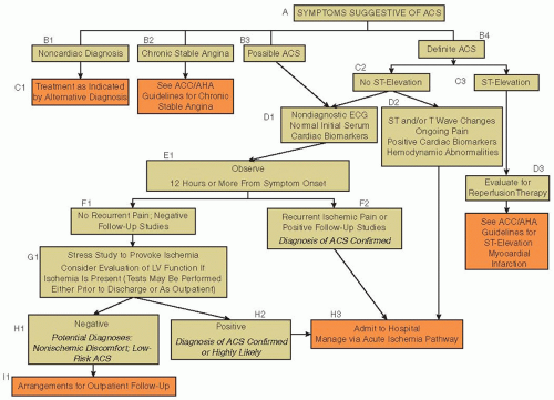
Because there is substantial overlap in the presenting symptoms of STEMI, NSTEMI, and UA, the initial evaluation of all patients presenting to a health care facility with symptoms concerning ACS should be the same (FIGURE 90.1). Patients should be evaluated in an acute care setting, with providers and staff equipped to perform critical care.

Because of the possibility of an immediate life-threatening condition, the initial evaluation must focus on rapidly identifying clinical instability and assessing for ST-segment elevation on an ECG if not done prior to arrival by emergency medical personnel. ST elevation in the setting of symptoms compatible with ACS should set the patient on a predefined treatment algorithm for urgent reperfusion therapy with fibrinolytics or primary PCI.32 The importance of the ECG in the initial evaluation has led the American Heart Association and the American College of Cardiology in its joint treatment guidelines to give its strongest recommendation possible (class I) to obtaining an ECG within 10 minutes of arrival. Rapid evaluation and implementation of treatment for ACS can translate into significant improvements in outcomes and ultimate cost savings.


THE ACUTE MANAGEMENT OF STEMI


Urgent Identification, Triage, and Reperfusion

In acute STEMI, there is complete occlusion of the epicardial coronary artery, leading to transmural myocardial ischemia and infarction. Generally, STEMI results from plaque rupture, leading to an occlusive thrombus composed of fibrin, platelets, and inflammatory cells. Acute STEMI represents a true medical emergency, and management strategies focus on urgent coronary reperfusion therapies to open the occluded coronary vessel and minimize myocardial necrosis. This may involve primary PCIs, fibrinolytic therapy, or a combination of both depending on patient characteristics and available resources and capabilities. The old adage of time is muscle holds true for STEMI patients,
with each minute of delay associated with an increase in mortality.33 Reperfusion therapy to open the occluded artery with either fibrinolytics or primary PCI should be implemented urgently.


FIGURE 90.1 Treatment algorithm for patients with suspected ACS. ACC/AHA, American College of Cardiology/American Heart Association; ECG, electrocardiogram; LV, left ventricular. (From Anderson JL, Adams CD, Antman EM, et al., ACC/AHA 2007 guidelines for the management of patients with unstable angina/non ST-elevation myocardial infarction. Circulation 2007;116(7):e148-e304.)

The diagnosis of STEMI is made in the right clinical context in conjunction with anginal symptoms when at least 1 mm of ST elevation is present in two contiguous leads or a new left bundle branch block is detected on the ECG. Acute ST-segment elevation is associated with the highest risk of early death, and the presence of such findings indicates the need for immediate reperfusion therapy. This diagnosis should trigger a preplanned algorithm to initiate a reperfusion strategy with either fibrinolytics or primary PCI. The initial assessment of a patient with STEMI should focus on evaluating for hemodynamic and arrhythmic complications and on rapidly evaluating for the optimal reperfusion strategy.

Delays in reperfusion can arise from a number of sources. Reasons cited for delay include a lack of initial recognition of the symptoms by the patient, inefficient transportation to medical centers, and delays in initiating reperfusion therapy once the patient arrives. A well-coordinated STEMI response team comprised of cardiologists a emergency departments, and the emergency medical services (EMS) is needed to minimize delays.

Often, the first medical contact with a patient is by EMS. EMS personnel need to be well trained in the recognition and acute management of STEMI. The ability by EMS to perform and interpret 12-lead ECGs can lead to an accelerated diagnosis of STEMI and a more rapid reperfusion.34 In addition, EMS should be equipped to handle complications that might arise from STEMI and have the ability to perform advanced cardiac life support, defibrillation, and intubation with mechanical ventilation when needed. Although not widely used, prehospital reperfusion with fibrinolytic agents given by appropriately trained personnel has been studied, and in a meta-analysis of six trials, it was shown that prehospital thrombolysis preceded the in-hospital thrombolysis by a mean of 58 minutes and reduced the all-cause hospital mortality with an odds ratio (OR) of 0.83 (95% confidence interval [CI], 0.70 to 0.98).35

Given the significant importance of time to reperfusion, benchmark guideline times have been put in place and are used to assess quality outcomes of health systems. Currently,
for fibrinolytic therapy, the recommended door-to-needle time is ≤30 minutes. This means that from the minute the patient arrives in the emergency room to the minute the fibrinolytic infusion is begun, the time elapsed is not >30 minutes. For primary PCI, the established benchmark is a door-to-balloon time of ≤90 minutes.32


Reperfusion Therapy for STEMI

Despite strong evidence for improved outcomes with early reperfusion therapy in STEMI, not all eligible patients receive this therapy. Currently, approximately 30% of STEMI patients receive no reperfusion therapy despite a lack of contraindications, and of those treated, <50% receive fibrinolytic agents within the recommended door-to-needle time of 30 minutes and <40% receive PCI within the recommended 90 minutes.36 However, more recent data in at least one study suggest improvement in this trend, with the percentage of eligible patients in contemporary practice not receiving reperfusion therapy being closer to 10%.37

Both fibrinolytic therapy and primary PCI are suitable options for revascularization in STEMI. Choosing between the two therapies is based on a number of factors, but the important common denominator is that the chosen strategy be implemented as rapidly as possible. In general, primary PCI is favored over fibrinolytic therapy when available in a timely fashion, especially in those who are at high risk or presenting late after symptom onset.38

A pooled analysis of 23 randomized trials comparing primary PCI to fibrinolysis for STEMI showed that primary PCI was better than fibrinolytic agents for nearly all endpoints, including mortality (OR, 0.73; 95% CI, 0.62 to 0.86), reinfarction (OR, 0.35; 95% CI, 0.27 to 0.45), and stroke (OR, 0.46; 95% CI, 0.30 to 0.72).39 However, when a patient presents to a hospital where PCI is not immediately available, the decision to transfer to a PCI-capable hospital versus immediate administration of fibrinolytic agents is a complex one.

Factors to consider when deciding between primary PCI and fibrinolysis include such factors as contraindications to fibrinolysis, the presence of cardiogenic shock, time from symptom onset, bleeding risk, diagnostic certainty of STEMI, and the time required to transfer for primary PCI.32,40 Contraindications to fibrinolytic therapy are listed in Table 90.3 and include both absolute and relative contraindications. Patients meeting any of the absolute contraindications should not receive fibrinolytics and should be considered for primary PCI. The risks of administering fibrinolytics to a patient with a relative contraindication to reperfusion must be weighed carefully.


Primary PCI

One of the most substantial systematic barriers to obtaining primary PCI as the reperfusion modality is the lack of facilities and the time required to transfer a patient from a non-PCI hospital to a PCI-capable hospital. In many rural areas, primary PCI is not readily available and either transfer to a PCI center must be arranged or fibrinolytic therapy administered. In general, if the added time to obtain primary PCI over fibrinolytic therapy is more than 60 minutes, the mortality advantage gained by primary PCI is felt to be largely lost.38 In many rural hospitals, fibrinolytic agents are the standard of care due to the long distances to the closest primary PCI centers. The focus at these institutions should be on initiating fibrinolytics as quickly as possible.


Table 90.3 Contraindications to fibrinolytic therapy in the management of STEMI


Absolute Contraindications


Any prior cerebral hemorrhage


Known structural central nervous system lesion (arteriovenous malformation, tumor, etc.)


Ischemic stroke within 3 mo UNLESS acute ischemic stroke of <3 h onset


Significant closed head or facial injury within 3 mo


Suspicion of aortic dissection


Active bleeding (excluding menses) or bleeding disorders


Relative Contraindications


History of chronic, severe, and poorly controlled hypertension, or severe hypertension on admission (systolic blood pressure > 180 mmHg or diastolic blood pressure > 100 mmHg)


Traumatic or prolonged (>10 min) CPR or noncompressible vascular punctures


Major surgery or internal bleeding within 3-4 wk


Other central nervous disease (structural or dementia) not noted above


Pregnancy


Active peptic ulcer disease


Current use of anticoagulants (the higher the INR, the higher the risk of bleeding)


Prior exposure to or prior allergic reaction to streptokinase or anistreplase, if using these agents


Adapted from Antman EM, Anbe DT, Armstrong PW, et al. ACC/AHA guidelines for the management of patients with ST-elevation myocardial infarction. Circulation 2004;110:588-636.


Fibrinolytic Therapy

Among the first studies to demonstrate the effectiveness of fibrinolytic therapy for the treatment of STEMI was the GISSI trial published in 1986.41 In this study, streptokinase used to treat STEMI was associated with a significant reduction in mortality at 21 days (10.7% vs. 13%; RR, 0.81; P = 0.0002). Early administration of fibrinolytic therapy is critical to maximizing its effectiveness in restoring coronary blood flow. In general, the earlier fibrinolytics are administered after the onset of symptoms, the larger the mortality benefit that is derived from this therapy.42 Currently, it is recommended that fibrinolytic therapy can be given to patients up to 12 hours after the onset of symptoms. Those receiving fibrinolytic agents more than 12 hours after the onset of symptoms derive little benefit, while those who receive fibrinolytic agents within 1 hour from the onset of symptoms have a significant reduction in mortality.


Several fibrinolytic agents are available and have been studied for the treatment of STEMI (see Chapter 112). These include streptokinase, tissue plasminogen activator (tPA; alteplase), reteplase, and tenecteplase (TNK). All agents can be used for this indication, but tenecteplase and reteplase are generally favored due to their relative ease of bolus dosing.

Overall, the choice of which fibrinolytic agent to use is less important than the early and rapid administration of fibrinolysis to appropriate patients without contraindications suffering from an acute STEMI (Table 90.3). In many parts of the world, fibrinolytics remain the reperfusion therapy of choice, largely due to the unavailability of primary PCI. For those that do not have resolution of anginal symptoms or at least 50% of the ST-segment elevation on ECG 1 hour after administration of fibrinolytics, plans should be made for urgent rescue PCI with a cardiac catheterization team.


THE ACUTE MANAGEMENT OF NSTEMI/UA


Assessing Risk in those with NSTEACS

After the initial triage, patients presenting with ACS without ST-segment elevation (non-ST-elevation ACS [NSTEACS]) or hemodynamic compromise can be evaluated in a more systematic manner, with the intent of determining the probability that the patient’s symptoms are due to coronary artery disease (CAD). This involves a careful review of the patient’s symptoms and physical findings, analyzing the ECG for ischemic changes such as ST-segment depression or T-wave inversion, reviewing the patient’s medical record for a personal history of CAD or risk factors for heart disease, and evaluating serial serum biomarkers such as troponin to assess for MI.

The initial history is critical to assessing the probability of ACS. Typical angina is usually described as a deep substernal chest discomfort that can radiate to the arm and jaw and is associated with shortness of breath or diaphoresis. In ACS, these symptoms may present with rest pain or accelerating angina that occurs with lessening degrees of activity. For older patients, women, and diabetics, these typical features may not be present and are unreliable. Despite the widely held belief to the contrary, the relief of chest pain by sublingual nitroglycerin does not predict the presence of ACS.43

The ECG provides important prognostic and diagnostic information at the time of initial triage. It is an essential component of the early triage algorithm. Findings of old Q waves on the ECG are important because they indicate likely prior MI and a higher risk for CAD. A normal ECG does not exclude the diagnosis of ACS, but patients who have an unchanged ECG when compared to a prior one have a low risk of adverse events.44


Cardiac Biomarkers

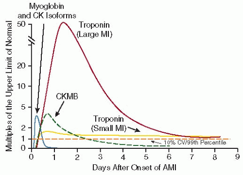
The evaluation of cardiac biomarkers is an essential component to early triage and risk stratification. The clinical diagnosis of MI hinges on the detectable elevation of these cardiac biomarkers in conjunction with other findings of ischemia such as symptoms or ECG changes. Biomarkers are released into the peripheral circulation from infarcted cardiac myocytes when necrosis occurs (Table 90.4). The most widely used cardiac biomarkers currently are CK-MB and the cardiac troponins I and T (TnI and TnT). Myoglobin is rarely used clinically today. These proteins are found in high concentrations within the cardiac myocytes, making them specific for myocardial cell damage, and circulate at low levels within the peripheral circulation in the absence of cardiac myocyte necrosis (FIGURE 90.2). The cardiac troponins are highly sensitive and specific for MI and may be detectable in the blood as soon as 2 hours after the onset of symptoms and may stay elevated for as long as 14 days after an infarct.

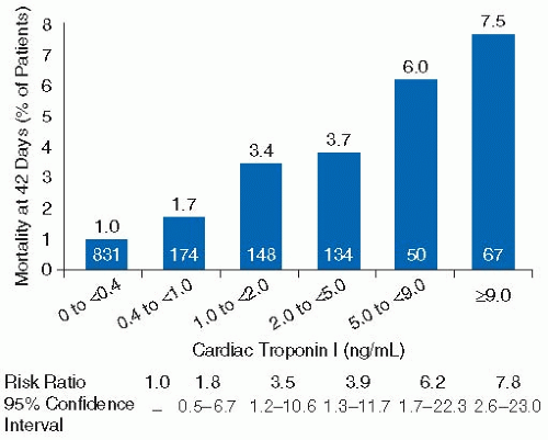
The elevation of cardiac biomarkers has important and well-validated prognostic significance beyond that obtainable from the ECG and the patient’s clinical parameters. An elevated troponin level is independently associated with an increased risk of death, and there is a direct and proportional relationship between the degree of elevation and this risk of death45,46,47 (FIGURE 90.3).


Risk Stratification in NSTEACS Using Risk Scores

Once other causes of the patient’s symptoms have been excluded and ACS is felt to be the diagnosis, early risk stratification is important to determining appropriate management. Doing this can aid in selecting the appropriate level of care needed for the patient, guiding the choices of the most appropriate medical therapies, and assisting in deciding between an early invasive strategy in the catheterization laboratory and a more conservative strategy initially with medical therapy.

Early risk stratification can help differentiate patients with NSTEMI and UA who often present with very heterogeneous symptoms. Multiple factors determine the likelihood of obstructive CAD and prognosis. Several risk scores have been developed to help clinicians identify high-risk patients rapidly. These include the Thrombolysis in Myocardial Infarction (TIMI) risk score and the Global Registry of Acute Coronary Events (GRACE) risk score (FIGURE 90.4 and Table 90.5).48,49

The TIMI risk score was developed as a simple tool comprising seven questions rating risk on presentation designed to assess patients with suspected UA or NSTEMI. For details on the score, see legend to Table 90.5. The TIMI risk score provides an estimate of death, MI, or recurrent ischemia requiring urgent revascularization within 14 days from presentation. It has been validated in several studies and is a quick and relatively easy bedside tool to help clinicians stratify patients based on risk.

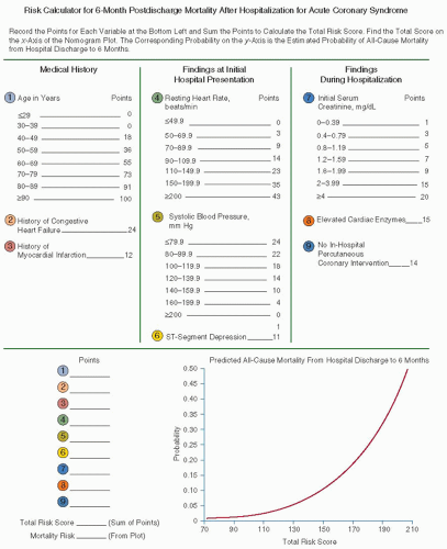
The GRACE prediction score is a model that can be used to predict in-hospital mortality and mortality from discharge to 6 months in patients presenting with ACS.49,50 The GRACE model for predicting 6-month postdischarge mortality uses factors such as age, personal history of heart failure and MI, initial blood pressure and pulse, ECG, and laboratory findings (FIGURE 90.4). It is a useful clinical tool for patients with NSTEMI, STEMI, and UA to predict 6-month mortality.


Initial Invasive Versus Initial Conservative Therapy

For patients presenting with NSTEMI or UA, one of two treatment pathways is usually decided upon based on patient risk and clinical stability. One pathway is labeled the “early invasive” strategy, while the other pathway for treatment is called the “initial conservative” or “selective invasive” strategy.3 This terminology refers to the chosen angiographic strategy for the patient and the intended timing of diagnostic angiography with planned PCI. In both pathways, patients receive the usual

medical therapy for NSTEMI and UA that includes antianginal, antiplatelet, and anticoagulant therapy (FIGURES 90.5 and 90.6).


Table 90.4 Cardiac biomarkers


Marker


Advantages


Disadvantages


Point-of-Care Test Available?


Comment


Clinical Recommendation


Cardiac troponins


Powerful tool for risk stratification
Greater sensitivity and specificity than CK-MB
Detection of recent MI up to 2 wk after onset
Useful for selection of therapy
Detection of reperfusion


Low sensitivity in very early phase of MI (<6 h after symptom onset) and requires repeat measurement at 8 to 12 h, if negative
Limited ability to detect late minor reinfarction


Yes


Data on diagnostic performance and potential therapeutic implications increasingly available from clinical trials


Useful as a single test to efficiently diagnose NSTEMI (including minor myocardial damage), with serial measurements. Clinicians should familiarize themselves with diagnostic “cutoffs” used in their local hospital laboratory


CK-MB


Rapid, cost-efficient, accurate assays
Ability to detect early reinfarction


Loss of specificity in setting of skeletal muscle disease or injury, including surgery
Low sensitivity during very early MI (<6 h after symptom onset) or later after symptom onset (more than 36 h) and for minor myocardial damage (detectable with troponins)


Yes


Familiar to majority of clinicians


Prior standard and still acceptable diagnostic test in most clinical circumstances


Myoglobin


High sensitivity Useful in the early detection of MI
Detection of reperfusion
Most useful in ruling out MI


Very low specificity in the setting of skeletal muscle injury or disease
Rapid return to normal range limits sensitivity for later presentations


Yes


More convenient early marker than CK-MB isoforms because of greater availability of assays for myoglobin; rapid-release kinetics make myoglobin useful for noninvasive monitoring of reperfusion in patients with established MI


ACS, acute coronary syndrome; CK-MB, MB fraction of creatine kinase; ECG, electrocardiogram; MI, myocardial infarction; NSTEMI, non-ST-elevation MI.


Adapted from Anderson JL, Adams CD, Antman EM, et al. ACC/AHA 2007 guidelines for the management of patients with unstable angina/non ST-elevation myocardial infarction: a report of the American College of Cardiology/American Heart Association Task Force on Practice Guidelines (Writing Committee to Revise the 2002 Guidelines for the Management of Patients With Unstable Angina/Non ST-Elevation Myocardial Infarction). Circulation 2007;116(7):e148-e304.


FIGURE 90.2 Release of biomarkers into the serum after acute myocardial infarction. The biomarkers are plotted showing the multiples of the cutoff for acute myocardial infarction (AMI) over time. The dashed horizontal line shows the upper limit of normal (ULN; defined as the 99th percentile from a normal reference population without myocardial necrosis; the coefficient of variation of the assay should be 10% or less). The earliest rising biomarkers are myoglobin and CK isoforms (leftmost curve). CK-MB (dashed curve) rises to a peak of 2 to 5 times the ULN and typically returns to the normal range within 2 to 3 days after AMI. The cardiacspecific troponins show small elevations above the ULN in small infarctions (e.g., as is often the case with NSTEMI) but rise to 20 to 50 times the ULN in the setting of large infarctions (e.g., as is typically the case in STEMI). The troponin levels may stay elevated above the ULN for 7 days or more after AMI. CK, creatine kinase; CKMB, MB fraction of creatine kinase; CV, coefficient of variation; MI, myocardial infarction. (Modified from Shapiro BP, Jaffe AS. Cardiac biomarkers. In: Murphy JG, Lloyd MA, eds. Mayo clinic cardiology: concise textbook, 3rd ed. Rochester, MN: Mayo Clinic Scientific Press and New York: Informa Healthcare USA, 2007:773-780. Used with permission of Mayo Foundation for Medical Education and Research. Also adapted from Anderson JL, Adams CD, Antman EM, et al. ACC/AHA 2007 guidelines for the management of patients with unstable angina/non ST-elevation myocardial infarction: a report of the American College of Cardiology/American Heart Association Task Force on Practice Guidelines (Writing Committee to Revise the 2002 Guidelines for the Management of Patients With Unstable Angina/Non ST-Elevation Myocardial Infarction). Circulation 2007;116(7):e148-e304.)

In the early invasive strategy, patients usually undergo coronary angiography within 24 to 48 hours after admission. In the initial conservative strategy, patients do not undergo coronary angiography initially unless they have failed medical therapy or have documented ischemia, such as that demonstrated by a stress test, despite optimal medical therapy.

Deciding between an early invasive and conservative strategy is based largely on patient characteristics. High-risk features favor an early invasive strategy. These may include refractory ischemia, elevated cardiac biomarkers, dynamic ECG changes, hemodynamic or arrhythmic instability, prior revascularization, an elevated risk score (TIMI or GRACE), or left ventricular dysfunction (Table 90.6). In addition, patients treated with an early invasive strategy include those without ST-segment elevation who undergo urgent cardiac catheterization due to clinical instability or refractory symptoms despite medical therapy. Patients at lower risk, or those with extensive comorbidities, where the risks of angiography may be considered to be too high, are typically treated with an initial conservative strategy.


FIGURE 90.3 Stepwise association between elevations in troponin levels and death. (From Anderson JL, Adams CD, Antman EM, et al. ACC/AHA 2007 guidelines for the management of patients with unstable angina/non ST-elevation myocardial infarction: a report of the American College of Cardiology/American Heart Association Task Force on Practice Guidelines (Writing Committee to Revise the 2002 Guidelines for the Management of Patients With Unstable Angina/Non ST-Elevation Myocardial Infarction). Circulation 2007;116(7):e148-e304. Originally from Ohman EM, et al. Cardiac troponin T levels for risk stratification in acute myocardial ischemia. GUSTO IIA Investigators. N Engl J Med 1996;335(18): 1333-1341.)

For patients being treated with a conservative strategy, medical therapy is pursued with the intent of delaying the decision to proceed to coronary angiography until further clinical data are obtained. Generally in this group, the decision to go to diagnostic angiography is based upon results of a functional stress test showing objective evidence of ischemia, or upon persistent symptoms despite medical therapy.

The reasoning behind a conservative strategy is to avoid an invasive procedure and its inherent risk until the patient demonstrates objective evidence for ischemia or has not responded to medical therapy. The benefits to this strategy include the fact that many patients will stabilize on medical therapy and not require coronary angiography, thus reducing the use of an invasive and costly procedure. Patients treated in this pathway should have a stress test prior to discharge as well as an assessment for left ventricular function.

The early invasive strategy provides an angiographic method for risk stratification and a definitive evaluation for CAD. This strategy can differentiate those patients with no CAD from
those with severe CAD who may benefit from revascularization.51,52 Furthermore, percutaneous revascularization of the culprit lesion may lead to a reduced risk for rehospitalization and reduced angina after discharge.53


FIGURE 90.4 GRACE risk prediction model. (Adapted from Anderson JL, Adams CD, Antman EM, et al. ACC/AHA 2007 guidelines for the management of patients with unstable angina/non ST-elevation myocardial infarction: a report of the American College of Cardiology/American Heart Association Task Force on Practice Guidelines (Writing Committee to Revise the 2002 Guidelines for the Management of Patients With Unstable Angina/Non ST-Elevation Myocardial Infarction). Circulation 2007;116(7):e148-e304. Originally from Eagle KA, Lim MJ, Dabbous OH, et al. A validated prediction model for all forms of acute coronary syndrome: estimating the risk of 6-month postdischarge death in an international registry. JAMA 2004;291:2727-2733. Copyright © 2004 American Medical Association.)

Several trials have compared the early invasive strategy to the initial conservative approach. A meta-analysis of these trials concluded that a routine invasive approach is superior to a conservative strategy in reducing death or MI (14.4% vs. 12.2%;
OR = 0.82; 95% CI, 0.72 to 0.93).54 This analysis showed an 18% relative reduction in death or MI, as well as a sustained reduction in mortality following discharge from the hospital with the invasive approach. In addition, the invasive strategy led to a reduction in angina, fewer hospitalizations, and improvements in quality of life.


Table 90.5 Thrombolysis in myocardial infarction risk scorea


TIMI Risk Score


All-Cause Mortality, New or Recurrent MI, or Severe Recurrent Ischemia Requiring Urgent Revascularization through 14 d After Randomization (%)


0-1


4.7


2


8.3


3


13.2


4


19.9


5


26.2


6-7


40.9


a The TIMI risk score is determined by the sum of the presence of seven variables at admission: 1 point is given for each of the following variables: age 65 years or older; at least three risk factors for CAD; prior coronary stenosis of 50% or more; ST-segment deviation on ECG presentation; at least two anginal events in prior 24 hours; use of aspirin in prior 7 days; elevated serum cardiac biomarkers. Prior coronary stenosis of 50% or more remained relatively insensitive to missing information and remained a significant predictor of events.


MI, myocardial infarction.


Adapted from Antam EM, Cohen M, Bemink PH, et al. The TIMI risk score for unstable angina/non-ST elevation MI: a method for prognostication and therapeutic decision making. JAMA 2000;284:835-842; Anderson JL, Adams CD, Antman EM, et al. ACC/AHA 2007 guidelines for the management of patients with unstable angina/non ST-elevation myocardial infarction: a report of the American College of Cardiology/American Heart Association Task Force on Practice Guidelines (Writing Committee to Revise the 2002 Guidelines for the Management of Patients With Unstable Angina/Non ST-Elevation Myocardial Infarction). Circulation 2007;116(7):e148-e304.


A second meta-analysis of more contemporary randomized trials in NSTEMI also showed benefit with an early invasive approach.55 The results showed a long-term improvement in mortality and morbidity with the invasive strategy, including a reduction in MI and rehospitalizations (FIGURES 90.7 and 90.8). These improvements seem to be sustained for years after the event. An analysis from FRISC-II (FRagmin and fast Revascularization during InStability in Coronary artery disease-II) showed that the reduction in death or nonfatal MI is sustained for up to 5 years, and data from RITA-3 (Third Randomized Intervention Treatment of Angina) also demonstrated a reduction in death and MI at 5 years.56,57

Overall, the objective is to identify those patients who are most likely to benefit from an aggressive early invasive approach and minimize the exposure to potential harm in those less likely to benefit. The decision to proceed to early coronary angiography, versus a delayed and more conservative approach, is an individual one that should consider the wishes of the patient and family as well as the patient’s comorbidities. It must also be emphasized that ACS is a dynamic process where the clinical stability of a patient and the perceived benefit of an aggressive approach may change rapidly.


Mechanical Reperfusion

Mechanical reperfusion of the coronary arteries can be performed percutaneously or surgically. PCI can restore coronary blood flow through a number of approaches that include balloon angioplasty, intracoronary stenting, or removal of existing thrombus with thrombectomy devices. Used in the appropriate settings, such as for the treatment of acute MI, these therapies can be life saving. Surgical revascularization with coronary artery bypass grafting (CABG) involves the surgical implantation of venous or arterial grafts to the coronary arteries. This procedure is generally reserved for those patients with severe diffuse CAD not easily approached percutaneously.

Only gold members can continue reading. Log In or Register to continue

Stay updated, free articles. Join our Telegram channel

Jun 21, 2016 | Posted by in HEMATOLOGY | Comments Off on Acute Coronary Syndromes: Manifestations and Management

Full access? Get Clinical Tree

Get Clinical Tree app for offline access