Acquired Nonimmune Thrombocytopenia


Acquired Nonimmune Thrombocytopenia


Andreas Greinacher

Theodore E. Warkentin


Acquired thrombocytopenic disorders can be divided into five categories, plus the spurious entity of pseudothrombocytopenia (Table 64.1): hemodilution, sequestration (usually due to hypersplenism), decreased platelet production, increased platelet consumption (by pathologically increased “physiologic” pathways of platelet activation and clearance), and platelet destruction (e.g., phagocytosis of antibody-coated platelets). This chapter summarizes the nonimmune differential diagnosis of acquired thrombocytopenic disorders. We also discuss thrombocytopenia in patients in intensive care units, as thrombocytopenia is very frequent in these patients, and provide an approach to separate the different causes of a low platelet count.


PERIOPERATIVE THROMBOCYTOPENIA

Early postoperative thrombocytopenia is a combined effect of hemodilution and platelet consumption. It is one of the most common events prompting consultation with a hematologist. A platelet count decline of 30% to 70% occurs universally following major surgery. Such hemodilution-associated thrombocytopenia is most prominent after cardiac surgery1 and is roughly proportional to the amount of crystalloid, colloid, or blood products administered. Most of this platelet count decline is abrupt and occurs within a few hours following surgery. Dilutional coagulopathy generally accompanies the thrombocytopenia,2 accounting for the minor-to-moderate increases in prothrombin time and activated partial thromboplastin time that are commonly seen immediately after surgery.

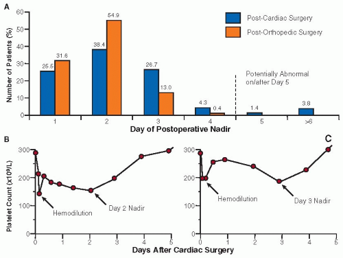
Perioperative hemodilution is accompanied by platelet consumption, an effect that correlates with the magnitude of tissue trauma. An important characteristic of the postsurgery platelet count is that it usually continues to decline over the next 1 or 2 days, with the postoperative nadir usually occurring at a median of postoperative day 2, with a range between postoperative days 1 and 4 (FIGURE 64.1A).3,4 Subsequently, there is a rise in the platelet count to levels often two to three times the patient’s preoperative baseline, peaking at approximately day 14, before it returns to baseline over the next 2 weeks (˜day 28).

This platelet count profile can be explained by thrombopoietin physiology. Thrombopoietin, produced at a constant rate by the liver, is regulated by the circulating platelet mass. Platelets bind thrombopoietin, thereby decreasing the amount of circulating thrombopoietin. With perioperative platelet count declines, more thrombopoietin becomes available to stimulate megakaryocytes. However, there is a lag between megakaryocyte stimulation and release of new platelets. Studies of administering the thrombopoietin receptor analogue, romiplostim, to human volunteers showed an increased platelet count beginning after 3 to 5 days and peaking about 7 to 10 days later.5 As platelet stimulation necessarily has an approximate 3-day lag, it would be expected that platelet counts reach a nadir as late as 3 or even 4 days after major surgery (SEE FIGURE 64.1B). This time course is the most important feature differentiating the normal platelet count decrease after major surgery from immune-mediated thrombocytopenias, especially from heparin-induced thrombocytopenia (HIT) where the platelet count decrease typically begins on or after day 5.4,6

This pattern can be changed by perioperative platelet transfusions that produce a transient increase in platelet count, with subsequently clearing of the transfused platelets over the next 2 or 3 days, resulting in a shift of the platelet count nadir to the right.


PLATELET SEQUESTRATION


Hypersplenism

Hypersplenism denotes the reduction in one or more of the peripheral blood counts due to sequestration within an enlarged spleen. Unlike postoperative hemodilution, hypersplenism is characterized by subacute or chronic, rather than acute, thrombocytopenia.

Normally, approximately one-third of the total body platelet mass resides within the spleen, even though the spleen receives only 5% of the cardiac output.7 This discrepancy occurs because the transit time of a platelet through the spleen averages 20 minutes, much longer than the time required for a platelet to return to the heart after passing through other organs (˜1 minute). In hypersplenism, the proportion of platelets within the spleen can be as high as approximately 90%.7

Characteristic clinical features of hypersplenism include splenomegaly (measured objectively as >11 cm by imaging studies) and mild-to-moderate pancytopenia. Often, the degree of anemia is not as marked as neutropenia and thrombocytopenia. In patients with advanced cirrhosis, thrombocytopenia is further aggravated by reduced platelet production due to decreased thrombopoietin levels8 or the toxic effects of alcohol and/or increased platelet consumption due to enhanced thrombin generation and hyperfibrinolysis.

Table 64.2 lists various causes of splenomegaly.9 In Western societies, cirrhosis (usually secondary to alcohol consumption or chronic viral hepatitis) is a common cause, whereas other explanations are more common elsewhere (e.g., schistosomiasis, malaria).

Treatment is usually not required, as thrombocytopenia rarely reaches clinically important levels and splenectomy is not without risk.


Table 64.1 Classification of thrombocytopenia


Six general explanations for thrombocytopenia, including pseudothrombocytopenia.


Explanation


Comment


Examples


Pseudothrombocytopenia


Antibody-mediated platelet clumping that occurs ex vivo, that is, spurious thrombocytopenia


EDTA-induced pseudothrombocytopenia; platelet satellitism (platelet binding to leukocytes)


Hemodilution


Abrupt platelet count fall due to administration of fluids or blood products


Early postoperative thrombocytopenia


Hypersplenism


Splenomegaly may require imaging studies to detect; concomitant leukopenia and (to lesser extent) anemia are usually present


Cirrhosis-associated portal hypertension


Decreased platelet production


Usually there is concomitant leukopenia and anemia (a notable exception is certain types of hereditary thrombocytopenia)


MDS; aplastic anemia; marrow infiltration by metastatic cancer; alcohol-induced thrombocytopenia


Platelet consumption


Accelerated platelet clearance due to pathologically increased platelet activation or other poorly defined mechanisms of increased platelet clearance


DIC; septicemia; multiorgan system failure; hemophagocytic syndrome


Platelet destruction


Accelerated platelet clearance due to pathological mechanisms that target platelets, especially due to platelet-reactive antibodies


Autoimmune thrombocytopenic purpura; HIT; drug-induced immune thrombocytopenia; TTP/HUS


EDTA, ethylenediaminetetraacetic acid; HIT, heparin-induced thrombocytopenia; TTP/HUS, thrombotic thrombocytopenic purpura/hemolytic uremic syndrome.


Hypothermia

Hypothermia can be accompanied by mild-to-moderate thrombocytopenia.10 The thrombocytopenia is transient and corrects with rewarming. Platelet kinetic studies in cooled dogs demonstrated reversible hepatic and splenic platelet sequestration.11 Indirect evidence suggests that decreased platelet production may also contribute.12


DECREASED PLATELET PRODUCTION

Thrombocytopenia often is caused by decreased platelet production, which can be divided into three categories: acquired, generalized marrow disorders; acquired, isolated megakaryocytic disorders (e.g., alcohol-associated thrombocytopenia); and hereditary thrombocytopenic disorders.


Acquired, Generalized Marrow Disorders

By far the most common cause of decreased platelet production is acquired, generalized bone marrow disease. The pathologic process usually perturbs all cell lines, and pancytopenia typically results. Occasionally, however, isolated thrombocytopenia is the first abnormality detected during the early stages of a panmyelopathy, for example, myelodysplasia (MDS).

Nonneoplastic Hypoproliferative Stem Cell Disorders— Thrombocytopenia accompanies aplastic anemia, chemotherapyinduced pancytopenia, and idiosyncratic drug-induced aplastic anemia. Sometimes, isolated thrombocytopenia is a precursor of aplastic anemia.

Paroxysmal Nocturnal Hemoglobinuria (PNH)—PNH is a stem cell defect resulting in the loss of phosphatidylinositol-anchored proteins, resulting in intravascular hemolysis due to increased susceptibility of red blood cells to complement-mediated lysis; PNH is also associated with thrombocytopenia.13 The low platelet count is a composite of a dysplastic platelet production defect, enhanced platelet consumption due to the higher susceptibility of PNH platelets to thrombin, and enhanced platelet destruction due to increased generation of lytic C5b9 complexes on platelet membranes.14 Patients are at increased risk for thrombotic complications, perhaps induced by the production of procoagulant microparticles from affected blood cells. Diagnosis is made by showing by flow cytometry a population of blood cells with decreased/absent phosphatidylinositol-anchored proteins. A treatment option in severe PNH is the monoclonal antibody, eculizumab.15,16

Myelodysplasia—Isolated, refractory thrombocytopenia is the initial presentation of MDS in 1% to 3% of patients and can be confused with immune thrombocytopenic purpura (ITP).17 Bleeding can result from thrombocytopenia or defective platelet function or from both. Findings that aid in differentiating MDS from ITP include red cell macrocytosis, a left-shifted white cell series, leukocyte morphologic abnormalities (e.g., hypogranular neutrophils and Pelger-Huët anomaly), and hypogranular, giant platelets. Marrow examination reveals dysmegakaryocytopoiesis in half of cases. Typically, the megakaryocytes are abnormally
small, but large mononuclear and abnormal segmented megakaryocytes are found in some patients.18 Megakaryocyte numbers are usually normal, but increased megakaryocytes (resembling ITP) or amegakaryocytic thrombocytopenia19 have also been observed.


FIGURE 64.1 Early postoperative platelet count decreases. A: Distribution of early postoperative count nadirs. For both orthopedic and cardiac surgery patients, day 2 represents the most common day for the postoperative platelet count nadir to occur (data exclude day 0); beyond postoperative day 4, it is likely that a superimposed thrombocytopenic disorder is occurring. Orthopedic surgery data obtained from Warkentin TE, Levine MN, Hirsh J, et al. Heparin-induced thrombocytopenia in patients treated with low-molecular-weight heparin or unfractionated heparin. N Engl J Med 1995;332(20):1330-1335. and cardiac surgery data obtained from Greinacher A, Selleng K. Thrombocytopenia in the intensive care unit patient. Hematology Am Soc Hematol Educ Program 2010;2010:135-143. B,C: Representative postcardiac surgery platelet count decreases. Both patients illustrate early hemodilution effects (day 0) and subsequent additional early platelet count declines with nadirs of (B) day 2 and (C) day 3. Neither patient received platelet transfusions.

Neoplastic Stem Cell Disorders—Thrombocytopenia is common with primary neoplastic disorders of the bone marrow. Usually the presence of pancytopenia and circulating malignant cells points to the diagnosis; bone marrow aspirate and biopsy are confirmatory. These disorders include acute myeloid and acute lymphoid leukemia, as well as many chronic lymphoproliferative disorders, such as chronic lymphoid leukemia, hairy cell leukemia, non-Hodgkin’s lymphoma, adult T-cell leukemia and lymphoma, multiple myeloma, and Waldenström’s macroglobulinemia.

Infiltrative Bone Marrow Disorders—Thrombocytopenia can be secondary to infiltration of the bone marrow by nonhematologic metastatic malignancies, infectious organisms, storagecell disorders, myelofibrosis, and osteopetrosis. When bone marrow replacement occurs from malignancies (e.g., breast cell carcinoma, prostate carcinoma), peripheral blood features include normoblasts (nucleated red cells), teardrop red cells, and leukopenia usually with leukocyte left shift. This constellation of findings is called a “myelophthisic” blood picture.20

Megaloblastic Anemia—Megaloblastic anemia, caused by vitamin B12 or folic acid deficiency, is often associated with mild thrombocytopenia. Vitamin deficiency-induced defective DNA synthesis results in megakaryocytes of low ploidy and small platelets.21 Thus, the combination of large, heterogeneous red cells (oval macrocytes) together with small platelets (low mean platelet volume) is characteristic of megaloblastic anemia.


Isolated Thrombocytopenia Due to Drugs

Alcohol-associated thrombocytopenia is associated with heavy and prolonged alcohol consumption and can cause an acute, self-limited thrombocytopenia.22 The platelet count increases 2 to 3 days after cessation of alcohol consumption, with “rebound” thrombocytosis. Bone marrow aspirates early in the course of the thrombocytopenia reveal reduced megakaryocyte numbers and sometimes vacuolation of normoblasts and promyelocytes.


Table 64.2 Causes of splenomegaly


Congestive Splenomegaly


Intrahepatic


Cirrhosisa


Extrahepatic


Portal vein obstruction


Splenic vein obstruction


Hepatic vein occlusion (Budd-Chiari syndrome)


Chronic passive congestion


Heart failure


Infections


Acute


Viral (viral hepatitis, infectious mononucleosis, cytomegalovirus infection)


Bacterial (septicemia, salmonellosis, brucellosis, splenic abscess)


Parasite (toxoplasmosis)


Subacute and chronic


Subacute bacterial endocarditis


Tuberculosis


Malaria


Kala-azar


Fungal disease


Inflammation


Felty syndrome


SLE


Serum sickness


Rheumatic fever


Sarcoidosis


Hematologic Disorders


Red cell disorders: hemolytic anemias, thalassemia, sickle cell disorders


Macrophage activation (hemophagocytic) syndrome


Neoplasia


Malignant


Myeloproliferative neoplasms


Myeloid metaplasia


Polycythemia rubra vera


Essential thrombocythemia


Chronic myeloid leukemia


Chronic lymphocytic leukemia


Hairy cell leukemia


Lymphoma


Acute leukemia


Malignant histiocytosis


Benign


Hamartoma


Hemangioma


Lymphangioma


Fibroma


Storage Diseases


Gaucher disease


Niemann-Pick disease


Miscellaneous


Amyloidosis


Cysts


Nontropical idiopathic splenomegaly (Dacie syndrome)


a The most common cause of hypersplenism in North America.


Interferon used for the treatment of hepatitis C causes a 10% to 50% drop in the platelet count in patients on combination therapy and 4% to 6% of patients require dose modification secondary to thrombocytopenia.23 Interferon is thought to alter thrombopoietin production, thereby reducing platelet production. In most cases, thrombocytopenia is clinically insignificant, but reduction of the interferon dose is necessary when platelet counts decrease below 50 × 109/L and cessation of treatment is required if platelet counts decrease below 30 × 109/L.

Drugs targeting RUNX1, a transcription factor important in leukemia, have the potential to cause decreased platelet production,24 as RUNX1 plays an important role in thrombopoiesis.25

Iron repletion: Mild-to-moderate thrombocytopenia can be observed also approximately 1 week after beginning iron replacement therapy26; this transient platelet count decline is most likely the result of a shift of stem cells to erythropoiesis.

Fatty acid-induced thrombocytopenia is most prominent in patients with adrenoleukodystrophy, who are treated with Lorenzo’s oil (a mixture of glycerol trioleate and trierucate).27 These oils change the composition of the platelet membrane, causing membrane anisotropy the production of enlarged and giant platelets, as well as platelet function defects.28


Viral Infections

Viral infections commonly cause thrombocytopenia, in part through decreased platelet production,29 perhaps due to direct megakaryocyte infection as best illustrated by HIV infection.30 In addition, viral infection can trigger platelet-reactive autoantibodies which can produce severe immune thrombocytopenia (see Chapter 61). Similarly, MMR (measles-mumps-rubella) vaccine results in acute immune thrombocytopenia beginning approximately 10 to 14 days—and up to 6 weeks—postvaccination in approximately 1/40,000 exposures.31


Miscellaneous Acquired Thrombocytopenia

Iron depletion—Mild-to-moderate thrombocytopenia can be observed at presentation of severe iron deficiency anemia.32

Acquired amegakaryocytic thrombocytopenia—Acquired amegakaryocytic thrombocytopenia is a rare disorder characterized by isolated thrombocytopenia associated with a total or marked reduction in megakaryocytes.33,34 Sometimes, progression to aplastic anemia or MDS occurs. In other patients, there are autoimmune features (e.g., association with systemic lupus erythematosus (SLE) or a positive direct Coombs’ test). Cyclic amegakaryocytic thrombocytopenia has also been observed.35

The pathogenesis of acquired amegakaryocytic thrombocytopenia is heterogeneous and at least three different mechanisms have been implicated: an intrinsic stem cell defect (defective growth of megakaryocyte precursors), autoantibodies directed against megakaryocyte precursors, and T-cell-mediated suppression of megakaryocyte development. Thrombopoietin levels are usually much greater than seen in patients with ITP.36

Treatment of these disorders have included corticosteroids, antithymocyte globulin, cyclosporine, cyclophosphamide, immunoglobulins, splenectomy, or allogenic bone marrow transplantation (BMT).33,34


Hereditary Thrombocytopenia

This topic is discussed in detail in Chapter 63; however, they are mentioned here because some hereditary thrombocytopenias with mild bleeding symptoms (e.g., MYH9-associated disorders) are often misconstrued to be an “acquired” disorder when previous platelet counts are not available and the associated macrothrombocytopenia suggests other diagnoses such as ITP or MDS.37


INCREASED PLATELET CONSUMPTION

Reduced platelet survival arising from platelet activation or accelerated platelet clearance can be represented as “consumptive” disorders. Pathologically increased thrombin generation as occurs with “disseminated intravascular coagulation (DIC)” or “consumptive coagulopathy” often produces thrombocytopenia and is discussed in Chapter 98. Decreased platelet survival (with or without overt DIC) due to enhanced macrophage-mediated platelet clearance is common in sepsis.


Septicemia

Thrombocytopenia occurs in 50% to 75% of patients with systemic bacterial or fungal infections, and in almost all patients with septic shock38 (see Chapter 123). The thrombocytopenia is generally mild to moderate in severity and bleeding due to thrombocytopenia alone is infrequent. Thrombocytopenia in sepsis is associated with increased mortality. However, increased mortality is not attributable to thrombocytopenia-related bleeding, but rather as a marker of underlying disease severity, as exemplified by a study of post-liver transplant patients.39

The likelihood of laboratory evidence for DIC increases as the platelet count falls to <50 × 109/L.38 The mechanism of thrombocytopenia in septicemia in the absence of DIC is uncertain, but could include chemokine-induced macrophage ingestion of platelets (hemophagocytosis40) and platelet activation by endogenous mediators of inflammation (e.g., platelet-activating factor) or microbial products.41 In rare situations, plateletreactive autoantibodies have been implicated.42 A diagnosis of sepsis-related thrombocytopenia is corroborated by a positive blood culture. Prompt recognition and treatment of the responsible infection is the most important therapy, as recovery of the platelet count tends to parallel the resolution of the infection. Prophylactic platelet transfusions are generally not required unless the platelet count falls to <10 × 109/L, but bleeding might occur at higher platelet counts if comorbid clinical features are present (e.g., concomitant coagulopathy, an invasive procedure, renal failure). The use of heparin for patients with septic shock and DIC is controversial (see Chapter 98). However, heparin may benefit a subset of patients with clinical evidence of DIC and microvascular thrombosis (e.g., acral tissue ischemia or necrosis) if it can reduce progression of thrombosis.

Thrombocytopenia that occurs after 2 to 3 weeks of hospitalization, especially in a patient who has received antibiotics, suggests fungemia (e.g., candidemia). Worldwide, malaria is the most common parasite causing thrombocytopenia.


HIV Infection

Thrombocytopenia in patients infected with HIV poses a special diagnostic problem, because there are many potential and sometimes coexisting explanations for the thrombocytopenia.43 These include immune-mediated platelet destruction, impaired platelet production secondary to HIV infection of megakaryocytes,29,30 drug-induced myelosuppression (commonly implicated drugs include zidovudine, ganciclovir, and trimethoprim-sulfamethoxazole), HIV-associated thrombotic microangiopathy, hypersplenism, and marrow infiltration by tumor or opportunistic infections. Platelet kinetic studies have found a complex interaction of decreased platelet production, increased platelet destruction, and splenic platelet sequestration.44 Immune mechanisms for platelet destruction include antibodies that cross-react with αIIbβ3 complexes (“molecular mimicry”)45 and induce platelet apoptosis,46 as well as immune complexes containing IgM antiidiotype antibodies (which could explain the paradox of high levels of platelet-associated IgG and IgM with low serum levels of platelet-reactive antibodies).47 Anti-HIV chemotherapy (e.g., zidovudine, HAART) often raises the platelet count in patients with HIV-associated thrombocytopenia.48 Most patients with HIV-associated thrombocytopenia respond to conventional treatments for ITP, including corticosteroids, splenectomy, intravenous immunoglobulin, and, particularly, anti-D.49

Only gold members can continue reading. Log In or Register to continue

Stay updated, free articles. Join our Telegram channel

Jun 21, 2016 | Posted by in HEMATOLOGY | Comments Off on Acquired Nonimmune Thrombocytopenia

Full access? Get Clinical Tree

Get Clinical Tree app for offline access