Fig. 14.1
Patient outcome on his mid upper back lesion – first day of treatment
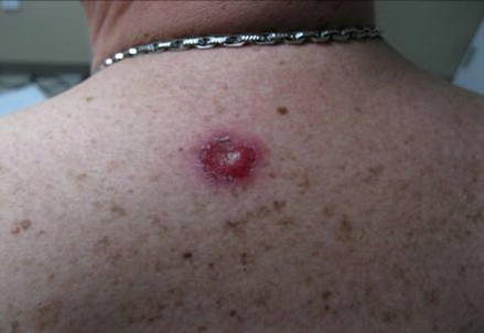
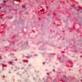
Fig. 14.2
Patient outcome on his mid upper back lesion – tenth day of treatment
Treatment Parameters for the Upper Scalp Lesion
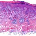
The upper scalp lesion was identified and circled. A 5–7 mm border was drawn around the lesion. The tumor depth was estimated to be <5 mm. A 0.762 mm thick led shield was utilized over a 2 cm field and placed over the lesion and extended field. Superficial radiotherapy was administered with a 3 cm cone for 15 treatments of 300 cGy at 70 kVp, 10 ma. Treatments were delivered Monday through Friday for three weeks, for a total dose of 4500 cGy (Figs. 14.3, 14.4, and 14.5).
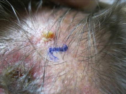
Fig. 14.3
 Topical Treatment of Skin Cancers and the Risks of ‘Fighting Fire with Fire’
 Amelanotic Malignant Melanoma of the Toe Presenting as an Ulcer: Management and Biopsy Guidelines
 Zosteriform Cutaneous Metastasis
 Metastatic Cutaneous Adenocarcinoma
 The Modified Rhomboid Flap: An Improvement on the Traditional Technique and Its Use in Defects of the Ala Nasi
 Balloon Cell Nevi and Balloon Cell Melanomas: What Are They?


Patient outcome on his upper scalp lesion – first day of treatment
Related posts:
Topical Treatment of Skin Cancers and the Risks of ‘Fighting Fire with Fire’
Amelanotic Malignant Melanoma of the Toe Presenting as an Ulcer: Management and Biopsy Guidelines
Zosteriform Cutaneous Metastasis
Metastatic Cutaneous Adenocarcinoma
The Modified Rhomboid Flap: An Improvement on the Traditional Technique and Its Use in Defects of the Ala Nasi
Balloon Cell Nevi and Balloon Cell Melanomas: What Are They?
Stay updated, free articles. Join our Telegram channel
Full access? Get Clinical Tree
