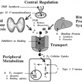
COMPUTED TOMOGRAPHY OF THE THYROID
The characterization of tissue and organs by CT is accomplished by computer-assisted analysis of the attenuation of x-rays that are transmitted through the patient. For some purposes, the images may be examined directly because the thyroid gland is somewhat more radiopaque than the rest of
the soft-tissue structures of the neck due to its high iodine content. More often, the contrast must be enhanced by the intravenous administration of iodinated material. Unfortunately, however, the iodine in the dye may be counterproductive to further diagnostic testing and to the treatment of thyroid disorders. Therefore, in most centers, CT has assumed a role in the diagnosis and management of thyroid problems that is complementary to that of MRI.14,17 CT should be used selectively and only in response to a specific clinical question that cannot be answered in a more cost-effective way. CT is more sensitive than MRI in detecting small metastases to cervical or mediastinal lymph nodes (stage I, 1.5 cm in diameter),25 is more reliable than MRI in the detection of small pulmonary nodules,26 is more appropriate for the unstable or claustrophobic patient, and is the only study possible in those patients with cardiac pacemakers or other biomechanical devices, who require sectional
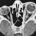
imaging.27 Pragmatically, access to CT is greater than access to MRI because of the larger number of CT scanners available and because the cost of a CT examination is lower than that of an MRI examination. Therefore, in some centers, CT is used in a manner similar to that described for MRI. An example of CT imaging for a patient with extensive thyroid cancer is shown in Figure 35-11.
the soft-tissue structures of the neck due to its high iodine content. More often, the contrast must be enhanced by the intravenous administration of iodinated material. Unfortunately, however, the iodine in the dye may be counterproductive to further diagnostic testing and to the treatment of thyroid disorders. Therefore, in most centers, CT has assumed a role in the diagnosis and management of thyroid problems that is complementary to that of MRI.14,17 CT should be used selectively and only in response to a specific clinical question that cannot be answered in a more cost-effective way. CT is more sensitive than MRI in detecting small metastases to cervical or mediastinal lymph nodes (stage I, 1.5 cm in diameter),25 is more reliable than MRI in the detection of small pulmonary nodules,26 is more appropriate for the unstable or claustrophobic patient, and is the only study possible in those patients with cardiac pacemakers or other biomechanical devices, who require sectional
imaging.27 Pragmatically, access to CT is greater than access to MRI because of the larger number of CT scanners available and because the cost of a CT examination is lower than that of an MRI examination. Therefore, in some centers, CT is used in a manner similar to that described for MRI. An example of CT imaging for a patient with extensive thyroid cancer is shown in Figure 35-11.
Related posts:

Stay updated, free articles. Join our Telegram channel

Full access? Get Clinical Tree


