MISCELLANEOUS ENTITIES
PITUITARY APOPLEXY
Pituitary apoplexy is the result of necrosis of the anterior lobe of the pituitary. When seen in a postpartum woman, it is referred to as Sheehan syndrome. It is usually the result of hemorrhage into the gland, but may also be nonhemorrhagic in
nature.30 Pituitary apoplexy is usually associated with hemorrhage into a preexisting adenoma, although it can occur in a normal gland (Fig. 20-20).30 Although CT in general is sensitive in making the diagnosis of acute hemorrhage, the changes in the pituitary gland may be difficult to appreciate. The imaging modality of choice for evaluation of hemorrhage in the pituitary is MRI. Also, this allows for evaluation of the optic chiasm, which may be compressed.
nature.30 Pituitary apoplexy is usually associated with hemorrhage into a preexisting adenoma, although it can occur in a normal gland (Fig. 20-20).30 Although CT in general is sensitive in making the diagnosis of acute hemorrhage, the changes in the pituitary gland may be difficult to appreciate. The imaging modality of choice for evaluation of hemorrhage in the pituitary is MRI. Also, this allows for evaluation of the optic chiasm, which may be compressed.
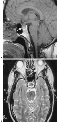 FIGURE 20-20. Hemorrhagic pituitary apoplexy. A, Sagittal T1-weighted spin-echo midline magnetic resonance imaging (MRI) demonstrates a high-signal-intensity abnormality of the sella (white arrow). It demonstrates bulging of the superior contour of the pituitary gland without major deformity of the optic chiasm. The high signal intensity without contrast suggests subacute hemorrhage. B, The pituitary apoplexy hemorrhage (white arrow) demonstrates low signal intensity on the T2-weighted axial image. This suggests that the hemorrhage is in the acute phase. Note the high-signal-intensity cerebro spinal fluid (CSF) spaces. |
SPHENOID SINUS DISEASE
The sphenoid sinus can be involved with expansile (mucocele), inflammatory (Wegener granulomatosis), infectious (pyocele), or neoplastic (squamous cell carcinoma or lymphoma) processes. The inflammatory disease may cause irritation or may directly extend to involve the adjacent sella turcica. This is most severe with necrotizing infections (such as mucormycosis) in immunocompromised and diabetic patients. In this setting, it may be difficult to differentiate inflammatory from neoplastic disease, such as squamous cell or adenocarcinoma of the sphenoid sinus and chordoma of the clivus. Endocrine symptoms are rare.
ANEURYSM
Aneurysms may involve any of the intracranial vessels and are usually found at branch points. Aneurysms of the cavernous supraclinoid carotid and anterior cerebral arteries may be located in the sella or suprasellar region (Fig. 20-21). Compressive symptoms of aneurysms depend on their location. Aneurysms that extend into the suprasellar region may compress the optic chiasm; thus, patients may present with bitemporal hemi-anopia. Aneurysms may also compress the pituitary or infundibulum and present with diabetes insipidus or other endocrine abnormalities. If there has been a subarachnoid hemorrhage, the patient may present with severe headache or with a neurologic deficit related to vasospasm.
Related posts:


Stay updated, free articles. Join our Telegram channel

Full access? Get Clinical Tree


