Vascular Lesions
Sean V. McGarry
C. Parker Gibbs
Hemangiomas are benign lesions originating from blood vessels. There is some controversy in the distinction between hemangiomas and vascular malformations. While histologically they appear identical under the microscope, vascular malformations are developmental irregularities that are usually latent and probably present at birth. Hemangiomas are true neoplasms that can be either active or latent. There are many histological subtypes of hemangiomas that have subtle microscopic differences, but these are beyond the scope of this text. Hemangiomas may involve one or more organs, including bone, the central nervous system, the liver, the skeleton, and skin and soft tissues. This chapter will discuss only lesions seen in bone. Bone hemangiomas usually occur as isolated lesions, although bone involvement may occur in conjunction with other organ systems when hemangiomas occur as part of a regional or systemic disease process, such as hemangiomatosis or skeletal/extraskeletal hemangiomatosis. Maffucci’s syndrome is a combination of hemangiomas and multiple enchondromas.
Pathogenesis
Epidemiology
Age: most common in fifth decade but may occur at any age
Gender: slight female predominance
Location: most frequently in the spine (vertebral body) and cranium
Frequency: Asymptomatic hemangiomas of the spine are thought to occur in up to 10% of the population.
Pathophysiology (Also see Fig. 11-32)
Most simply stated, hemangiomas are capillary-sized proliferations of blood vessels occurring in bone. Hemangiomas of bone are generally asymptomatic. They are often found incidentally on radiographs taken in the work-up of other problems. When they occur in the spine they can rarely expand the vertebral body and cause neurological compromise. Pathologic fracture in the spine can also cause neurological symptoms by impinging on the cord. Hemangiomas have no potential for malignant degeneration.
Classification
Solitary osseous hemangiomas follow Enneking’s classification of benign tumors (see Chapter 4.2, Treatment Principles).
There are numerous histologic subtypes of solitary hemangiomas, and the World Health Organization recognizes various forms of more disseminated hemangioma-related lesions as well as osseous glomus tumors and lymphangiomas (Box 5.7-1).
Box 5.7-1 Classification of Hemangiomas and Hemangioma-Related Lesions
|
Diagnosis
Physical Examination and History
Clinical Features
Asymptomatic or an incidental finding
In a symptomatic patient, vague, achy pain
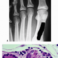
In a subcutaneous bone, expansion of the bone may produce a mass or localized pain (Fig. 5.7-1)
Stay updated, free articles. Join our Telegram channel

Full access? Get Clinical Tree


