Tendon and Ligament Anatomy, Biology, and Biomechanics
Brian J. Harley
Joseph W. Bergman
Tendons and ligaments act as the bonds that tie the body together. Ligaments connect one bone to another at a joint, and tendons connect bone to muscle. While the specific natures of their tasks differ, tendons and ligaments share a great many features in their construction and function.
Tendon
Tendon Anatomy, Structure, and Composition
Gross Anatomy
Macrostructure
Variable sizes and shapes: wide and flat to round and narrow
The larger the muscle unit, and therefore the potential force, the larger diameter the corresponding tendon
Unique features in regions of compression
Sheaths and bursa
Shield the tendon from abrasion and friction
Tendon assumes more of a cartilage-like appearance in these areas.
Synovial sheath
Encloses the path of the tendon
Provides a reservoir of fluid to hydrate and lubricate the tendon
Ultrastructure
While variety is seen in tendon macrostructure, all tendons are organized with a similar ultrastructure (Fig. 24-1).
Musculotendinous junction
Collagenous structure of the tendon blends with the muscle.
As the tendon fans out into the muscle, the collagen fibrils connect to the myocytes, allowing for the transmission of force from muscle to tendon.
Epitenon
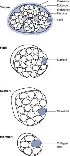
Figure 24-1 Tendon ultrastructure.
Layer of organized tissue that tightly encircles the entire surface of the tendon
Endotenon
Layer of loose connective tissue between fascicles within a tendon
Paratenon
Layer of loose areolar tissue that substitutes for a tendon sheath; serves a nutritional role
Present in tendons not enclosed within a fibrous sheath (e.g., Achilles tendon)
Fibrils
Collagen molecules organized into microfibrils
Microfibrils assembled into subfibrils, fibrils, and fascicles
One or more fascicles compose a tendon.
Biochemistry
Collagen
Primary component of tendon
Type I collagen predominates, typically representing up to 85% of the dry weight.
Other collagens in tendon: III, IV, V, VI
Collagen provides a strong molecule capable of transmitting tensile loads.
Constructed from tropocollagen, a triple-helical polypeptide molecule
Ends of tropocollagen overlap at regular intervals, giving the collagen fiber a banded appearance.
Primarily oriented in the direction of expected tensile force
Variety of fibers that travel in an oblique or perpendicular direction and tie the longitudinally oriented fascicles together
Noncollagenous substances
Proteoglycans, proteins, and various supporting molecules, including elastin
Small variations in the concentrations of all these components are seen in tendons in differing locations and functions.
Matrix
Proteoglycans and glycosaminoglycans
Ionic charge on the side chains of these molecules causes them to spread out and occupy as large a volume as possible.
Water molecules are strongly attracted by this structure and are strongly restrained within the tendon.
Presence of water bound to the proteoglycan molecules allows the tendon to resist compressive loads.
Compressive load passes through the water in much the way that the fluid inside a can of soda supports the thin walls surrounding it.
Resistance to fluid flow through the tendon also contributes greatly to its viscoelastic biomechanics.
Cellular Population
As a consequence of the mechanical demands placed on the tendon, much of the structure of the tendon is composed of mechanical elements.
As a result, the tendon has a high structure to cellularity ratio.
Tenocytes and tenoblasts
Spindle-shaped cells
Subjected to significant mechanical loads
Poorly supplied with blood and nutrients
Stimuli triggering cellular responses
Mechanical load
Electric potential
Various chemical and cytokine messengers
Transforming growth factor (TGF) and interleukin (IL) groups, as well as a variety of extracellular molecular fragments
Function: responsible for maintaining the collagen and proteoglycan matrix
Normal tendon
Constant turnover of extracellular matrix in response to chemical and mechanical damage that occurs from normal day-to-day function
Acute tendon injury
Inflammatory response (including lysis and removal of damaged molecules), followed by a regenerative, depositional phase (by tenocytes and tenoblasts)
Due to the paucicellular nature of tendon combined with areas of poor blood supply, it is possible for the cellular repair mechanism (tenocytes and tenoblasts) to become overwhelmed.
Chronic tendon conditions
Inflammatory and lysis responses predominate.
Reparative response by tenocytes muted
Chronic tendinitis, tendinosis, and even tendon failure are manifestations of this state.
Blood Supply
Vascular anatomy of tendons
Blood vessels exist within the epitenon and endotenon.
Fine areolar structures that surround tendon bundles and supply individual fibrils and fibers
Variable blood supply depending upon location within tendon
Variable amount of blood supply at origin and insertion
Myotendinous junction allows an increased microperforating blood supply to nourish a portion of the tendon extending away from the junction.
Factors decreasing vascular incursion
Intense mechanical environment
Confined geometry of the tendon
Increased tensile and compressive forces within the extracellular matrix
Secondary path for nutrition in poorly vascularized regions
Diffusion of nutrients and oxygen from the adjacent synovial layers
Innervation
Neural elements within tendons
Two predominant mechanoreceptors, both of which sense pressure and tension within tendon
Rapidly adapting receptors: Pacinian corpuscles
Slow-adapting receptors: Ruffini endings
Free nerve endings less common
Sympathetic and parasympathetic innervation
Importance of tendon innervation
Important for normal function
Proprioceptive receptors communicate with gamma-muscle-spindle system to modulate joint position.
Recovery of mechanical function after an injury is not always accompanied by return of nervous function.
Unknown what effect this defect has on rehabilitation and musculotendinous action
Tendon Biomechanics
Tendon acts as a relatively rigid connector between the motor unit of the muscle and the bone. Its first role is to transmit tensile forces created within the muscle to initiate and modulate motion. To accomplish this, tendon has one of the highest tensile strengths of any material in the body. Collagen has a high tensile strength (and high ultimate stress) along its longitudinal axis, and the parallel orientation of the collagen fibrils within tendon takes advantage of this strength. The clinical importance of these properties is evident when the biomechanical properties of the healing tissue in a tendon or ligament are compared to the intact state. The scar tissue is weaker, therefore reducing the material properties of a tendon. Increasing the total cross-sectional area of the tendon with this scar tissue may allow for the same structural properties, however.
Structural Properties of Tendons
Describes the properties of the whole tissue complex, as in the entire tendon–bone insertion unit, in terms of force and displacement (Fig. 24-2)
Strength: overall force transmitted
Stiffness: ability of the structure to resist deformation when force (or load) is passed through it
Material Properties of Tendons
Describes properties according to cross-sectional area, in terms of stress and strain (Fig. 24-3)
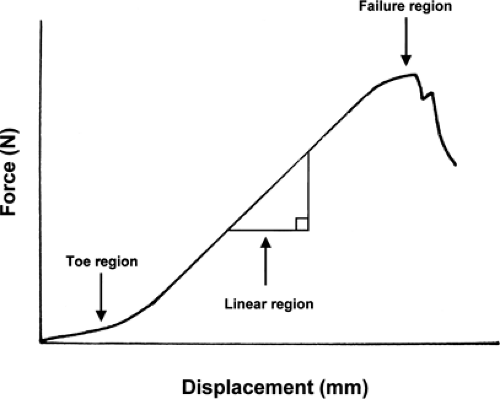
Figure 24-2 Typical force–displacement curve during low-load tensile testing of a tendon. A toe region exists representing the changes from crimp. The linear region represents the high stiffness obtained from full recruitment of collagen fibers. The failure region occurs with sequential loss of continuity of fibers.
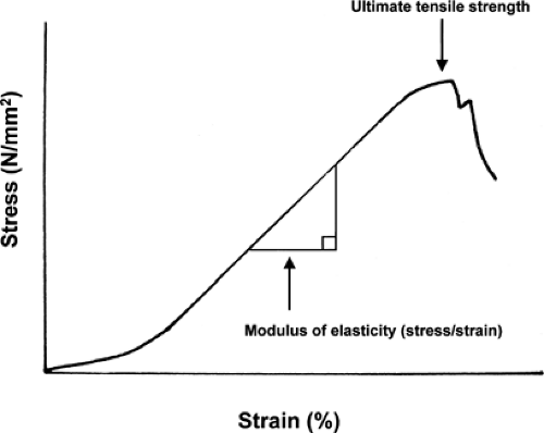
Figure 24-3 Typical stress–strain curve during low-load tensile testing of a tendon. The slope of the linear region represents the modulus of elasticity of the tendon. The ultimate tensile strength represents the stress at failure of continuity of the tendon.
Stress: force divided by cross-sectional area
Strain: amount of deformation of the structure divided by length over which the deformation takes place
Useful for comparing tendons at various anatomic locations or comparing a healing tendon to an intact tendon, or when relating tendons to ligaments
Low-Load Tendon Mechanics
The behavior of tendon at low loads reveals a degree of laxity that allows some movement through the tendon before load is passed to muscle.
Crimp in tendons
At low loads, this “crimp” present in the collagen fibers straightens out before the collagen begins to conduct load (see Fig. 24-2).
Steepening of force–deformation curve
Not all of the collagen is crimped equally, so there is a steepening of the force-deformation curve as more and more fibrils are recruited.
Effect of loading rate
Most of the work performed on the mechanics of tendon has loaded the tissue at a very slow rate of loading, and in this quasi-static state the curve slowly steepens until the elastic modulus of the tendon is reached (see Fig. 24-3).
At higher loading rates, which more accurately represent the condition in vivo, the structure displays a viscoelastic behavior and acts as a stiffer structure.
Elastic Biomechanics
Structural and Material Properties
An elastic substance is one in which a force and displacement and stress and strain are linearly related.
Doubling a certain force will double the deformation.
Doesn’t matter if the force (load) is arrived at during loading or unloading
Elastic Properties
Tendon demonstrates very important elastic behavior.
Any force generated by a muscle is transferred to the intended bone with virtually no energy wasted.
Cycling between loaded and unloaded states does not waste energy.
Whatever energy is stored during loading of the material is released during its unloading.
Viscous Properties
A purely viscous substance is one that will deform infinitely given a particular force.
This is seen with fluids where rate of motion through the fluid is dependent on the force.
Viscoelastic Behavior
A time-dependent property of material behavior whereby the tendon behaves partly as a viscous substance and partly as an elastic substance
Interaction of the viscous and elastic properties during loading
The collagen fibers take up most of the force and the loaded fibers tend to squeeze together.
The water in the extracellular matrix resists this inward movement of the collagen fibers.
Typically the water would quickly be squeezed out of the tissue by the tension on the tendon.
The proteoglycans and glycosaminoglycans within the matrix act as a colloid attraction force to keep water within the tendon.
The rate at which a tendon is loaded is very important.
The more quickly a tensile load is placed on the tendon, the more effective is the resistance to outward flow of the water.
Advantages of viscoelastic behavior in tendons
Load “sharing” between the elastic and viscous portions of the matrix
During very large loads, the viscous nature of the tendon absorbs and dissipates energy. This helps to protect the collagen from irreparable damage.
At lower, cyclic loads, the tendon behaves more as an energy-conserving, elastic material.
Energy conservation
One of the important functions of tendon is efficient transmission of energy. The most efficient way to transmit force is with a very stiff structure.
Within short time frames, the viscoelastic behavior of tendon causes it to act like a much stiffer and energy-conserving structure.
Loss of energy
Due to the properties of creep, force-relaxation, and mechanical hysteresis, upwards of 10% of total work is lost during normal cycles of loading and unloading.
Tendon Injury, Healing, and Repair
Tendon dysfunction adversely affects overall orthopaedic health. Degenerative and/or traumatic injuries to tendons throughout the musculoskeletal system are a common cause of presentation to an orthopaedic surgeon.
Injury Mechanisms
Direct Trauma or Laceration
Common example: laceration of the finger flexor tendon
Acute Application of Tensile Loads Exceeding the Strength of the Tendon
Results in partial or complete ruptures of the tendon
Distinct locations common
Avulsion of the tendon from its bone insertion
Healthy tendons can easily withstand tensile forces larger than the maximum force generated by the muscles or tolerated by the bones, so failures tend to occur at bony insertions.
Common example: distal biceps tendon avulsion
Midsubstance rupture
Generally pre-existing pathology at the site of a midsubstance rupture
Common example: Achilles tendon rupture
Disruption at the musculotendinous junction
Typically caused by a very forceful eccentric muscle contraction
Partial loss of continuity of the muscle fibers close to the junction (complete loss of continuity of tendon is uncommon)Related posts:

Stay updated, free articles. Join our Telegram channel

Full access? Get Clinical Tree


