Fig. 17.1
Different macroscopic appearance of intestinal hemangiomas
Although intussusception caused by a hemangioma of the small bowel is a rare condition (commonly founded in blue rubber bleb nevus syndrome), it should be taken into consideration in the differential diagnosis of abdominal pain of doubtful origin.
Only 1 % of patients with minor rectal bleeding examined by sigmoidoscopy have hemangiomas.
Hemangiomas of the greater or lesser omentum are not exceptional and very often asymptomatic in the context of diffuse neonatal hemangiomatosis.
Unfortunately many patients still undergo emergency laparotomies and intestinal resections in the context of gastrointestinal bleeding without having the option of systemic propranolol therapy.
3.
Liver: Hemangiomas are the most common benign liver tumors in children. They can be difficult to diagnose and complex to treat. Differential diagnosis must be accurate in any patient with atypical presentation of liver hemangioma [2]. An important differentiating factor in the evaluation of pediatric hepatic masses is the age of the patient. Hepatoblastomas, mesenchymal hamartomas, and metastatic disease from Wilms tumor or neuroblastoma are usually seen in the first 3 years of life, whereas hepatocellular carcinoma, focal nodular hyperplasia, hepatic adenoma, and metastases from lymphoma are more common in older children.
Several current studies attempt to determine how common liver hemangiomas are in children with infantile hemangiomas by comparing liver ultrasound results in patients with one to four cutaneous hemangiomas, five or more cutaneous hemangiomas, or at least one large hemangioma versus ultrasound results in children without hemangiomas identifying at the same time specific risk factors in patients who have liver hemangiomas.
Vascular anomaly specialists must be able to distinguish hemangiomas from various vascular malformations as well as appreciate their dynamic course with time. Several attempts made to classify pediatric liver vascular tumors led to an unhelpful plethora of confusing terminology. Immunohistochemistry has been used in the study of hemangiomas and supports a new classification of these tumors based on the expression of GLUT-1 as a substitution of the old and confusing term of hepatic infantile hemangioendothelioma:
1.
GLUT-1-positive expression is usually demonstrated in multifocal and diffuse hepatic infantile hemangioma, which shares clinical and morphological features with cutaneous infantile hemangioma (Fig. 17.2). Diffuse neonatal hemangiomatosis is a frequently fatal disorder characterized by multiple cutaneous and visceral hemangiomas. Complications include high-output cardiac failure, hemorrhage, hepatic failure, and consumption coagulopathy. The diagnosis of consumptive hypothyroidism as a result of increased type 3 iodothyronine deiodinase activity in the hemangiomas has to be done in every patient with multifocal or diffuse lesions. Myocardial depression secondary to hypothyroidism in children with hepatic hemangioma has been reported. Coincident with the involution of the hemangiomas, the child’s hypothyroidism improves.
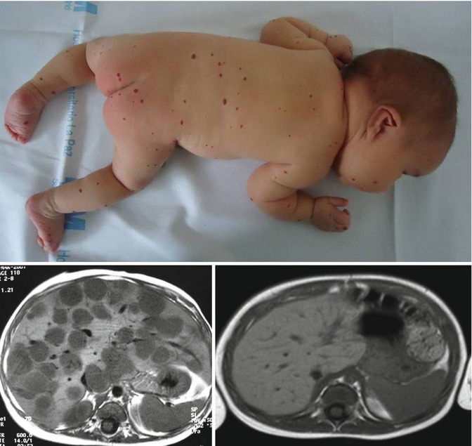
Fig. 17.2
Neonatal hemangiomatosis with multifocal liver hemangioma. Response to a 6-month course of propranolol
2.
GLUT-1-negative vascular liver tumors occur in neonates with unique clinical, imaging, and pathological features. They differ from diffuse hemangioma in terms of earlier presentation as solitary masses with central necrosis, rapid involution, and pathologic features showing a notable, often prolific, lymphatic compliment immunoreacting positively with the monoclonal antibody D2-40.
Following the parallelism with cutaneous anomalies, the term hepatic congenital hemangioma has been suggested comparing their behavior with rapid involuting congenital hemangiomas (RICH) described in skin and subcutaneous locations (Fig. 17.3).
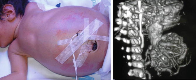
Fig. 17.3
Hepatic congenital hemangioma (RICH). Angioarchitecture on CT
To sum up, hepatic hemangiomas represent three different categories of lesions: focal, multifocal, and diffuse [3].
The natural history for focal hemangiomas is spontaneous regression in the first year of life; however, shunt embolization or complete surgical excision is required in case of cardiac failure.
Related posts:

Stay updated, free articles. Join our Telegram channel

Full access? Get Clinical Tree


