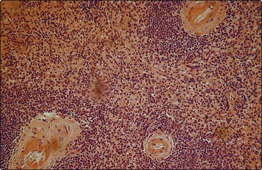
5 The splenic artery penetrates the thick capsule which invests the organ (Fig 5.1). Branches of the splenic artery are surrounded by a highly organised aggregate of lymphoid tissue which is termed the ‘white pulp’ (Fig 5.2). Intimate to the central arteriole is the ‘periarteriolar lymphatic sheath’ – an area mainly populated by T-lymphocytes. Among these T-lymphocytes are non-phagocytic, antigen-presenting cells known as ‘interdigitating cells’. Spaced at intervals in the periarteriolar lymphatic sheath are lymphoid follicles (‘Malpighian bodies’). In an inactive state these follicles are composed of recirculating B-lymphocytes intertwined with cytoplasmic processes of follicular dendritic cells. The latter cells may play a role in long-term antibody production. When contact with antigen stimulates B-cell activation, a germinal centre of rapidly dividing cells forms in the follicle. This is a key area in the normal B-lymphocyte proliferative response and development of B-cell memory (see p. 8 for discussion of lymphocytes). Fig 5.1 Structure of the spleen.
The spleen
Structure
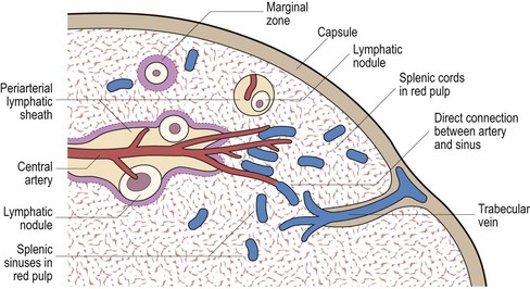
The white pulp is composed of the periarteriolar lymphatic sheath and lymphatic nodules. The red pulp contains the splenic cords and sinuses and is separated from the white pulp by the marginal zone. See text for full discussion.
Oncohema Key
Fastest Oncology & Hematology Insight Engine