(1)
Department of Surgery, Dar A lAlafia Medical Company, Qatif, Saudi Arabia
5.1 Introduction
The word spleen comes from the Greek σπλήν (splḗn) and is the idiomatic equivalent of the heart in English, i.e., to be good-spleened (εὔσπλαγχνος, eúsplankhnos) means to be good-hearted or compassionate.
The spleen is considered part of the lymphatic system and resembles a large lymph node.
It is the largest lymphoid tissue of the human body, accounting for 25 % of total body lymphocytes.
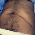
The spleen is a valuable organ and should not be removed unless it becomes pathological (Figs. 5.1 and 5.2).
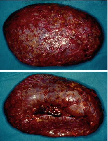
Figs. 5.1 and 5.2
Clinical photographs showing a pathological spleen in a patient with sickle cell anemia that was removed
The spleen, in healthy adults, is approximately 11 cm (4.3 in) in length.
The spleen weight normally ranges from 150 g (5.3 oz.) to 200 g (7.1 oz.).
The role of 1 × 3 × 5 × 7 × 9 × 11. This is an easy way to remember the anatomy of the spleen.
1, 3, 5: the spleen is 1″ by 3″ by 5″ in size.
7: the spleen weighs approximately 7 oz.
9, 11: the spleen lies between the 9th and 11th ribs on the left side.
The spleen is one of the commonly organs to be affected in patients with SCA.
It enlarges early and then undergoes progressive atrophy as a result of repeated attacks of vaso-occlusive crisis leading to autosplenectomy. The spleen becomes shrunken and nonfunctional. The spleen may disappear completely or form a small fibrotic nodule. Sometimes the remnant of the spleen becomes calcified and takes different shapes (Figs. 5.3, 5.4, and 5.5).
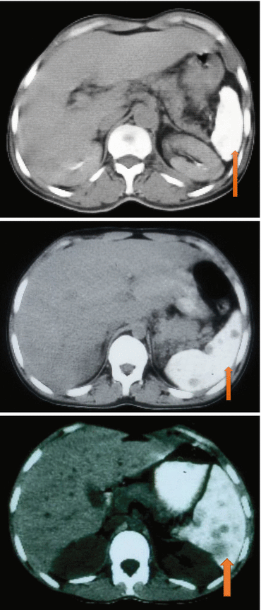
Figs. 5.3, 5.4, and 5.5
CT scan showing a small calcified spleen in patients with SCA
Splenomegaly however persists in some of these patients to an older age group and sometimes to adults.
The reason for this is not known, but an elevated level of HbF is known to contribute to the persistence of splenomegaly in these patients (Figs. 5.6 and 5.7).
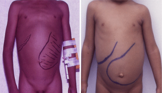
Figs. 5.6 and 5.7
Clinical photographs showing persistent splenomegaly in two patients with SCA
Persistence of splenomegaly in these patients is known to be associated with complications necessitating splenectomy.
These complications include:
Acute splenic sequestration crisis
Splenic abscess
Massive splenic infarction
Hypersplenism
5.2 Embryology
Normally, the gastrointestinal tract is derived from endoderm. The spleen is unique as it is derived from mesenchymal tissue.
The spleen however shares the same blood supply where it receives its blood supply from the celiac trunk via the splenic artery and also the short gastric blood vessels.
Embryologically, the spleen develops within and from the dorsal mesentery.
The spleen appears about the 5th week of intrauterine life as a localized thickening of the mesoderm in the dorsal mesogastrium above the tail of the pancreas.
As the stomach changes its position, the spleen moves to the left and comes to lie behind the stomach and in contact with the left kidney.
The part of the dorsal mesogastrium which intervened between the spleen and the greater curvature of the stomach forms the gastrosplenic ligament.
Histologically, the spleen is made of two parts:
The red pulp
The white pulp separated by the marginal zone
Each part of the spleen has separate and important functions.
Splenectomy should always be avoided, and every attempt should be made to preserve the spleen even traumatic rupture of the spleen.
5.3 Functions of the Spleen
The red pulp:
This is made up of thin-walled venous sinusoids which are lined by special endothelial cells with a discontinuous wall allowing the passage of red blood cells between the sinuses and splenic cords.
The sinuses are separated by splenic cords (cords of Billroth) which contain macrophages.
The red pulp has the following important functions:
It filters red blood cells and ingests old RBC, damaged RBCs such as sickle cells or spherocytes, and antibody-coated RBCs.
It removes Heinz bodies and other RBC inclusions such as Howell-Jolly bodies.
The white pulp:
This is made up of sheaths of lymphoid cells located around arteries (periarteriolar lymphatic sheath).
These sheaths are composed of T cells and lymphoid follicles (B cells) which traps antigens for processing.
The white pulp has the following functions:
It is responsible for the active immune response through both humoral- and cell-mediated pathways.
Other functions of the spleen:
Production of opsonins, properdin, and tuftsin.
Natural antibodies are required for phagocytosis. These are immunoglobulins that facilitate phagocytosis either directly or by complement deposition on the capsule. They are produced by IgM memory B cells in the marginal zone of the spleen.
Production of red blood cells:
Normally, the bone marrow is the primary site of hematopoiesis, but the spleen has important hematopoietic functions up until the 5th month of gestation.
After birth, erythropoietic functions cease, except in some hematologic disorders.
As a major lymphoid organ and a central player in the reticuloendothelial system, the spleen retains the ability to produce lymphocytes and, as such, remains a hematopoietic organ.
Storage of red blood cells, lymphocytes, and other formed elements.
The red blood cells can be released when needed.
It has been estimated that in humans, up to a cup (236.5 ml) of red blood cells can be stored in the spleen and released in cases of hypovolemia.
It can store platelets in case of an emergency.
Up to a quarter of lymphocytes can be stored in the spleen at any one time.
The presence of Howell-Jolly bodies and less commonly Heinz bodies in the blood smears is an indication of decreased or absence of splenic function, but an isotope scan (technetium sulfur colloid scan) is more important to evaluate the function of the liver and spleen (Fig. 5.8).
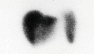
Fig. 5.8
A technetium sulfur colloid scan showing a normally functioning spleen in a patient with sickle cell anemia
5.4 Post-splenectomy Complications
An increase in white blood cells.
An increase in platelets. The post-splenectomy platelet count may rise to abnormally high levels (thrombocytosis), leading to an increased risk of potentially fatal clot formation or portal vein thrombosis.
An increase in the number of Howell-Jolly bodies and less commonly Heinz bodies in the blood smears. Heinz bodies are usually found in cases of G6PD (glucose-6-phosphate dehydrogenase) deficiency and chronic liver disease.
A diminished responsiveness to some vaccines.
An increased susceptibility to infections by bacteria and protozoa; in particular, there is an increased risk of post-splenectomy sepsis from polysaccharide encapsulated bacteria such as pneumococci.
5.5 Immunizations and Splenectomy
Splenectomy is known to be associated with an increased risk of sepsis due to encapsulated organisms (such as S. pneumoniae and Haemophilus influenzae).
These patients especially children should receive immunization prior to splenectomy.
These vaccines include:
Pneumococcal vaccine
Meningococcal vaccines
Haemophilus influenza type b vaccine
These vaccines should be given at least 2 weeks prior to splenectomy and followed subsequently by poster doses.
If splenectomy is done for an emergency reason, these vaccines can be given postoperatively.
Children who undergo splenectomy should also receive prophylactic antibiotics for a minimum of 2 years depending on their age at the time of splenectomy.
This is in the form of penicillin prophylaxis either orally or intramuscularly. Oral penicillin is preferable.
This is because overwhelming post-splenectomy sepsis is commonly seen within the first two years post-splenectomy.
5.6 Splenic Complications of Sickle Cell Anemia
SCA is known to affect any part of the body and poses diagnostic and therapeutic dilemmas to the treating physicians.
The spleen is one of the most common and early organs to be affected in SCA.
It is commonly enlarged during the first decade of life but then undergoes progressive atrophy as a result of repeated attacks of vaso-occlusion and infarction leading to autosplenectomy.
This however is not the case always, and sometimes splenomegaly persists into an older age group or even into adulthood necessitating splenectomy for a variety of reasons including (Figs. 5.9 and 5.10):
Acute splenic sequestration crisis
Hypersplenism
Massive splenic infarction
Splenic abscess
Splenomegaly with a nonfunctional spleen
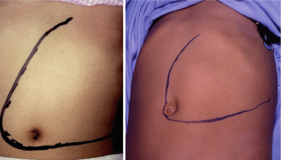
Figs. 5.9 and 5.10
Clinical photographs showing persistent splenomegaly in two patients with sickle cell anemia
Splenic Complications of Sickle Cell Anemia
Acute splenic sequestration crisis (major and minor)
Hypersplenism
Massive splenic infarction
Splenic abscess
Persistent splenomegaly with a nonfunctional spleen
Splenic complications of SCA are associated with an increased morbidity, and in some it may lead to mortality.
To obviate this, splenectomy is an essential part of their management (Figs. 5.11 and 5.12).
Obviously, splenectomy does not cure SCA, but it is valuable and in certain patients may be an essential part of their management.
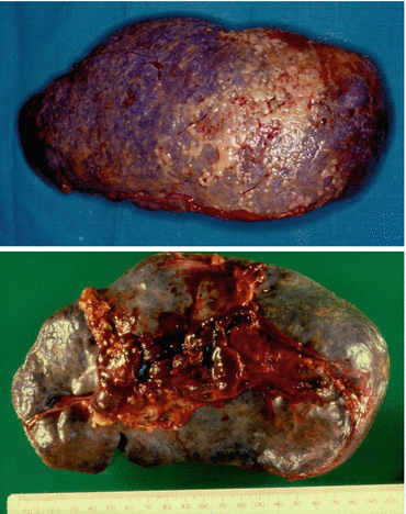
Figs. 5.11 and 5.12
A clinical photograph showing a removed spleen in a patient with sickle cell anemia. Note also the multiple infarcts
5.7 Acute Splenic Sequestration Crisis
Acute splenic sequestration crisis (ASSC) results from the rapid sequestration of red blood cells in the spleen (Fig. 5.13, 5.14 and 5.15).
In ASSC, there is good inflow of blood into the spleen, but the blood outflow from the spleen is obstructed as result of occlusion of the splenic vein by sickled RBCs. This will lead to pooling of blood into the spleen sinusoids leading to anemia and sometimes hypovolemia.
It is a serious complication of SCA and considered the second leading cause of death after infection in the first decade of life.
ASSCs are usually seen in infants and young children commonly between 5 months and 2 years of age.
This however is not the case always, and ASSC can be seen in older patients with persistent splenomegaly. The exact reason for this is not known. One contributing factor is persistence of splenomegaly in these patients into an older age group. This is partly attributed to persistently high hemoglobin F level in these patients.
Acute splenic sequestration crisis is characterized by:
Sudden onset of anemia.
Sudden splenomegaly or an enlargement of an already enlarged spleen.
Evidence of active bone marrow.
The spleen size regresses after blood transfusion (Fig. 5.16).
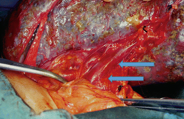
Fig. 5.13
A clinical intraoperative photograph showing enlarged spleen with multiple small infarcts in a patient with SCA and acute splenic sequestration crisis. Note the enlarged congested splenic vein
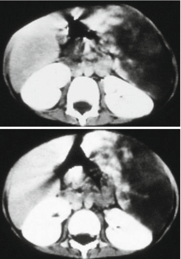
Figs. 5.14 and 5.15
CT scan showing acute splenic sequestration crisis. Note the spleen enlarged and filled with sequestered blood
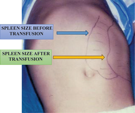
Fig. 5.16
A clinical photograph of a child with sickle cell anemia and acute splenic sequestration crisis. Note the regression of the size of the spleen following blood transfusion
Characteristics of Acute Splenic Sequestration Crisis
Sudden onset of life-threatening anemia
Sudden enlargement of the spleen
High reticulocyte count
Regression of the spleen size after blood transfusion
This differentiates ASSC from hypersplenism where the spleen size is chronically enlarged and does not regress after blood transfusion.
Acute splenic sequestration crisis is divided depending on the severity of the attack into:
Minor attack
Major attack
Minor attacks are characterized by:
A moderate increase in the size of the spleen
A sudden drop of hemoglobin of 2–3 g/dl
Major attacks are characterized by:
A significant sudden increase in spleen size
A greater drop of hemoglobin sometimes decreasing to as low as 2–3 g/dl
Hypovolemia
The cause of ASSC is not known, but an association with upper respiratory tract infection suggesting a precipitating viral cause was proposed. This includes infection with human Parvovirus B19.
The diagnosis of ASSC is clinical and based on sudden onset of anemia, sudden enlargement of the spleen, and regression of spleen size after blood transfusion. In these patients, it is important to keep a record of the spleen size in each visit to the clinic or hospital. This will help to recognize a sudden enlargement of the spleen.
CBC will show anemia. The severity of anemia depends on the type of ASSC. CBC will also show reticulocytosis. The WBC and platelets may also decrease.
The treatment of acute splenic sequestration crisis is initially supportive and includes:
Clinical evaluation and close monitoring.
Patients with major attacks should be admitted to ICU.
Hydration with intravenous fluids.
Blood transfusion with packed red blood cells. It is important to avoid over transfusing these patients as this with lead to increase in blood viscosity with its associated risks. It is also important to remember that blood is stored in the spleen and once blood transfusion is given, some of this blood will be released into the circulation which can also lead to an increase in blood viscosity. The aim should be to increase the hemoglobin level to around 10–11 g/dl.
It is important to teach the parents how to palpate the spleen in their affected children and recognize sudden enlargement. They should report to the hospital early if they recognize sudden enlargement.
These patients should be immunized with pneumococcal, meningococcal, and H. influenzae vaccines.
Types of Acute Splenic Sequestration Crisis
ASSC is divided depending on the severity of the attack into:
Minor attack
Major attack
Minor attacks are characterized by:
A moderate increase in the size of the spleen
A sudden drop of hemoglobin of 2–3 g/dl
Major attacks are characterized by:
A significant sudden increase in spleen size
A greater drop of hemoglobin sometimes decreasing to as low as 2–3 g/dl
Hypovolemia
The indication for splenectomy in major attacks is well established. Splenectomy is indicated if the child develops one major attack.
Major ASSC is a serious complication that is associated with a high mortality. To obviate this, the parents should be taught how to palpate the spleen and report quickly to the hospital if they recognize sudden enlargement.
The role of splenectomy in minor attacks is still controversial.
Some advocate chronic blood transfusion as a therapy to prevent recurrent attacks. This is effective on the short term, but it is not without risks including allosensitization, hepatitis, iron overload, and other blood-borne infections. The duration of chronic blood transfusion is not known. Add to this the fact that a significant number of patients on chronic blood transfusion therapy developed ASSC when attempts were made to stop blood transfusion or shortly after completing the program. The fact that blood is not readily available, it is not without complications, and poor compliance of parents makes chronic blood transfusion unsuitable form of therapy in these patients.
Others advocate splenectomy if the child develops two minor attacks. This is to obviate a recurrence risk of 40–50 % and a mortality rate as high as 20 %.
5.8 Hypersplenism
Hypersplenism is arbitrarily defined as:
Splenomegaly with:
Anemia (when the transfusion requirements exceed 250 ml/kg of packed red blood cells per year)
Thrombocytopenia (when the platelet level is less than 100,000/mm3)
Neutropenia (when the white blood cell count is less than 4000/mm3)
Either singly or in combination
Splenomegaly in hypersplenism is persistent to differentiate it from acute splenic sequestration crisis.
These hematological changes normalize following splenectomy.
The incidence of hypersplenism is relatively low in patients with sickle cell anemia. The reason for this low incidence of hypersplenism in is not known, but early autosplenectomy in these patients is a contributing factor.
In the past minor ASSCs were confused with hypersplenism, but when strict definition criteria are applied, hypersplenism is relatively rare in patients with sickle cell anemia.
Hypersplenism is more commonly seen in sickle B+ thalassemia patients. This confirms the observation that patients with sickle B+ thalassemia tend to behave more like thalassemia.
To obviate the risk of overwhelming post-splenectomy infection (OPSI), a variety of treatment modalities other than total splenectomy have been proposed to treat patients with hypersplenism. These include:
Chronic blood transfusions
Partial splenectomy
Percutaneous intraluminal occlusion of the splenic artery
Embolic therapy

