Figure 7.1
Steps of the Bezier island flap
The Keystone Design Perforator Island Flap
This flap was first described by Felix Behan [10]. In Roman architecture, it was necessary to design a stone called a keystone – a way of locking arches in a building using gravity. Behan felt that the shape of this flap seemed to lock into the defect.
The keystone flap is a curvilinear shaped trapezoidal design flap and the curvilinear shape of the flap fits well into body contours especially in the lower limb. The flap has a ratio of 1:1 for the width of the defect to the width of the flap. The length of the flap is determined by the size of defect that is excised and a 90° angle is created at the limits of the excision as shown in Fig. 7.2. Blunt dissection allows mobilization of the surrounding tissue while the flap advances to close the defect. We can see this is an island flap –being designed within dermatomal segments, and the longitudinal design allows nerves and veins to be incorporated in the flap design – therefore it is limited to certain bodily sites as illustrated in Fig. 7.3.
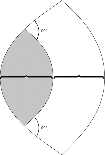
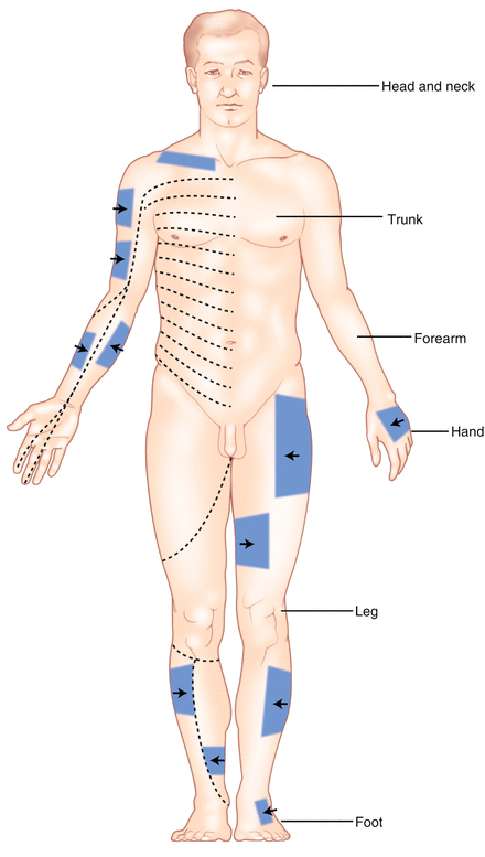

Figure 7.2
Keystone flap design

Figure 7.3
Sites for the Keystone flap
Additionally, Behan et al. [8] classified the keystone flap into several subtypes:
Type I: The deep fascia is left intact for smaller lesions up to 2 cm (Type I keystone) The trapezoidal shaped flap is contoured along the side of the defect with 90° angle at the limits of the island flap (Fig. 7.4).
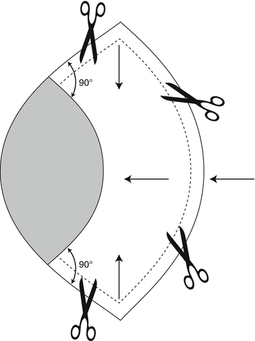
Figure 7.4
Type I Keystone island flap
Type II: For larger areas >2 cm, located over muscular compartments, the deep fascia is divided along the outer curvature of flap to permit further mobilization of the flap
Type IIA: Division of the deep fascia along the outer curvilinear line (in one case, our patient ended up with a troublesome seroma after division of fascia was performed as part of a keystone flap. However, this resolved after 4–6 weeks with compression stockings)
Type IIB: Skin graft to the secondary defect when undue tension exists (In type IIB a graft is often needed where tissue has limited elastic stretch on the lower one-third of the lower limb – which is why I personally avoid this type as if a skin graft is needed, it is simpler to just do a skin graft)
Type III: Double keystone flap (Fig. 7.5) – this is reserved for considerably larger defects (5–10 cm) where a double keystone design can exploit maximum laxity of the surrounding tissues. (I’ve found this useful in sacral region for closure of pilonidal sinuses as well after wide excision of deep melanomas)
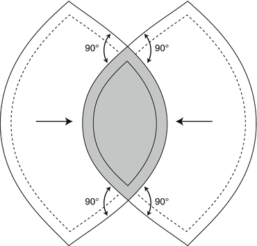
Figure 7.5
Double Keystone flap
Type IV: Rotational keystone flap – this is more useful in joint contractures and I shall not detail this here.
For closure after wide excision of melanoma, the Type I and Type II (with division of fascia) keystone flaps are especially useful.
Case Study
A 60-year-old woman presented with a 3 cm basal cell cancer on the lower limb. This was over the pretibial region and he had notable varicose veins. I elected to perform a keystone flap. In the image (Fig. 7.6) I have marked the direction of the RSTL to show why an elliptical closure would not work. A skin graft was an option but less ideal as the lesion was right on the pre-tibial region overlying bone. The keystone flap was raised and in this case a Type II flap was used with fascial division needed to achieve closure. The end result at 3 weeks post-operatively shows a well-healed flap with no contour defect (Figs. 7.7, 7.8, and 7.9). While most of this discussion has been for island flaps post-melanoma excision, as in this case it can be also used for non-melanoma skin cancers on the lower limb.
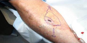
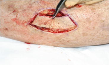

Figure 7.6
Keystone flap planning

Figure 7.7


Keystone flap raised; in this case fascia was released to help closure
Stay updated, free articles. Join our Telegram channel

Full access? Get Clinical Tree


