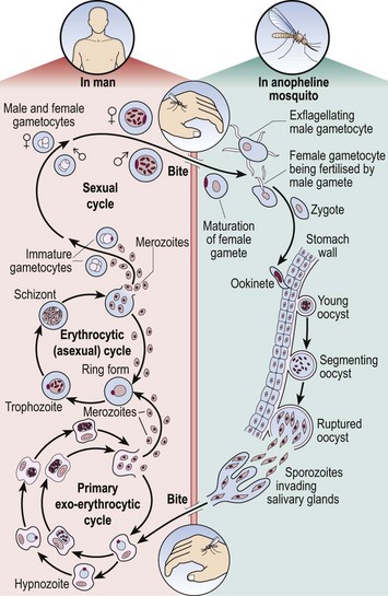
The developing world

49


The developing world


The term ‘developing world’ is used to describe the majority of tropical countries which are ‘hot, humid and poor’. An alternative term is the ‘less economically sound’ nations, as these countries are often advanced in human and cultural resources. Haematological practice is different to that in most developed countries. Genetic diseases such as the haemoglobinopathies and red cell enzymopathies are frequent in many tropical regions. Deficiency anaemia and haemolytic anaemia are often secondary to infections such as ancylostomiasis (hookworm) and malaria. Medical treatment regarded as routine in the developed countries is commonly unavailable. For instance, only about 20% of the world’s haemophiliac population has access to factor VIII replacement therapy.


With the ever-increasing availability of ‘exotic’ holidays and regular foreign travel within immigrant populations, doctors in the developed world are seeing more tropical diseases. In the patient with unexplained symptoms such as malaise and fever, or signs such as splenomegaly, a history of travel should not be overlooked.


Malaria


Malaria is a protozoal disease, the infectious agent being Plasmodium falciparum, P. vivax, P. ovale or P. malariae. Mortality from the disease has fallen over the last decade but it remains a serious health risk throughout the tropics and subtropics where insecticide resistance of anopheline mosquitoes and multiple drug resistance of malarial parasites make control and treatment challenging. According to the World Health Organization, there were 216 million cases of malaria and an estimated 655 000 deaths in 2010. Most deaths occur in children living in Africa where the disease accounts for 20% of all childhood mortality.


Pathogenesis


The life cycle of the malaria parasite is illustrated in Figure 49.1. When taking a meal of blood an infected mosquito initiates human infection by the inoculation of malarial sporozoites. These rapidly pass to the liver where they enter hepatocytes and divide. After several days, enormously increased numbers of parasites (merozoites) depart the liver and invade red cells. Here the merozoites develop via ring forms and trophozoites into schizonts. Rupture of the schizont releases 12–20 merozoites back into the blood, thus perpetuating the cycle. The duration of the blood cycle varies between malarial species, explaining the different periodicity of fever in each type. A further mosquito becomes infected when it feeds on blood containing gametocytes, the sexual form of the parasite.


Diagnosis


Although malarial parasites may be detected in normal blood films, their identification is generally easier in Leishmann or Giemsa stain at a higher pH. A thick film is best for detection and a thin film for determination of the species. Prolonged inspection of the film is sometimes necessary to spot malarial parasites as there can be a low level of parasitaemia. Where malaria is suspected on clinical grounds repeated samples may be needed to make or exclude the diagnosis. P. falciparum

Stay updated, free articles. Join our Telegram channel

Jun 12, 2016 | Posted by in HEMATOLOGY | Comments Off on The developing world

Full access? Get Clinical Tree

Get Clinical Tree app for offline access