“True” thrombocytopenia, to a variable degree, affects all types of ICU patients in all parts of the world; adult medical ICU patients are mostly affected, but it is also observed in surgical and pediatric patients. These observations underlie the comment made by R.I. Parker in his recent review [1] that thrombocytopenia in ICU patients is “a truly universal occurrence.”
Although a threshold value of 150*109/L is generally accepted to indicate thrombocytopenia, stable platelet counts between 150 and 100*109/L are not necessarily considered pathological. Moreover, it is now recognized that the risk of clinically spontaneous bleeding is significantly high when platelet counts fall below 20–10*109/L [3].
The two main mechanisms responsible for thrombocytopenia are reduced production and increased destruction of platelets; less frequently, a reduced platelet count may also be due to sequestration and hemodilution [1, 2].
Table 6.1 summarizes the main classification criteria for thrombocytopenia, the most frequent pathological mechanisms and the associated clinical conditions. The table does not include causes of thrombocytopenia in pregnancy and postpartum, since these conditions go beyond the scope of this chapter.
Table 6.1
Causes of thrombocytopenia
Main classification criteria | Pathological mechanism | Examples of clinical conditions | References |
|---|---|---|---|
Decreased production | Primary bone-marrow failure | Myelodisplastic disorders, Fanconi’s anemia, congenital amegakaryocytic thrombocytopenia | |
Secondary bone-marrow failure | Sepsis, severe idiopathic aplastic anemia, severe malnutrition | [8] | |
Infiltration of bone marrow due to neoplastic diseases | Acute leukemia, widespread marrow metastases | [9] | |
Infiltration of bone marrow due to storage disorders | Gaucher’s disease | [10] | |
Drug-related marrow suppression | Chemotherapy, other drugs | [11] | |
Marrow failure due to radiation therapy | Internal radiation, external radiation | [12] | |
Enhanced destruction | Nonimmune: mechanical | Intravascular devices such as central venous catheters, intraaortic balloon pump | [13] |
Nonimmune: microangiopathic | Thrombotic thrombocytopenic purpura (TTP), disseminated intravascular coagulation (DIC), subacute bacterial endocarditis (SBE), vasculitis | ||
Nonimmune: platelet aggregation | Drugs | [11] | |
Immune: platelet specific auto-antibodies | Immune thrombocytopenic purpura (ITP) | [16] | |
Immune: immune complexes | Autoimmune disorders | [17] | |
Immune: cell-mediated | Hypersplenism, hemophagocytic lymphohistiocytosis | [18] | |
Immune: platelet-specific allo-antibodies | Posttransfusion purpura | [19] | |
Immune: Drugs | Drug related immune thrombocytopenia: antiepileptics, gold compunds, vancomycin, thiazides, quinine/quinidine | [11] | |
Immune: Heparin-induced | Heparin-Induced Thrombocytopenia (HIT) | [20] | |
Immune: sepsis | Sepsis | [8] | |
Sequestration | Congestive splenomegaly | Portal hypertension leads to the redistribution of platelets from the circulating pool to the splenic pool | [21] |
Hemodilution | Secondary to fluid infusion in case of massive hemorrhage | Transfusion of platelet-poor blood products, infusion of colloids and crystalloids | [22] |
It should always be remembered that in a significant number of cases, thrombocytopenia is due to multiple factors, such as for example in sepsis.
The diagnostic workup for thrombocytopenia must include, in addition to laboratory tests discussed in this chapter, a family history for thrombocytopenia, the evaluation of its “dynamics,” meaning if it is a new finding, if it is chronic or whether it has a relapsing presentation. Information on bleeding episodes is also very important, as is the history of concomitant diseases such as infections, tumors, or autoimmune diseases. Finally, it is of paramount importance to collect the history related to recent medication (heparin, antibiotics) and blood transfusion since especially for hospitalized patients, drug-induced thrombocytopenia (DITP) is among the most common causes of low platelet counts. Since the aim of this chapter is to discuss thrombocytopenia in critically ill patients, it goes without saying that it is challenging to understand this condition in these patients also because a complete history may be difficult to obtain.
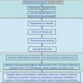
Whereas by definition, the Whole Blood Count is the basic laboratory test for diagnosing thrombocytopenia, the microscopic examination of the blood smear gives additional, important information on the pathogenetic mechanism involved [3]. Figure 6.1 illustrates an algorithm that guides the hematologist in the diagnosis of isolated thrombocytopenia. Other tests employed in the diagnosis of the causes of thrombocytopenia are liver and renal function tests, coagulation tests including d-dimers, lactate dehydrogenase, and bone marrow aspirate and biopsy.
Platelet antibody assays and other tests such as reticulated platelets have a limited specificity and therefore their use is debatable [16].
Before describing the clinical conditions associated with thrombocytopenia, the importance of the rate of decline in platelet counts must be pointed out. When a constant, slow reduction in platelet number is observed with minimum (nadir) counts falling below 20*109/L, a DITP due to marrow inhibition is the probable cause. On the other hand, when there is a fast rate of decline (24–48 h) in platelet numbers, an immune mechanism is suspected. A variable rate in platelet reduction is suggestive of consumptive coagulopathy [1].
6.1.1 Thrombocytopenia Due to Reduced Production
Thrombocytopenia caused by bone marrow suppression may be due to acquired or congenital conditions. In the latter category are comprised Fanconi’s anemia, congenital amegakaryocytic thrombocytopenia, thrombocytopenia, and absent radii syndrome; a comprehensive review of these clinical conditions has been recently published by Parikh and Bessler [4]. The inherited bone marrow failure syndromes are genetic disorders affecting blood cell lineages. They are characterized by a wide spectrum of symptoms ranging from aplastic anemia to symptoms related to the suppression of one or two cell lines. Congenital amegakaryocytic thrombocytopenia is an inherited bone marrow failure syndrome usually diagnosed at birth, and characterized by insufficient production of megakaryocytes due to a defect in the thrombopoietin receptor [5].
Acquired bone marrow failure is often due to myelodysplastic syndromes, a heterogeneous group of clonal bone marrow disorders characterized by ineffective hematopoiesis, morphological and functional abnormalities of hematopoietic cells, and increased risk of malignant transformation. The prevalence of thrombocytopenia in these diseases varies from 40 to 65 % [6], and together with platelet dysfunction, is responsible for the increased hemorrhagic risk in these patients.
Sepsis is a condition affecting a significant number of patients admitted to hospitals; a recent review reports that in the USA, 2 % of patients corresponding to 750,000 per year are septic, half of which are admitted to ICUs [8]. Clinical signs of sepsis are diverse and depend on the microorganism, site of original infection, and health condition of the patient. Thrombocytopenia in sepsis is a common finding and severe forms of sepsis are associated with coagulation disorders that can lead to disseminated intravascular coagulation (DIC).
Thrombocytopenia can also be caused by drugs that suppress the bone marrow, and in particular megakaryocyte proliferation and maturation. Whereas antimetabolytes, cytotoxic drugs, and alkylating agents exert a toxic effect on all bone marrow cell lines, some antibiotics such as linezolid, may cause a selective suppression of platelet cell lines [11].
Other causes of thrombocytopenia due to decreased production (Table 6.1) are storage disorders [10], infiltration of bone marrow due to neoplastic diseases [9] and radiation therapy [12].
Thrombocytopenia due to reduced production is not a frequent cause of admission to the ICUs, since it is more often preexistent.
6.1.2 Thrombocytopenia Due to Enhanced Destruction or Consumption
6.1.2.1 Thrombocytopenia Due to Enhanced Destruction: Nonimmune Mechanisms
Medical devices such as mechanical heart valves, left-ventricular assistance devices, and aortic balloon pumps may be responsible for the destruction of platelets. In a study on 1,302 patients who underwent percutaneous coronary intervention (PCI) with baseline normal platelet counts (≥150*109/L), 3.1 % developed post-PCI thrombocytopenia. Multivariate analysis showed that the use of intra-aortic balloon pump was an independent predictor of thrombocytopenia, with an odds ratio of 2.8, confidence intervals 1.1–6.8, p = 0.024. Post-PCI thrombocytopenia was significantly associated with major adverse cardiovascular events at 6 months (hazard ratio 2.7, CI 1.3–5.5, p = 0.0069) [13].
Microangiopathic processes such as thrombotic thrombocytopenic purpura (TTP), hemolytic uremic syndrome (HUS), and disseminated intravascular coagulation (DIC) may be responsible for thrombocytopenia due to enhanced platelet destruction.
TTP is characterized by microvascular platelet clumping, which leads to thrombocytopenia and microangiopathic hemolytic anemia. Common findings are “broken” erythrocytes or schistocytes (see algorithm reported in Fig. 6.1), neurological disorders, renal failure, and fever [14]. The disease is due to a congenital or acquired deficiency in ADAMTS13, a metalloprotease which cleaves von Willebrand factor. ADAMTS13 deficiency is responsible for microvascular thrombosis and thrombocytopenia. Plasma exchange is the optimal therapy, and its effectiveness is probably due to the removal of anti-ADAMTS13 autoantibodies and large von Willebrand factor multimers.
HUS is similar to TTP in that microvascular thrombosis, thrombocytopenia, microangiopathic hemolytic anemia, renal insufficiency, and altered mental status are common features. However, ADAMTS13 is normal and the disease is generally due to endothelial cell damage caused by a toxin produced by pathogenic strains of Escherichia or Shigella. In HUS, thrombocytopenia is usually not severe but dialysis may be required to treat renal insufficiency [23].
DIC does not occur as an isolated event but is practically always associated with an underlying condition such as tissue damage (trauma, burns, hemolytic transfusion reaction, acute transplant rejection), neoplasia, systemic infection, obstetric conditions (abruption placentae, placenta previa, amniotic fluid embolism), and other clinical conditions such as shock, cardiac arrest, and aortic aneurysm. DIC is the result of an overstimulation of the coagulation system and its clinical presentation varies from severe hemorrhage to thrombosis (or both simultaneously). Thrombocytopenia, abnormal prothrombin time and activated partial thromboplastin time (PT and aPTT), decreased fibrinogen and elevated fibrinogen degradation products are common laboratory features of DIC. DIC-associated mortality is mostly due to the original disease, which is complicated by hemorrhage or thrombosis. Multiorgan dysfunction syndrome is a frequent consequence of DIC and is due to hemorrhagic or thrombotic events in liver, heart, kidneys, central nervous system, and lungs [15].
The main therapeutic goal in DIC is that of treating the underlying condition. As far as transfusion of blood products is concerned, there has been a lot of debate on its benefit and potential harm; generally, platelet counts should be kept more than 20*109/L in presence of mild bleeding and more than 50*109/L when there is active bleeding. Plasma or cryoprecipitate should be considered when bleeding is associated with low fibrinogen levels. The aim of fibrinogen replacement is to maintain levels more than 100 mg/dl to prevent or treat bleeding [24].
6.1.2.2 Thrombocytopenia Due to Enhanced Destruction: Immune Mechanisms (Except HIT)
In addition to Heparin-Induced Thrombocytopenia (HIT) which will be discussed in the following section, primary Immune Thrombocytopenia (ITP), post-transfusion purpura (PTP), and drugs may lead to immune platelet destruction.
ITP is an acquired disorder mediated by immunological mechanism, characterized by low platelet counts in the absence of any possible known cause of thrombocytopenia. It affects children and adults (with a slight prevalence in women) and symptoms range from massive bleeding (gastrointestinal, skin–mucosal, and intracranial) to minimal bruising or only alterations in whole blood count. Evaluation of the blood smear is important in the diagnosis of ITP (Fig. 6.1) and antiplatelet antibody assays are not routinely performed due to the low specificity of this test. Adult ITP is treated with corticosteroids or IVIg and platelet transfusions are recommended only for emergency cases in presence of active bleeding [16].
PTP is a rare complication of transfusion occurring 7–10 days after a red blood cell or platelet transfusion and is characterized by a dramatic fall in platelet count reaching a nadir less than 10*109/L. Thrombocytopenia is caused by platelet alloantibodies in the recipient which at first destroy the transfused platelets, but successively also react with self-platelets. PTP is managed by administering IVIg or if available, compatible platelets (usually HPA-1a negative) [19].
Drug-induced thrombocytopenia (DITP) may either be caused by drugs suppressing bone marrow (see previous section) or by drugs eliciting diverse types of antibodies. Table 6.2 summarizes the main types of antibodies implicated in DITP [11]. DITP may be hard to diagnose in critically ill patients, since thrombocytopenia may become evident several days after the beginning of therapy, and has to be distinguished from other causes of thrombocytopenia.
Table 6.2


Antibodies and mechanisms implicated immune drug-induced thrombocytopenia
Stay updated, free articles. Join our Telegram channel

Full access? Get Clinical Tree


