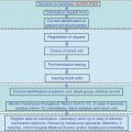
Fig. 2.1
Diagnostic algorythm for anemia
The first question is whether anemia is associated with other hematological abnormalities such as low platelet levels and/or low leukocyte counts and/or presence of abnormal leukocytes (blasts) on blood smear. If this is the case, the presence of bone marrow failure (aplastic anemia) or of malignant hematological disorders such as acute leukemias or myelodysplastic syndromes is likely. In these cases, the bone marrow biopsy and the appropriate cytometric studies of marrow and peripheral blood are mandatory.
The second question is whether anemia determined is associated with an appropriate reticulocyte response. The reticulocyte count is important to evaluate the new red cell production and is very helpful in determining the marrow response to anemia. Very often the reticulocyte count is lacking for the evaluation of the anemic conditions, while this test has a crucial role in the diagnostic process. Until a few years ago, the red blood cells were stained with brilliant cresyl blue, which allows the visualization of ribosomes and reticulin network, thereafter the blood smear was examined by microscope with manual count of stained cells. This method was time-consuming and often the responses were delayed, thus reducing the clinical impact of the test. Lately, automated reticulocyte analyzers are available; these counters have a higher degree of precision than can be achieved manually and, in addition, the responses are immediate. These automated reticulocyte counters may show errors in few rare conditions as the case of presence of Heinz or Howell-Jolly bodies inside red cells. Much more important than the percentage of reticulocytes is their absolute count, which can be easily determined starting from the red cell count: absolute reticulocytes count = % of reticulocytes × red cells count/L3. The value over 100 × 109/L is indicative of a bone marrow responding normally to hemolysis or blood loss. If the anemia is associated with a poor reticulocyte count (less than 25 × 109/L), an impaired red cell production is likely.
2.1 Anemias with High Reticulocyte Count
In the case of high reticulocyte count, the subsequent question is: Is there evidence of hemolysis or not? The laboratory tests used to identify a hemolytic process are available easily in any hospital: Serum unconjugated bilirubin, serum lactic dehydrogenase (LDH), and serum aptoglobin. These tests are related to the red cell increased destruction rate and, in most patients, are indicative of a hemolytic process, but in critically ill patients may be misleading. An increased level of total and unconjugated bilirubin is a common finding in intensive care units for several reasons: prolonged fasting or artificial nutrition, hypotension or shock with reduced liver blood flow, heart failure or tamponade with secondary liver venous stasis, hepatosplenic blood flow modification by endotoxemia or peritonitis, portal thrombosis, preexisting chronic liver diseases, and other less common causes. LDH is an enzyme not specific to the red cells, and it can be found in any organ and tissue; therefore, any cytolitic process is able to increase LDH serum levels. In critically ill patients, high of very high level of serum LDH can be found very easily due to crush syndrome with muscle necrosis, lung inflammatory processes, chronic and acute viral liver diseases or acute cholestasis, fatty liver, sepsis, myocardial ischemia, bone fractures, and others. In addition, high LDH levels without evidence of disease can be found in about 3 % of normal people. The LDH isoenzymes could be useful for determining the involved tissue, but this test is not available in most hospitals and it is used for research purposes only. In conclusion, LDH is not trustworthy in the context of the critically ill patient. The haptoglobin is a protein synthesized by the liver, and it is able to bind to Hb when this molecule is released in the plasma (like occurs in hemolysis). The complex haptoglobin-Hb is removed by the hepatocytes. Despite the presence of haptoglobin in serum only, this protein decreases or becomes undetectable in case of both intravascular and extravascular hemolysis. Serum haptoglobin determination is useful in the diagnostic path of the majority of patients, but in the intensive care units the interpretation of its levels is complicated and its diagnostic power is significantly reduced. In fact, haptoglobin is an acute-phase protein, therefore, its synthesis increases in response to inflammation, infections, or malignant diseases. Taking into account these characteristics, in critically ill patients, the increased synthesis of this protein due to sepsis, infections, inflammatory states of various etiologies, may overcome the decrease induced by hemolytic process. Conversely, abnormal low levels of haptoglobin can be found in the absence of hemolysis in the case of malnutrition or of the other clinical situations characterized by abnormal protein loss like occurs after extensive burns or for nephritic syndrome; by preexisting chronic liver disease; or by the impossibility of a normal aliment absorption like occurs in large intestine resections for vascular disease or for accident perforation, events not uncommon in the intensive care units. In conclusion, the usual laboratory tests used to identify a hemolytic process are have a limited diagnostic value in the intensive care setting and, often, additional tests and a careful follow-up of the patient are needed for a correct diagnosis. Even the diagnosis of posthemorrhagic anemia may be difficult in these patients. In fact, after an acute blood loss, the plasma volume and red cell mass are reduced in proportional amount; consequently, the Hb concentration does not change. Therefore, the amount of blood loss can be underestimated by the degree of anemia, especially early. In the days following the blood loss, the reticulocyte count is normal and increases only after 6–10 days; in this “window,” even the iron stores are unmodified, and mean corpuscular volume is still normal. An external hemorrhage sufficient to determine anemia is usually evident, but internal bleeding may be less apparent. If the hemorrhage occurs in retroperitoneal space, into a body cavity or in a cyst, the decrease of Hb level may be a diagnostic problem. In addition, the breakdown and the absorption of red cell in the tissues are able to increase indirect bilirubinemia, and this picture, along with high reticulocyte count, can be confused with a hemolytic anemia. Therefore, a careful follow-up of the patient and appropriate tests are mandatory for a correct diagnosis.
If repeated tests confirm high reticulocyte counts (in the absence of blood loss) and a possible hemolytic process is suspected, the main causes of hemolysis should be carefully checked. Since in the adult patients the most common acquired hemolytic disorders are the immune-mediated processes, the direct anti-globulin test (Coomb’s test) should be determined. Thereafter, the diagnostic process can be separated for the patients with positive and negative direct anti-globulin test.
2.1.1 Patients Positive for Direct Anti-globulin Test
These cases have presumably an immune-hemolytic anemia and can undergo immediate glucocorticoids therapy, which remains the treatment of choice of this immune disorder. Intravenously administered doses of 1.0 mg/kg b.w. of methyl-prednisolone daily are efficacious in most cases. The response may not be evident for several days and an increase of Hb level can be noticeable only after 7 days of treatment. A further delay in the response is expected in critically ill patients since many acute factors may interfere in the red cell production like prolonged fasting or artificial nutrition, hypotension, reduced liver blood flow, acute renal failure with reduced erythropoietin production, endotoxemia or other acute stress situations. In the rare cases of lack of response or in the case of worsening of the hemolytic process, high-dose i.v. immunoglobulin administration (1 g/kg b.w.) can be useful in decreasing the clearance of the red cells by the monocyte macrophage system. This therapy can be repeated after 1 or 2 weeks if required.
2.1.2 Patients Negative for Direct Anti-globulin Test
In these cases, the clinical history (when available) is helpful to exclude the exposure to chemical or physical agents; thereafter, some infections (malaria, leishmaniasis, trypanosomiasis, bartonellosis) should be taken into consideration in white people back from recent adventure travels in the third world or in people shortly after arriving from Africa or from other underdeveloped countries. In critically ill patients, the septicemia of Clostridium perfrigens should be taken into consideration, in fact it may occur after traumatic wound infections, necrotizing enterocolitis, genitourinary or gastrointestinal surgery, and other acute severe conditions. In this case, a severe, often-fatal, hemolytic anemia occurs with a massive hemolysis, and hemoglobin concentration may fall to a very low level in a matter of hours. The diagnosis is suspected when high fever, jaundice, and anemia occur together in a patient of the intensive care unit. The clostridial infection responds well to antibiotics therapy but the treatment must be started as quickly as possible, even before the blood culture results are available.
After the exclusion of these infective causes with appropriate tests, the other causes of nonimmune hemolytic anemia should be considered. For the diagnosis of the most common diseases, a few laboratory investigations are needed:
1.
Hb electrophoresis
2.
Osmotic fragility test
3.
Red cell enzyme determination
4.
Blood smear examination
The Hb electrophoresis may indicate the presence of genetic diseases like sickle cell anemia, or thalassemia or of the rare conditions associated with abnormal Hb (Hb C, SC, D, SD, and E). The osmotic fragility test is able to discover the spherocytic anemia and related disorders, and, finally, the enzyme determination is useful to detect the glucose-6-phosphate deficiency (G6PD), known as favism, or pyruvate kinase deficiency. All these conditions are inherited diseases; some of these are common in Italy like thalassemias or favism, while others are very rare in Europe, like sickle cell anemia or the unstable Hb diseases. All these diseases worsen the degree of anemia in patients in critical medical conditions and should be recognized to avoid unnecessary support treatments or delay in discharging the patient fearing covert bleeding.
Stay updated, free articles. Join our Telegram channel

Full access? Get Clinical Tree


