The Cardiovascular System in Hypothyroidism
Irwin L. Klein
Sara Danzi
The changes in cardiovascular function that occur in patients with hypothyroidism are diametrically opposite to those of thyrotoxicosis (see Chapter 25) (1). Symptoms and signs and cardiovascular hemodynamic changes are much less prominent in patients with hypothyroidism than in those with thyrotoxicosis. This chapter reviews the pathophysiologic basis for the cardiac findings of hypothyroidism, the changes in intermediary metabolism and adrenergic activity and the potential of hypothyroidism as a risk factor for accelerated atherosclerosis.
Cardiovascular Hemodynamics
The hemodynamic changes associated with hypothyroidism are listed in Table 40.1. There is an increase of 50% to 60% in peripheral vascular resistance in hypothyroid patients; this increase is accompanied by a 30% to 50% decrease in resting cardiac output (2,3). The net effect of the decrease in blood flow and the decrease in tissue oxygen consumption that occurs in hypothyroidism is that arteriovenous oxygen extraction across major organs is similar in patients with hypothyroidism and normal subjects (2). Studies of the molecular mechanism underlying the increase in systemic vascular resistance suggest that thyroid hormone, specifically triiodothyronine (T3), may have a direct vasodilator action on vascular smooth muscle cells (4,5). In addition endothelium-derived vascular relaxation, which involves nitric oxide release and action, is impaired in both mild and overt hypothyroidism (6,7). Thus, in the absence of T3, vascular smooth muscle tone and systemic vascular resistance may increase. This increase can in part be reversed by acute T3 administration (8).
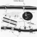
All measurements of left ventricular contractility and performance are lower in patients with both acute and chronic hypothyroidism (9,10,11,12). Cardiac output (index) is decreased as a result of decreases in both stroke volume and, to a lesser extent, heart rate (1,12). The duration of both the preejection period and the isovolumic contraction time is prolonged compared with normal subjects, and these measures return to normal in response to thyroid hormone replacement therapy (9,12). In addition, and perhaps more important, the rate of ventricular relaxation during diastole is slower, and diastolic filling and compliance are impaired (3,12). The rate of diastolic filling may decrease from normal values of 400 mL/second to less than 300 mL/second. The mechanisms underlying the impaired ventricular performance in patients with hypothyroidism are multifactorial and appear to result from changes in the expression of important myocyte-specific contractile and regulatory proteins which mediate contractile measures involving both pre-and afterload (1,12,13,14). As noted in Chapter 25, the expression of many cardiac genes is modulated by thyroid hormone, and in experimental animals, lowering serum concentrations of thyroxine (T4) and T3 alters the expression of the heavy chain isoforms of myosin and in humans an important change in the calcium regulatory proteins (15,16,17). It appears that the cardiac myocyte is unique from many other cell types in that it does not transport T4 but is selective for T3 (18). Myocyte nuclei contain T3 receptors that bind to specific DNA sequences in several genes, and thus many of the cardiac actions of T3 occur at the level of gene transcription (1,13). Decreases in the expression of the genes for the sarcoplasmic reticulum calcium adenosine triphosphatase (SERCA2), Na+K+-ATPase, β-adrenergic receptors, and voltage-gated potassium channels (Kv) all play a role in the altered cardiac physiology in patients with hypothyroidism (see Chapters 8 and 25) (1,13,17).
Table 40.1 Cardiovascular Hemodynamics in Hypothyroidism | ||||||||||||||||||||||||||||||
|---|---|---|---|---|---|---|---|---|---|---|---|---|---|---|---|---|---|---|---|---|---|---|---|---|---|---|---|---|---|---|
|
The rate of tension development during cardiac systole is determined in part by the rate at which myosin catalyzes the hydrolysis of ATP. There are two isoforms of myosin ATPase, α and β, in cardiac myocytes. The predominant form in human ventricular tissue is β-myosin (19). In one hypothyroid patient with severe left ventricular dysfunction, the content of α-myosin in ventricular muscle increased slightly during treatment with T4, but β-myosin predominated at both times (17). The patient’s left ventricular contractile function improved during treatment, most probably a result of improved calcium handling (cycling) in cardiac muscle cells (13,20). The activities of several enzymes involved in regulating calcium fluxes in the heart are controlled by thyroid hormone. These include SERCA2 and phospholamban (21,22). In addition, in hypothyroidism, the increase in systemic vascular resistance (afterload) and decrease in blood volume and venous return (preload) impair cardiovascular hemodynamics (1,11,12) all resulting in a lowered cardiac output.
As a result of the increase in systemic vascular resistance and decrease in cardiac output, mean blood pressure is largely unaltered in hypothyroidism (1,2). There may be an increase in diastolic pressure and a decrease in systolic pressure, so the pulse pressure is low. Hypothyroid patients may have diastolic hypertension that declines, sometimes to normal, during treatment with thyroid hormone as systemic vascular resistance decreases (23,24,25). Plasma volume is decreased in patients with hypothyroidism (11), an unexpected finding in light of the clinical finding that some patients have pitting edema suggestive of fluid overload. Capillary permeability is increased, and therefore the albumin and water content in the interstitial space is increased. Fluid may accumulate not only in gravity-dependent regions, but also in the pericardial, pleural, and other spaces (26). Patients with hypothyroidism also have nonpitting edema (myxedema), caused by the accumulation of hydrophilic glycosaminoglycans in the interstitial space.
The response of hypothyroid patients to exercise supports the hypothesis that peripheral circulatory changes mediate many of the changes in cardiac performance. Exercise lowers systemic vascular resistance in patients with hypothyroidism, causing an increase in cardiac index, heart rate, and stroke volume to 85% to 90% of the responses in normal subjects (10). These observations indicate that the reduced left ventricular function in hypothyroid patients at rest is at least partially the result of a reversible decrease in hemodynamic loading (27).
Thyroid Hormone and Intermediary Metabolism
In addition to the characteristic cardiovascular changes there are several metabolic abnormalities in hypothyroid patients. These include a low basal metabolic rate, increased total and low-density lipoprotein (LDL) cholesterol and hypertriglyceridemia. It can be viewed that the lowered cardiac output is an appropriate response to the decrease in fuel and oxygen needs for the deceleration in many biologic processes (2,10,27,28,29). Protein synthesis is decreased in hypothyroid animals and responds to thyroid hormone supplementation in a dose-dependent manner (29). A component of the anabolic response to TH is probably mediated by growth hormone and IGF-1 (30). As predicted, protein turnover and amino oxidation are reduced in hypothyroidism (31). The effects of TH on carbohydrate metabolism are not of major clinical significance. Fasting blood glucose may be reduced in severe hypothyroidism due to impaired gluconeogenesis which in turn results from lowered cytosolic phosphoeno/pyruvate carboxykinase (PEPCK) activity. This enzyme in turn is stimulated by glucagon, norepinephrine, glucocorticoids and T3, the latter through a transcriptional mechanism of action (32). Similar to protein and carbohydrate metabolism, T3 stimulates both the de novo synthesis of fatty acids (lipogenesis) in liver and the degradation of lipids (lipolysis and fatty acid oxidation) in peripheral tissues. The studies of thyroid hormone on lipid metabolism derive primarily from hypothyroid animals but have also been demonstrated in humans (33,34). Serum triglyceride levels may be increased in hypothyroid patients (35). It has been suggested that this is the result of an increase in lipoprotein lipase activity (36).
Serum cholesterol levels vary inversely across the entire spectrum of thyroid disease (see Chapter 26). Total serum and LDL cholesterol is reduced in thyrotoxicosis and increased in hypothyroidism. This effect is the result of variations in the rate of the removal of cholesterol by the liver via the stimulation of
the expression of sterol regulatory element binding protein-2 (SREBP-2) (37).
the expression of sterol regulatory element binding protein-2 (SREBP-2) (37).
The Sympathoadrenal System in Hypothyroidism
Many clinical and basic animal studies have indentified an interaction between catecholamines and the sympathoadrenal system and thyroid disease and these are best seen in measures of cardiovascular hemodynamics. Hypothyroidism influences the clinical signs of adrenergic activity in a direction opposite that of thyrotoxicosis (38). However, in contrast to the measures of cardiovascular hemodynamics, studies in hypothyroid patients show that the overall production rate of norepinephrine is faster than normal (39).
Severely hypothyroid patients may develop hypothermia and myxedema coma after exposure to low environmental temperatures (see Chapter 44). In humans with hypothyroidism, evidence has been found that the responses to adrenergic stimulation such as glycogenolysis, lipolysis, gluconeogenesis, and intracellular calcium mobilization are reduced (40). This includes skin and subcutaneous tissue vasoconstriction (41) perhaps resulting from a loss of the effects of T3 on vascular smooth muscle and nitric oxide production (6,42). Even though serum levels of catecholamines are increased in hypothyroidism (39), heart rate and contractility, both systolic and diastolic, are often impaired (3,9).
Clinical Manifestations
Most patients with hypothyroidism have few symptoms directly referable to the cardiovascular system (43). Although fatigue, lack of energy, exertional dyspnea, and exercise intolerance have been attributed to impaired cardiac performance, they are more likely to be related to psychological or skeletal muscle dysfunction (44). Whether or not heart failure with orthopnea and paroxysmal nocturnal dyspnea occurs solely as a result of hypothyroidism is discussed later.
Patients with hypothyroidism can have angina-like pain (27), and many have hypercholesterolemia. Various metabolic changes and cardiac risk factors predisposing to atherosclerosis are present in patients even with mild hypothyroidism (Table 40.2) (45). The majority of these risk factors respond to thyroid hormone therapy in a positive manner. An occasional patient with normal coronary arteries has angina that resolves with thyroid hormone therapy (27).
Table 40.2 Cardiovascular Risk Associated with Hypothyroidism and Effect of Thyroid Hormone Therapy | ||||||||||||||||||||||
|---|---|---|---|---|---|---|---|---|---|---|---|---|---|---|---|---|---|---|---|---|---|---|
| ||||||||||||||||||||||
Physical Examination
The abnormalities that may be found on physical examination of the cardiovascular system in patients with hypothyroidism include bradycardia, narrow pulse pressure, low-amplitude pulse, diminished apical impulse, and distant heart sounds (1,2,3,10) (Table 40.1). The bradycardia may result from a loss of the chronotropic action of thyroid hormone (T3) directly on the sinoatrial pacemaker cells (25); heart rate analysis reveals changes in both sympathetic and parasympathetic tone (46). The abnormal heart sounds may be caused by decreased left ventricular contractility or pericardial effusion (9,10,26,47). From 20% to 40% of patients with hypothyroidism have a diastolic blood pressure greater than 90 mm Hg (11,23,48,49). Patients not only complain of cold intolerance but also may have cold feet and hands because of decreased skin blood flow (43).
Stay updated, free articles. Join our Telegram channel

Full access? Get Clinical Tree


