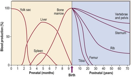
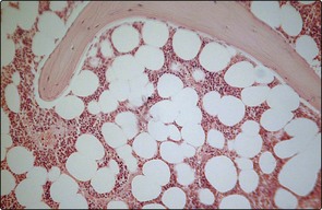
1 The bone marrow In early fetal life, blood is produced in the mesoderm of the yolk sac. During the second to seventh months the liver and spleen take over. Only in the last 2 months of fetal development does the bone marrow become the predominant site of blood formation. During childhood, marrow in the more peripheral bones becomes gradually replaced by fat, so that in adult life over 70% is located in the pelvis, vertebrae and sternum (Fig 1.1). This explains the sites used for bone marrow sampling (see p. 106). Fig 1.1 Sites of blood production in the fetus and after birth. The structure of the bone marrow A trephine biopsy allows a two-dimensional view of the bone marrow down the light microscope (Fig 1.2). Haematopoietic cells of varying lineage and maturity are packed between fat spaces and bony trabeculae. Ultrastructural studies reveal clusters of haematopoietic cells surrounding vascular sinuses which allow eventual discharge of mature cells into the blood. Different lineages are compartmentalised; for example, the most immature myeloid precursors lie deep in the marrow parenchyma while more mature forms migrate towards the sinus wall. Lymphocytes tend to surround small radial arteries while erythrocytes form islands around the sinus walls. Fig 1.2 Normal bone marrow.Light microscopy of bone marrow trephine biopsy. Blood precursor cells in the marrow exist in close proximity to stromal cells. Stromal cells are those cells which do not mature into the three main types of peripheral blood cells – thus they include macrophages, fat cells, endothelial cells and reticulum cells. Immature blood cells are attached to these stromal cells by multiple cellular adhesion molecules (e.g. fibronectin and collagen). Adhesive molecules have specific receptors on stromal and haematopoietic cells. As blood cells mature, the receptors down-regulate and the cells become less adherent and commence the journey through the sinus wall and into the bloodstream. Haematopoiesis: the stem cell hierarchy Haematopoiesis means the formation of blood. A number of transcription factors (e.g. GATA-1, MLL) are critical both for stem cell formation and function and lineage-specific differentiation. The first adult haematopoietic stem cells (HSCs) are generated in the aorto-gonad-mesonephros (AGM) region of the embryo. The classical hierarchy diagram (Fig 1.3 Only gold members can continue reading. Log In or Register to continue You may also needLymphocytesMyelomaAcute myeloid leukaemiaHaematology in the elderlyAplastic anaemiaNeutrophils, eosinophils, basophils and monocytesExamining the patientChronic lymphocytic leukaemia Share this:Click to share on Twitter (Opens in new window)Click to share on Facebook (Opens in new window)Click to share on Google+ (Opens in new window) Related Tags: Haematology An Illustrated Colour Text Jun 12, 2016 | Posted by admin in HEMATOLOGY | Comments Off on The bone marrow