The Antiphospholipid Syndrome


The Antiphospholipid Syndrome


Jacob H. Rand

Lucia R. Wolgast


HISTORICAL BACKGROUND

The antiphospholipid syndrome (APS) is an autoimmune thrombophilic condition that is defined by a constellation of clinical and laboratory manifestations. In retrospect, antiphospholipid (aPL) tests were first described in 1952 with the reports of the biologic false-positive serologic test for syphilis (STS)1 and an inhibitor of the partial thromboplastin time.2 In 1957, an association among the biologic false-positive STS, a circulating anticoagulant, and recurrent pregnancy loss was described.3 The absence of bleeding and a paradoxical association between these coagulation inhibitors and thrombosis were recognized in 1963.4 The term lupus anticoagulant (LA) for these inhibitors of phospholipid-dependent coagulation reactions was introduced,5 because most of the initial patients appeared to have systemic lupus erythematosus (SLE), although this finding was not sustained by later studies. In the 1980s, immunoassays were developed for anticardiolipin (aCL) antibodies,6, 7 and a new “syndrome of thrombosis, abortion, and neurologic disease”8 was named the “anticardiolipin syndrome,”9 subsequently revised to the “antiphospholipid syndrome.”10 In the early 1990s, it was discovered that the antibodies in this autoimmune thrombotic condition did not recognize phospholipids without a protein cofactor identified as β2-glycoprotein I (β2GPI)11, 12 and that other phospholipid-binding proteins13 could also fulfill this role. These antibodies were therefore referred to in literature as being “cofactor-dependent,” or “β2GPI-dependent” aPL antibodies, in contrast to the antibodies generated in patients with syphilis and other infections that recognized phospholipid directly and classified as “cofactor independent.” As explained below, it is now accepted that β2GPI itself is the major target of antibodies in APS.


CURRENT DEFINITION—DIAGNOSTIC CRITERIA AND THEIR LIMITATIONS

The current investigational criteria for APS (Table 101.1), referred to as the Sydney Criteria,14 were not intended as absolute requirements for the clinical diagnosis. Rather, they were meant to characterize patients whom investigators could agree have “definite APS” for clinical studies.” These criteria include histories of documented thrombosis and/or defined pregnancy complications together with specific laboratory tests. However, the syndrome is incompletely understood, and many patients appear to have other clinical abnormalities (Table 101.2) that are referred to as “noncriteria clinical manifestations.”15 Also, as described in the historical background above, the current laboratory assays used for defining the syndrome were derived from empiric observations and were not designed to measure disease mechanisms. Newer laboratory tests that have been developed but not yet accepted by consensus are referred to as “noncriteria laboratory tests.” To complicate matters, some patients may have typical clinical manifestations of APS but entirely negative aPL antibody assays and/or LA test, a situation referred to as seronegative APS.16 Rare patients with aPL antibodies may develop disseminated thrombosis in large and small vessels with resulting multiorgan failure, known as catastrophic antiphospholipid syndrome (CAPS). Preliminary criteria that define CAPS for clinical studies have been published (Table 101.3).17


PREVALENCE AND ANTIGENIC SPECIFICITIES OF “ANTIPHOSPHOLIPID ANTIBODIES”

A significant proportion of patients with venous thrombosis have elevated levels of aPL antibodies, with prevalence rates estimated at 5% to 30% for any aPL antibody, 1% to 16% for LA, and 4% to 24% for aCL antibodies. In arterial thrombosis, the prevalence is 0.1% to 10% for aCL antibodies and 0.02% to 0.3% for anti-β2GPI antibodies. In patients with recurrent spontaneous pregnancy losses, the prevalence is estimated to be 11% for LA and 29% for aCL antibodies.18 The prevalence of aPL antibodies in the asymptomatic “normal” population has generally been estimated at 3% to 10%,19, 20 with a prevalence of 1% to 5% for LA, 1% to 5% for aCL antibodies, and 3% for anti-β2GPI antibodies.18 In one group of healthy young women, who served as controls in a study, 18% had elevated aCL antibodies and 13% tested LA positive.21 aPL antibodies that occur after infections are generally not associated with thrombosis.22

A significant proportion of SLE patients have elevated aPL antibodies; estimates range between 12% and 30% for aCL antibodies and 15% and 34% for LA antibodies.23 aPL antibodies have been associated with virtually all other autoimmune conditions.24, 25, 26, 27, 28, 29, 30, 31, 32 In some patients, the presence of aPL antibodies may herald the development of SLE; one study reported that 18% of SLE patients had positive aCL antibodies prior to diagnosis.33


Antigenic Targets for aPL Antibodies

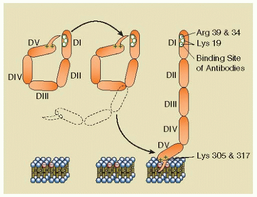
The main antigen targeted by the APS disease process, β2GPI, is a 50-kd glycoprotein comprised of five 60-amino acid domains with homology to other members of the complement control protein superfamily34 (FIGURE 101.1). The biologic function of β2GPI has not yet been established. β2GPI binds to apoptotic cells and may aid their clearance.35, 36 The protein also binds to platelet microvesicles and promote their phagocytosis by macrophages.37 β2GPI binds as well to oxidized low-density lipoprotein (LDL) and may promote its clearance.38


Table 101.1 Sydney investigational criteria for the diagnosis of the APSa


Clinical


Vascular thrombosis (one or more episodes of arterial, venous, or small-vessel thrombosis). For histopathologic diagnosis, there should be no evidence of inflammation in the vessel wall


Pregnancy morbidities attributable to placental insufficiency, including (a) three or more otherwise unexplained recurrent spontaneous miscarriages, before 10 weeks of gestation, (b) one or more fetal losses after the 10th week of gestation, (c) stillbirth, and (d) episode of preeclampsia, preterm labor, placental abruption, intrauterine growth restriction, or oligohydramnios that are otherwise unexplained


Laboratory


Medium- or high-titer aCL or anti -β2GPI IgG and/or IgM antibody present on two or more occasions, at least 12 weeks apart, measured by standard ELISAs


LA in plasma, on two or more occasions, at least 12 weeks apart, detected according to the guidelines of the ISTH SSC Subcommittee on Lupus Anticoagulants and Phospholipid-Dependent Antibodies


“Definite APS” is considered to be present if at least one of the clinical criteria and one of the laboratory criteria are met


aCL, anticardiolipin; aPL, antiphospholipid; β2GPI, β2-glycoprotein I; ELISA, enzyme-linked immunosorbent assay; Ig, immunoglobulin.


aModified from Miyakis et al. International consensus statement on an update of the classification criteria for definite antiphospholipid syndrome. Thromb Haemost 2006;4:295-306.


Table 101.2 Clinical manifestations associated with the APS


Systemic vascular thrombosis


DVT of lower extremity, thoracic, abdominal, or pelvic veins, pulmonary embolism, arterial thrombosis with downstream ischemia and infarction


Reproductive manifestations


Recurrent fetal loss, preeclampsia, abruption placentae, miscarriage, prematurity, intrauterine fetal demise, intrauterine growth restriction, and oligohydramnios


Neurologic manifestations


Stroke, TIA, migraines, seizures, chorea, Guillain-Barrè Syndrome, transient global amnesia, dementia, diabetic peripheral neuropathy, and orthostatic hypotension


Cardiovascular manifestations


Thrombotic occlusion of nonatherosclerotic coronary artery, thrombotic myocardial infarction, coronary artery disease, premature atherosclerosis, atherosclerotic myocardial infarction, peripheral vascular disease, valvular abnormalities


Pulmonary manifestations


Pulmonary hypertension attributable to thrombotic occlusion, other pulmonary hypertension, acute diffuse alveolar damage


Hepatic and gastrointestinal manifestations


Esophageal necrosis with perforation, intestinal ischemia/infarction, pancreatitis, colonic ulceration, acalculous acute cholecystitis attributable to biliary vascular occlusion, mesenteric venoocclusive disease, primary biliary cirrhosis


Renal manifestations


Thrombosis of renal arteries or veins, renal infarction, APS nephropathy, renal artery stenosis, minimal change disease/focal segmental glomerulonephritis, membranous nephropathy, mesangial C3 nephropathy, pauciimmune crescentic glomerulonephritis


Skin manifestations


Ulcerations attributable to microvascular infarction, livedo reticularis, skin ulcerations, skin necrosis, necrotizing vasculitis, livedoid vasculitis, thrombophlebitis, erythematous macules, purpura, ecchymoses, painful nodules, subungual splinter hemorrhages, Anetoderma, discoid lupus erythematosus, cutaneous T-cell lymphoma


Retinal abnormalities


Retinal vein occlusion, cilioretinal artery occlusion, optic neuropathy


Other organ manifestations


Acute adrenal failure due to bilateral adrenal hemorrhagic infarction, osteonecrosis without histologic evidence for microvascular occlusion or vasculitis, acute sensorineural hearing loss


Other coagulation abnormalities


Thrombocytopenia, ITP, hypoprothrombinemia, protein S deficiency, acquired APC resistance, acquired inhibitors to specific coagulation factors, and AvWS


Manifestations that are not included in consensus-based diagnostic criteria are italicized.


Table 101.3 Proposed criteria for the classification of CAPS


1.


Evidence of involvement of three or more organs, systems, and/or tissuesa


2.


Development of manifestations simultaneously or in less than a week


3.


Confirmation by histopathology of small-vessel occlusion in at least one organ or tissueb


4.


Laboratory confirmation of the presence of aPL antibodies (LA and/or aCL antibodies)c


Definite CAPS


All four criteria


Probable CAPS


All four criteria, except for only two organs, systems, and/or tissues involvement


All four criteria, except for the absence of laboratory confirmation at least 6 weeks apart due to the early death of a patient never previously tested for aPL prior to the CAPS event


Criteria 1, 2, and 4


Criteria 1, 3, and 4 and the development of a third event in more than a week but less than a month, despite anticoagulation


a Usually, clinical evidence of vessel occlusions, confirmed by imaging techniques when appropriate. Renal involvement is defined by a 50% rise in serum creatinine, severe systemic hypertension (N180/100 mm Hg), and/or proteinuria (N500 mg/24 h).

b For histopathologic confirmation, significant evidence of thrombosis must be present, although, in contrast to Sydney criteria, vasculitis may coexist occasionally.

c If the patient had not been previously diagnosed as having an APS, the laboratory confirmation requires that the presence of antiphospholipid antibodies must be detected on two or more occasions at least 6 weeks apart (not necessarily at the time of the event), according to the proposed preliminary criteria for the classification of definite APS.


Modified from Asherson RA, Cevera R, de Groot PG, et al. Catastrophic antiphospholipid syndrome: international consensus statement on classification criteria and treatment guidelines. Lupus 2003;12:530-534.


Anti-β2GPI antibodies from different patient samples are heterogeneous in their epitope specificities and recognize all five domains of the protein. IgG antibodies against an epitope comprising Gly40-Arg43 in the domain I of β2GPI have been reported to have a stronger correlation with thrombosis than antibodies against other epitopes.39 Recent data have shed light on interesting changes in the conformation of β2GPI that may have a role in the APS disease process. Circulating plasma β2GPI was found to have a closed circular conformation due to the affinity of a portion of carboxyterminal domain V for the protein’s aminoterminal domain I, where the phospholipid binding site is located.40 The affinity for anionic phospholipids derives from (a) cationic residues from its aminoterminus that have affinity for anionic polar heads of phospholipids and (b) a hydrophobic loop which inserts into the lipid bilayer. Binding to anionic phospholipid membranes requires the protein to open from its circular conformation, which exposes an epitope in domain I that had been cryptic in the circular conformation (FIGURE 101.1).


FIGURE 101.1 Schematic representation of β2GPI conformational change. (Reprinted from Rand JH. A snappy new concept for APS. Blood 2010;116:1193-1194, with permission. Inside Blood commentary on Agar C, van Os GM, Morgelin M, et al. β2-Glycoprotein I can exist in 2 conformations: implications for our understanding of the antiphospholipid syndrome. Blood 2010;116:1336-1343, with permission. Professional illustration by Paulette Dennis.)

Several other antigenic targets for aPL antibodies include prothrombin, coagulation factor V, protein C, protein S, annexin A2, annexin A5, high and low molecular weight kininogens, heparin, factor VII/VIIa,41, 42, 43, 44 oxidized LDLs,45 vimentin complexed to cardiolipin,46 lyso(bis)phosphatidic acid,47 sulfatides, and acidic glycosphingolipids.48


PATHOPHYSIOLOGIC MECHANISMS


Overview

A host of mechanisms have been postulated to explain the thrombotic tendency in APS (Table 101.4). The APS disease entity is probably composed of subcategories that are attributable to specificities of the particular antibodies. aPL antibodies are highly heterogeneous even in individual patients, and these may exert different effects in different experimental
systems. In an effort to find additional clues, some investigators have recently pursued proteomic studies to identify candidate proteins that might be involved in the APS disease process.49


Table 101.4 Proposed pathogenic mechanisms of APS


I.


Inhibition of endogenous anticoagulant and fibrinolytic mechanisms


A.


Disruption of the annexin A5 anticoagulant shield


B.


Inhibition of the protein C pathway—decreased activation of protein C, barrier of APC proteolysis of factor Va and VIIIa, prevention of protein C and EPCR binding


C.


Interference with thrombin regulation—enhanced thrombin generation, decreased thrombin inactivation


D.


Inhibition of heparinoids, TFPI, and inactivation of factor IXa


E.


Interference with fibrinolysis via annexin A2, β2GPI cofactor activity, autoactivation of XIIa, direct inhibition of plasmin, and increase of PAI-1


II.


Activation of platelets


A.


Activation of platelets—via ApoER2 and via β2GPI-PF4 interaction


B.


Interference of vWF-mediated platelet adhesion


III.


Injury and activation of endothelial cells and monocytes


A.


Direct injury


B.


Signaling via annexin A2/TLR4/ApoER2-inducing proadhesive prothrombotic phenotype


IV.


Complement activation


Inhibition of Endogenous Anticoagulant and Fibrinolytic Mechanisms


Disruption of the Annexin A5 Anticoagulant Shield

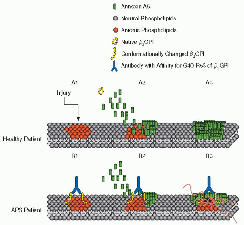
Annexin A5 is highly expressed by a variety of cells serving barrier functions including endothelial cells, where the protein binds to the surfaces and inhibits thrombin formation.50 It is also expressed on placental trophoblasts on the apical membranes of the syncytiotrophoblasts that interface with the maternal blood circulation.51 Annexin A5 forms two-dimensional crystal lattices over phospholipid bilayers52, 53 that shield them from availability for phospholipid-dependent coagulation enzyme reactions52 (FIGURE 101.2). The protein appears to be important for maintaining placental blood circulation; pregnant mice treated with antiannexin A5 antibodies developed placental necrosis, fibrosis, and pregnancy loss.54

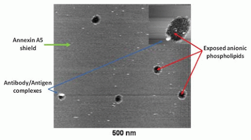
There is significant evidence that aPL antibodies interfere with the annexin A5 binding and crystallization and thereby promote coagulation reactions; this has been demonstrated through immunohistochemical studies of placental tissues,55 cell culture experiments,56 and atomic force microscopic imaging52 (FIGURE 101.3). The aPL antibody-mediated disruption of annexin A5 correlates with antibody recognition of the epitope on domain I of β2GPI, which in turn has been correlated with significantly increased risk of thrombosis.39, 57


Inhibition of the Protein C Pathway

aPL antibodies can interfere with the protein C pathway (see Chapter 19) in several ways (Table 101.4). Antibodies against protein C and against protein S have been described,58, 59, 60 with high levels of antiprotein C IgM correlating with increased risk of venous thrombosis.60 Antibodies against β2GPI may crossreact with activated protein C (APC),61 and APS patients often exhibit acquired APC resistance,62 an effect that has also been correlated with anti-β2GPI domain I antibodies.63, 64 Antiendothelial antibodies in patients with APS65, 66 may interfere with the positioning of protein C on the endothelial protein C receptor (EPCR) or bind to thrombomodulin and block the activation of protein C by thrombin. Antithrombomodulin antibodies can also interfere with the activation of protein C in SLE patients.67, 68 Antiprothrombin antibodies from APS patient also interfere with the protein C pathway by reducing availability of thrombin for binding to thrombomodulin.69, 70 Antibodies against EPCR have been identified in pregnant APS patients and associated with increased risk of fetal death.71


Interference with Thrombin Regulation

It has been proposed that antibodies against noncatalytic epitopes on prothrombin may provoke thrombosis by crosslinking prothrombin molecules, thus increasing their density and thrombin generation on the surface of endothelial cells.72, 73, 74 aPL antibodies and antiprothrombin antibodies can also interfere with inactivation of thrombin by antithrombin.75 Some antiprothrombin antibodies may themselves display a catalytic function and proteolyze prothrombin to generate a thrombinlike activity.76


Inhibition of Heparinoids, TFPI, and Inactivation of Factor IXa

aPL antibodies crossreact with heparin and heparinoid molecules and inhibit the acceleration of antithrombin activity.77 APS patients can have autoantibodies against tissue factor pathway inhibitor (TFPI) that reduce its activity.78 Some aPL antibodies interact with factor IXa and impair its inactivation by antithrombin.79


FIGURE 101.2 Proposed mechanism for thrombosis in APS. Proposed mechanism of anti-D1 IgG-mediated disruption of annexin A5 on cell surface. (A1-A3) Annexin A5 anticoagulant activity in absence of APS. When injury exposes anionic phospholipids on the cell surface (A1), annexin A5 assembles over damaged areas of endothelium, creating a shield over the endothelial surface and inhibiting anionic phospholipid-dependent coagulation reactions. (B1-B3) Annexin A5 disruption in the presence of APS. In the presence of antibodies against the G40-R43 epitope on domain I of β2GPI, there is a conformational change and dimerization of β2GPI (B1), and anti-β2GPI/β2GPI complexes displace annexin A5 on anionic phospholipid surfaces (B2). This creates gaps in the annexin A5 shield, exposing more anionic phospholipids to coagulation reactions and thrombosis (B3). (Modified from de Laat B, Wu XX, van Lummel M, et al. Correlation between antiphospholipid antibodies that recognize domain I of β2-glycoprotein I and a reduction in the anticoagulant activity of annexin A5. Blood 2007;109(4):1490-1494.)


Interference with Fibrinolysis

aPL antibodies can interfere with fibrinolysis in several ways. Antibodies against annexin A2, an endothelial surface receptor for tissue plasminogen activator and plasminogen, have been reported,80 and these can interfere with binding of plasminogen and tissue-plasminogen activator (t-PA) and thereby reduce plasmin formation and fibrinolysis.81, 82 Monoclonal antibodies derived from APS patients can directly interfere with plasmin activity.83 β2GPI is a cofactor for t-PA-mediated activation of plasminogen,84 and antibodies against β2GPI can also interfere with this activity. Anti-β2GPI antibodies can inhibit the autoactivation of factor XII, with ensuing reductions of kallikrein and urokinase.85 Also the elevated plasminogen activator inhibitor-1 (PAI-1) levels observed in women with APS may impair fibrinolysis.58


Platelet Activation

aPL antibodies can activate platelets via signaling through ApoER2′, a member of the LDL-receptor family, which binds β2GPI via domain V.86 IgG-mediated dimerization of β2GPI and binding to ApoER2′ increases the sensitivity of platelets to agonists of aggregation.87 Antibodies directed against domain I of β2GPI can also activate platelets by binding to platelet GPIbα, which activates the phosphatidylinositol 3-kinase/Akt pathway.88 The aPL antibody-mediated interactions with ApoER2′ and with GPIbα may be related in vivo since ApoER2′ has been shown to complex with GPIbα on the platelet surface.89 β2GPI has been shown to dampen platelet adhesion by interfering with the platelet-von Willebrand factor (vWF) interaction, and aPL antibodies may interfere with this effect, thereby promoting platelet adhesion.90 The majority of a cohort of APS patients with neurologic manifestations were found to have activated platelets in their blood by flow cytometry measurement of CD62 positivity.91

Binding of anti-β2GPI antibodies to β2GPI complexed to homotetramers of platelet factor-4 (PF4), each of which can bind two molecules of β2GPI, activates platelets.92 Whereas heparininduced thrombocytopenia and thrombosis (see Chapter 108) is associated with antibodies against PF4 that activate platelets via their Fc domains, immune complexes of anti-β2GPI antibodies with PF4/β2GPI activate platelets through their F(ab)2 domains.


FIGURE 101.3 Disruption of annexin A5 shield by monoclonal aPL antibodies and β2GPI. Atomic force microscopy picture showing the effect of aPL mAB IS3 on a preformed annexin A5 crystal. The figure demonstrates the smooth lipid bilayer covered by the annexin A5 crystals, disrupted by antibody-β2GPI complexes (white circles) and exposing anionic phospholipids (black holes) to coagulation factors and accelerated coagulation. (Modified from Rand JH, Wu XX, Quinn AS, et al. Human monoclonal antiphospholipid antibodies disrupt the annexin A5 anticoagulant crystal shield on phospholipid bilayers: evidence from atomic force microscopy and functional assay. Am J Pathol 2003;163(3):1193-1200.)


Injury and Activation of Endothelial Cells and Monocytes

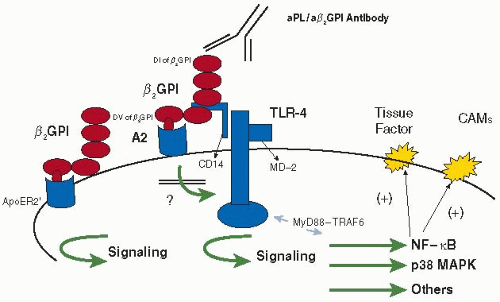
aPL antibodies can injure and/or activate cultured vascular endothelial cells.93, 94, 95, 96 An indicator that this may occur in vivo is that circulating levels of endothelial-derived microparticles are increased in the blood of patients with LAs.97 Antibody binding to β2GPI on the endothelial surface can also trigger signaling events that increase expression of tissue factor and adhesion molecules. This appears to occur with the involvement of annexin A2, which serves as a receptor for β2GPI on vascular endothelium.98 Binding of anti-β2GPI, which cross-links the β2GPI-annexin A2 complex, can activate signaling pathways through the Toll-like receptor-4 (TLR-4) adaptor molecule, also called myeloid differentiation factor 88.99 This leads to the upregulation of the transcription factor and nuclear factor-κB (NF-κB) and subsequent increase in the release of cytokines such as tumor necrosis factor-α (TNF-α), interleukin (IL)-1, IL-6, IL-8, and surface expression of tissue factor and cell adhesion molecules (FIGURE 101.4). A mutation in murine TLR-4 that results in loss of ability to bind lipopolysaccharide (LPS) attenuated the prothrombic state in wildtype mice injected with aPL antibodies.100 However, a direct interaction between TLR-4 and anti-β2GPI/β2GPI complexes has yet to be confirmed. This endothelial activating effect has also been described for antibodies that bind annexin A2 directly,81, 101— that is, without the involvement of β2GPI.


FIGURE 101.4 β2GPI receptors on the endothelial cell membranes and their signaling pathways. Anti-β2GPI/β2GPI complexes bind through domain V of β2GPI to ApoER2 and annexin A2 (A2) to activate downstream signaling pathways and activate p38 mitogen-activated protein kinase and NF-κB, leading to the upregulation of tissue factor and cellular adhesion molecules and a proinflammatory/prothrombotic phenotype. Note: annexin A2 does not have a transmembrane domain; therefore other membrane proteins such as TLR-4 and ApoER2 may act as accessory molecules. (Modified from Romay-Penabad Z, Montiel-Manzano MG, Shilagard T, et al. Annexin A2 is involved in antiphospholipid antibody-mediated pathogenic effects in vitro and in vivo. Blood 2009;114:3074-3083.)


Apolipoprotein E Receptor 2 on the Endothelial Surface

ApoER2′, whose role on platelets was described above, may also serve as receptor for anti-β2GPI/β2GPI complexes on endothelial cells102 and monocytes,103 where it can also trigger tissue factor and cell adhesion molecule expression (FIGURE 101.4). Evidence from ApoER2′-/--knockout mice has supported this mechanism in mediating the prothrombotic effects of aPL antibodies.104 In accordance with this hypothesis, a soluble binding domain I of ApoER2′ that interfered with the binding of dimerized β2GPI to ApoER2′ inhibited the effect of aPL antibodies on wildtype, that is, ApoER2′+/+, mice.104


Activation of Complement Cascade

There is evidence from an animal model that complement activation can play a role in the APS disease process. Blockade of complement activation using a C3 convertase inhibitor or genetic deletion of C3 protected mice from pregnancy complications induced by aPL antibodies.105, 106, 107 These effects on pregnancy appeared to be due to stimulation of tissue factor expression by myeloid cells108 and appear to involve proteinactivated receptor-2 signaling.109 Those results suggested the interesting concepts that thrombin can be pathogenic via both its stimulation of fibrin formation and via its stimulation of signaling pathways, and that complement activation can be pathogenic in two ways—via both direct injury and downstream signaling.


Insights into the Pathogenesis of Pregnancy Complications in APS

Although there has been some controversy whether the pregnancy complications of APS are attributable to increased coagulation, a significant body of evidence supports this concept. Prothrombin activation fragment F1.2, a marker for thrombin generation, is increased in pregnant patients with aPL antibodies and a previous history of pregnancy losses, compared to control healthy aPL antibody-negative pregnant women.110 Histologic abnormalities occur in many, but not all, placentas of APS patients.111 Evaluation of placentas from aPL-positive women revealed that about half of women with aPL antibodies but without a prior history of fetal loss had evidence for uteroplacental vascular pathology, about half had evidence of thrombotic occlusion, and about one-third had chronic villitis and/or decidual plasma cell infiltrates.112, 113 These events may be due to the effects of aPL antibodies on placental annexin A5, by the triggering of tissue factor expression by aPL antibodies, and as described above by complement activation.

aPL antibodies have been implicated in mechanisms that do not involve thrombotic or inflammatory pathways. The antibodies can directly target the maternal decidua and affect the expression of integrins and cadherins interfering with decidual adhesion and invasion.114 aPL antibodies were found to impair endometrial differentiation and reduce expression of complement regulatory proteins (DAF/CD55).115 The antibodies may impact upon endometrial endothelial cell angiogenesis by reducing vascular endothelial growth factor (VEGF) and matrix metalloproteinases as well as NF-κB DNA-binding activity.116 aPL antibodies may be involved in defective placentation; they were found to bind to trophoblast monolayers and reduce heparin-binding epidermal growth factor, which is important for blastocyte implantation, in placental tissue.117


Genetic Aspects of the APS Disease Process

Genetic factors are likely to be important determinants of susceptibility to development of aPL antibodies and APS. Several cases of familial APS have been described.118, 119 One study of seven families with 30 individuals, who met consensus criteria for APS, concluded that the inheritance pattern of aPL antibodies appeared to be autosomal dominant.120 In other studies, genes in the major histocompatibility complex (MHC) have been associated with autoimmune disorders including specific MHC antigens. In patients with SLE, HLA-DPB1 alleles have been associated with anti-β2GPI.121

Investigators have begun to apply genomic studies to identify specific pathogenic mechanisms. A recent genomic study done on peripheral blood mononuclear cells of aPL antibody-positive patients reported a gene-expression pattern that appeared to correlate with a predisposition toward developing thrombosis122. Some of the genes that were associated with APS encoded proteins are known to be involved in thrombosis—for example, apolipoprotein E (ApoE), coagulation factor X, and thromboxane-while others had no known connection to prothrombotic mechanisms.


CLINICAL MANIFESTATIONS OF THE ANTIPHOSPHOLIPID SYNDROME


Systemic Vascular Thrombosis

Although patients may present with venous and/or arterial thromboembolism in virtually any portion of the vasculature, the majority tend to have venous thromboembolic events. In one study, 59% of patients had thrombi limited to the venous circulation, 28% had solely arterial thrombi, and 13% had both types of events.123 Deep vein thrombosis (DVT) of the lower extremities was the most common finding, occurring in about half of the patients; other sites of venous thrombotic events included pulmonary embolism, thoracic veins (superior vena cava, subclavian vein, or jugular vein), and abdominal or pelvic veins.123 As in patients without APS, thrombosis may occur spontaneously or in the presence of risk factors such as estrogen hormone replacement therapy, oral contraceptives, pregnancy, the postpartum state,124, 125 vascular stasis, surgery, or trauma. Some patients with venous thrombosis—but generally not with arterial thrombosis126—also have concurrent hereditary thrombophilic conditions.126, 127, 128, 129 The most significant risk factor for a future thromboembolic event is having a clinical history of a thromboembolic event. The risk of recurrence increases to approximately 30% in patients with a first episode of venous thromboembolism (VTE) and aCL antibodies during 4 years of observation from the initial event.130 The risk of recurrence and death appears to be increased in patients who have increased levels of antibodies130, 131 and with the presence of an LA. Antibodies against β2GPI domain I significantly increase the risk of thrombosis, compared to antibodies and/or LA that are domain I independent.39


Catastrophic Antiphospholipid Syndrome

Rare patients develop a catastrophic form of APS, with widespread vascular occlusions and a high mortality (Table 101.3).132 An inciting cause, such as surgery or infection, is suspected in about half of the patients. CAPS is defined by severe multiorgan ischemia/infarction, usually with concurrent microvascular thrombosis. Patients can present with massive VTE, along with respiratory failure due to acute respiratory distress syndrome and diffuse alveolar hemorrhage, stroke, abnormal liver enzymes, renal impairment, adrenal insufficiency, and cutaneous infarction. For recent information on the clinical characteristics of CAPS, the interested reader is referred to (http://www.med.ub.es/mimmun/forum/caps.htm) “The Registry of the European Forum on Antiphospholipid Antibodies for Patients with CAPS.” At the time of writing, this registry included 280 patients of whom 72% were females, with a mean age of 37 (range 11 to 60); 46% had primary APS—that is, without other autoimmune disorders—and 40% had SLE. In about half of the patients, CAPS was the initial presentation for APS or thrombosis. Seventy percent of the patients had noninflammatory thrombotic microangiopathy as a main pathologic finding. In contrast to APS, which usually manifests as thrombosis in a large vein or artery, CAPS patients also had a susceptibility to intra-abdominal thrombotic complications affecting the kidneys, adrenal glands, splenic, intestinal, and mesenteric or pancreatic vasculature. Dermatologic abnormalities such as livedo reticularis, purpura, and necrosis occur in about half of CAPS patients. Finally, CAPS patients were more likely to have evidence of infarction at unusual sites—for example, testicular/ovarian infarction, prostate necrosis, acalculous cholecystitis, bone marrow infarction, esophageal rupture, gastric ulceration, colonic ulcerations, thrombotic pancreatitis, and adrenal infarction.133 Although the mortality of the disorder is high, patients who recover from CAPS rarely experience a recurrence.


Pediatric Antiphospholipid Syndrome

APS is increasingly recognized to be a significant cause of thrombosis in children134 and, as with adults, diverse clinical features are common. A European registry recently reviewed the initial 121 registered cases of pediatric APS and found thrombotic manifestations similar to those experienced by adults. Differences between primary and secondary APS were noted in the pediatric registry, with the primary APS patients being younger and having a higher frequency of arterial thrombotic events and the secondary APS patients being older and having a higher frequency of venous thrombotic events.135 CAPS appears to be much less common in children than in adults.136

Only gold members can continue reading. Log In or Register to continue

Stay updated, free articles. Join our Telegram channel

Jun 21, 2016 | Posted by in HEMATOLOGY | Comments Off on The Antiphospholipid Syndrome

Full access? Get Clinical Tree

Get Clinical Tree app for offline access