Testicular Cancer
Lawrence H. Einhorn
I. EPIDEMIOLOGY AND ETIOLOGY
A. Epidemiology
1. Incidence. Testicular cancer constitutes only 1% of all cancers in men but is the most common malignancy that develops in men between the ages of 20 and 40 years. About 8,000 new cases are diagnosed annually in the United States.
2. Racial predilection. The incidence of testicular cancer in blacks is one-sixth that in whites. Asians also have a lower incidence than whites.
3. Bilateral cancer of the testis occurs in about 2% of cases.
B. Etiology
1. Cryptorchidism. Male patients with cryptorchidism are 10 to 40 times more likely to develop testicular carcinoma than are those with normally descended testes. The risk for developing cancer in a testis is 1 in 80 if retained in the inguinal canal and 1 in 20 if retained in the abdomen. Surgical placement of an undescended testis into the scrotum before 6 years of age reduces the risk for cancer. However, 25% of cancers in patients with cryptorchidism occur in the normal, descended testis.
2. Testicular feminization syndromes increase the risk for cancer in the retained gonad by 40-fold. Tumors in these patients are often bilateral.
3. Other risk factors. The magnitude of other suggested risk factors, such as a history of orchitis, testicular trauma, or irradiation, is not known.
II. PATHOLOGY AND NATURAL HISTORY
A. Histology. Immunophenotypes of germ cell tumors are shown in Appendix C.4.VI.
1. Nearly all cancers of the testis in members of the younger age groups originate from germ cells (seminoma, embryonal cell, teratoma, and others). Other types, which account for <5% of cases, include rhabdomyosarcoma, lymphoma, and melanoma. Rarely, Sertoli cell tumors, Leydig cell tumors, or other mesodermal tumors develop.
2. In men >60 years of age, 75% of neoplasms are not germinal cancers. Lymphomas are the most common testicular tumors in this age group.
3. Metastatic cancer to the testis is most often associated with small cell lung cancer, melanoma, or leukemia.
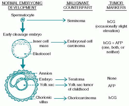
B. Histogenesis. Each type of germinal cancer is thought to be a counterpart of normal embryonic development (Fig. 12.1). Seminoma is the neoplastic counterpart of the spermatocyte. The tissues of the early cleavage stage are the most undifferentiated and pluripotential and give rise to both the embryo and the placenta; the malignant counterpart is embryonal cell carcinoma. Teratomas are the neoplastic counterparts of the developing embryo. Choriocarcinoma is actually a more highly undifferentiated cancer; its aggressive biologic behavior reflects the capacity of its normal counterpart (the placenta) to invade blood vessels. The histologic similarity between germ cell cancer and normal embryology is illustrated by the following observations:
1. Pure choriocarcinomas usually metastasize only as choriocarcinomas, especially hematogenous. However, patients with pure choriocarcinoma in
the orchiectomy specimen can have tumors in retroperitoneum comprised of both choriocarcinoma and teratoma.
the orchiectomy specimen can have tumors in retroperitoneum comprised of both choriocarcinoma and teratoma.
 Figure 12.1. Histogenesis of testicular neoplasms. Embryonic counterparts and tumor marker production are shown. (hCG, human chorionic gonadotropin; AFP, α-fetoprotein) |
2. Seminomas usually metastasize as seminomas; those that do not are believed to represent mixed tumors undetected on the original histologic examination.
3. Metastases from embryonal carcinomas may be found to consist of teratoma or choriocarcinoma elements.
4. In metastases from mixed tumors, chemotherapy destroys the rapidly growing, drug-sensitive cell elements. The drug-resistant teratomatous elements persist after chemotherapy and require surgical resection.
C. Natural history. The natural history of testis cancer varies with the histologic subtype. Both blood-borne and lymphatic metastases occur. Lymphatic drainage usually occurs in an orderly progression. A right-sided primary will spread lymphatically to the interaortocaval nodes and a left-sided primary to paraaortic retroperitoneal nodes. Previous surgery, such as scrotal contamination with scrotal orchiectomy, disrupts normal lymphatic drainage patterns and can cause inguinal nodal metastases.
1. Seminoma (40% to 50% of testicular cancers) occurs in an older age group than other germ cell neoplasms, most commonly after the age of 30 years. Sixty percent of patients with cryptorchidism who develop testicular cancer have seminoma. Seminomas tend to be large, show little hemorrhage or necrosis on gross inspection, and metastasize in an orderly, sequential manner along draining lymph node chains. About 25% of patients have lymphatic metastases, and 1% to 5% have visceral metastases at the time of diagnosis. Metastases to parenchymal organs (usually lung or bone) can occur late. Seminoma is the type of testicular cancer most likely to produce osseous metastases.
a. Spermatocytic seminoma (4% of seminomas) occurs mostly after the age of 50 years and is the most common germ cell tumor after the age of 70 years. It is more often bilateral (6% compared with 2%) and appears to have a much lower incidence of both lymphatic and parenchymal metastases (even to draining lymph nodes) when compared with typical seminoma. These patients are usually cured with orchiectomy alone.
2. Pure choriocarcinoma (<0.5% of testicular cancers) metastasizes rapidly through the bloodstream to lungs, liver, brain, and other visceral sites. They have very high serum human chorionic gonadotropin (hCG) levels with normal α-fetoprotein (AFP) levels.
3. Yolk sac tumors are common cancers in children and have a relatively unaggressive clinical course. Yolk sac elements in testicular cancer found in adult patients, on the other hand, portend a worse prognosis compared with that of children. Pure yolk sac tumors have elevated AFP and normal hCG.
4. Embryonal cell carcinoma can be associated with elevated serum hCG, AFP, both, or neither tumor marker. When patients have clinical stage I testicular cancer with predominantly embryonal cell carcinoma, they are more likely to have occult microscopic disease in the retroperitoneum or elsewhere. Vascular invasion also predicts metastatic spread.
5. Teratoma appears pathologically inert as it is associated with cartilage, glandular, and glial tissue. Teratoma of and by itself usually does not have the capacity to metastasize, but it is often associated with embryonal cell carcinoma, choriocarcinoma, yolk sac tumor, and seminoma in the testis and can metastasize as a template. In that situation, chemotherapy will often completely eliminate the nonteratomatous elements, but the teratoma will remain and require surgical resection for cure. When teratoma remains after chemotherapy, it can and will grow by local extension and can even cause death from teratoma alone. In addition, because teratoma is pluripotential tissue that can differentiate along endodermal, ectodermal, or mesoderm elements, it can undergo malignant transformation. The most common of these is mesodermal differentiation to sarcomatous elements associated with teratoma. Malignant transformation of teratoma does have the potential to metastasize. These elements can sometimes briefly respond to chemotherapy directed at the dominant cell type of malignant transformation.
6. Rare testicular tumors
a. Sertoli cell and Leydig cell tumors are not germ cell tumors and are not associated with elevated serum hCG or AFP. They vary in malignant potential, but all can metastasize. Size, necrosis, and mitotic index predict potential for spread and need for retroperitoneal lymph node dissection. Leydig cell tumors rarely respond to chemotherapy. Sertoli cell tumors may benefit from platinum combination chemotherapy. These tumors are inhibin positive on immunohistochemical stain.
b. Rhabdomyosarcoma of the testis occurs most often before 20 years of age. Its clinical behavior is similar to that of embryonal carcinoma; metastases to draining lymph nodes and lung are common at the time it first appears. They are usually paratesticular.
III. DIAGNOSIS
A. Symptoms and signs. Postorchiectomy, most patients will have an otherwise normal history and physical examination.
1. Symptoms
a. Mass and pain. The most common symptom of testicular cancer is a painless enlargement, usually noticed during bathing or after a minor trauma.
Painful enlargement of the testis occurs in 30% to 50% of patients and may be the result of bleeding or infarction in the tumor. Acute pain in a patient with a cryptorchid testis suggests the possibility of torsion of a testicular cancer.
Painful enlargement of the testis occurs in 30% to 50% of patients and may be the result of bleeding or infarction in the tumor. Acute pain in a patient with a cryptorchid testis suggests the possibility of torsion of a testicular cancer.
b. Acute epididymitis. Nearly 25% of patients with mixed teratoma and embryonal cell tumor present with findings indistinguishable from acute epididymitis. The testicular swelling from tumor may even decrease somewhat after antimicrobial therapy.
c. Gynecomastia due to high levels of serum hCG is rarely a presenting sign.
d. Infertility is the primary symptom in about 3% of patients.
e. Back pain from retroperitoneal node metastases is a presenting feature in 10% of patients.
f. Other presenting symptoms. Thoracic symptoms are rare even when extensive pulmonary metastases are present. When there is extensive replacement of pulmonary parenchyma, patients may develop hemoptysis, chest pain, or dyspnea.
2. Physical findings
a. Scrotum. A testicular mass is nearly always present. The testis should be palpated using bimanual technique; the finding of irregularity, induration, or nodularity is indication for further evaluation, including a testicular ultrasound to look for a hypoechogenic mass.
b. Lymph nodes. Patients must be carefully examined for lymphadenopathy, particularly in the supraclavicular region. Scrotal contamination, such as following testicular biopsy, vasectomy, or herniorrhaphy, alters the normal lymphatic drainage; as a result, ipsilateral inguinal nodes may become involved. Large retroperitoneal masses may be palpable on abdominal examination.
c. Breasts. Gynecomastia is associated with tumors that secrete high levels of hCG.
B. Differential diagnosis
1. Hydroceles are usually benign, but about 10% of testicular cancers are associated with coexisting hydroceles. The finding of a hydrocele in a young man should increase suspicion for an associated neoplasm.


