TECHNIQUE OF ILIAC CREST BIOPSY
PRETREATMENT OF THE PATIENT WITH TETRACYCLINE
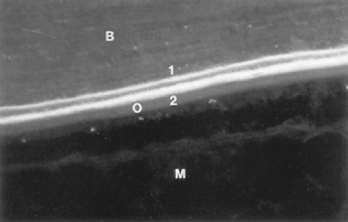
The information obtainable from an iliac crest biopsy is significantly increased by the use of in vivo markers of bone formation, such as the tetracycline antibiotics.1 Approximately 3 weeks before the scheduled biopsy, tetracycline is administered for 2 days. This is followed by an interval of 12 drug-free days and then a 4-day course of tetracycline (2:12:4 sequence). The tetracycline is incorporated into bone at sites of new bone formation, binding irreversibly to hydroxyapatite at the mineralization front. When thin biopsy sections are subsequently viewed under violet or blue light, the tetracycline fluoresces so that bright lines (“labels”) are visible at the formation sites (Fig. 55-1). At sites where bone formation was ongoing during the entire labeling sequence, two tetracycline labels are present, corresponding to the two discrete periods of tetracycline exposure. At sites where formation either commenced or ended in the interval with no tetracycline administration, only single fluorescent labels are visible. At least 5 days should elapse between the last day of tetracycline administration and the date of the biopsy to prevent the last label from diffusing out.
Demeclocycline, tetracycline, and oxytetracycline all are effective labeling agents, although demeclocycline appears to produce more intense fluorescent bands. Demeclocycline, 600 mg per day (4 × 150-mg tablets), is taken on an empty stomach; dairy products and antacids containing aluminum, calcium, or magnesium impair absorption. Tetracyclines should not be given to children younger than 8 years of age or to pregnant women because it is incorporated into growing teeth and causes discoloration. Although tetracycline is generally well tolerated, some patients react adversely to these drugs with nausea, vomiting, or diarrhea. All patients should avoid direct exposure to sunlight or ultraviolet light during the time of administration because of potential skin phototoxicity.
THE SITE AND THE SURGICAL PROCEDURE
The anterior iliac crest is the preferred site for several reasons: (a) it is easily accessible, making the surgical procedure simple and safe; (b) the bone sample obtained consists of both trabecular and cortical bone (Fig. 55-2A); (c) the amount of trabecular bone correlates with that of the axial skeleton (vertebral bodies); and (d) a large body of histomorphometric data has been accumulated at this site in both normal persons and patients with a wide variety of bone diseases.2, 3, 4 and 5
Stay updated, free articles. Join our Telegram channel

Full access? Get Clinical Tree


