CLINICAL PRESENTATION AND ETIOLOGY
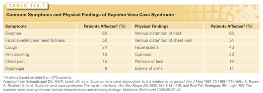
A subjective sensation of fullness in the head or objective facial swelling and dyspnea are common presenting symptoms (Table 119.1).6,7 The characteristic physical findings are venous distention of the neck (66%) and chest wall (54%), facial edema (46%), plethora (19%), and cyanosis (19%). These symptoms and signs may be aggravated by bending forward, stooping, or lying down. SVCS may be the earliest manifestation of invasive involvement of additional critical structures in the thorax such as the bronchi. Pleural effusions are common in SVCS. In one series, effusions were detected in 60% of patients, both in those with malignant etiology and in those with a nonmalignant cause.8 The majority are exudative and often chylous. Rarely do patients present with life-threatening symptoms like confusion, obtundation, stridor or syncope without precipitating factors, hypotension, or renal insufficiency.

Malignant disease is the most common cause of SVCS. The percentage of patients in different series with a confirmed diagnosis of malignancy varies from 60% to 86% (Table 119.2).7,9–12 In large series of lung cancer, SVCS was identified in 4% to 8.6% of the patients.13,14 Small-cell lung cancer (SCLC) and squamous cell carcinoma are the most common histologic subtypes because of their frequent central location.7,15,16 Lymphoma involving the mediastinum was the cause of SVCS in 2% to 21% (see Table 119.2). In a large series, most patients with lymphoma with SVCS had either diffuse large-cell lymphoma or lymphoblastic lymphoma.17 The presence of dysphagia, hoarseness, or stridor was a major adverse prognostic factor for patients with lymphoma who presented with SVCS. In a series of patients with primary mediastinal B-cell lymphoma with sclerosis, SVCS was present in 57% of patients.18 Other primary mediastinal malignancies that may cause SVCS are thymomas and germ cell tumors. SCLC, non-Hodgkin lymphoma (NHL), and germ cell tumors constitute almost half of the malignant causes of SVCS. Hodgkin lymphoma commonly involves the mediastinum, but it rarely causes SVCS. Breast cancer is the most common metastatic disease that causes SVCS in up to 11% of the cases.9,10 The prognosis of patients with SVCS strongly correlates with the prognosis of the underlying disease.

In recent years, nonmalignant conditions causing SVCS have been more often observed. When the data were collected from general hospitals, as many as 40% of patients had noncancerous causes of SVCS, most commonly due to thrombosis in the presence of central vein catheters or pacemakers.7,10,12,19 The increasing use of such catheters for the delivery of chemotherapy agents or hyperalimentation in conjunction with a common thromboembolic disposition in many patients with cancer contributes to the development of SVCS.19
Obstruction of SVC in the pediatric age group is rare and has a different etiologic spectrum, mainly iatrogenic, secondary to cardiovascular surgery for congenital heart disease, ventriculoatrial shunt for hydrocephalus, SVC catheterization for parenteral nutrition, and mediastinal fibrosis secondary to histoplasmosis.20,21
The modern treatment of SVCS has become disease-specific from the outset. Therefore, a clear diagnosis prior to initiation of emergency treatment such as mediastinal irradiation should be made. Mediastinal irradiation before a biopsy may preclude proper interpretation of the specimen in almost half of the patients and should be avoided.22 The safety of modern, minimally invasive diagnostic procedures such as bronchoscopy, mediastinoscopy, computed tomography (CT)-guided biopsy or supraclavicular lymph node biopsy has markedly improved, and concerns about risks from these procedures should not preclude proper diagnostic workup. In contrast to past opinions, there is little evidence to suggest that diagnostic procedures such as venographies, thoracotomies, bronchoscopies, mediastinoscopies, and lymph node biopsies carry an excessive risk in patients with SVCS.2 Therefore, patients should, if possible, undergo a complete staging workup prior to the initiation of treatment.
The most common radiographic abnormalities are superior mediastinal widening and pleural effusion apparent on chest X-ray. CT with intravenous contrast provides more detailed information about the SVC, its tributaries, and other critical structures, such as the trachea, the bronchi, the esophagus and the spinal cord.23 The additional anatomic information is necessary to more clearly determine the involvement of these structures that requires prompt action for relief of pressure. Although not fully evaluated, fluorodeoxyglucose-positron emission tomography scanning is useful in patients with SVCS caused by lymphoma or lung cancer, as it may significantly influence the design of the radiotherapy field and treatment intent (definitive versus palliative).2
Pathologic confirmation of a thoracic malignancy can be obtained as a cytologic specimen via endobronchial fine needle aspiration, or via CT-guided needle biopsy or mediastinoscopy. Endobronchial fine needle aspirations are as accurate as tissue diagnosis and supply sufficient malignant cells for cytologic evaluation in most cases of lung cancer.24 Mediastinoscopy has a very high success rate for providing a diagnosis and has a complication rate of approximately 5%.25 Reports by several authors on using mediastinoscopy for patients with SVCS whose histologic diagnosis could not be established with less invasive techniques confirmed the safety and high diagnostic yield of mediastinoscopy.25,26 No perioperative mortality was recorded, and the diagnostic yield was excellent. Percutaneous transthoracic CT-guided fine needle biopsy is an effective and safe alternative to an open biopsy or mediastinoscopy.27 In the presence of a pleural effusion, thoracocentesis can establish the diagnosis of malignancy in 71% of patients with malignancy.8 A thoracoscopic biopsy or thoracotomy is diagnostic if all other procedures have failed.
DISEASE-SPECIFIC MANAGEMENT AND OUTCOMES
The goal of treatment of SVCS depends on the cause and—in malignancies—the stage of the disease. Malignant tumors are potentially curable if they present at a nonmetastatic stage, even in the presence of SVCS. The treatment of SVCS should be selected according to the histologic disorder and stage of the primary process. During the diagnostic process, the patient can benefit from bed rest and oxygen administration. Some clinicians advocate the use of diuretics and/or corticosteroids if the patient is uncomfortably symptomatic. This should only be considered after pathologic confirmation of the cause of SVCS in symptomatic patients who require urgent palliation, as steroid initiation prior to pathologic confirmation can significantly hamper efforts for an accurate diagnosis, especially with lymphomas. Prophylactic anticoagulation in the absence of thrombosis is of no proven benefit and may interfere with diagnostic procedures.
When the therapeutic goal is only palliation of SVCS, or when urgent treatment of the venous obstruction is required, direct opening of the occlusion should be considered. Endovascular stenting and angioplasty with possible thrombolysis may provide prompt relief of symptoms before more cancer-specific therapy.3,28
Chemotherapy alone or in combination with thoracic irradiation therapy is the standard treatment for SCLC and is effective in rapidly improving the symptoms of SVCS.14–16,29 No significant difference in response rates to chemotherapy or radiation has been detected in most studies. Response rates to chemotherapy range from 73% to 93% and to radiation from 43% to 94%.14,29 Relief of SVCS typically occurs within 7 to 10 days after initiation of therapy. In patients with SCLC with recurrent or persistent SVCS, additional chemotherapy and/or radiotherapy is still likely to relieve symptoms.29
A review of SVCS in lung cancer indicated that chemotherapy relieved SVCS in 59% of patients with non-SCLS (NSCLC); radiotherapy relieved the obstruction in 63% of patients with NSCLC.28 Nevertheless, in almost 20% of the patients the obstruction recurred. Response to radiotherapy was higher in patients who had received prior therapy (94% versus 70%).28
The primary treatment for NHL is chemotherapy, as it has both local and systemic activity. Local consolidation with radiation therapy is beneficial in patients with early stage diffuse large-cell lymphoma, particularly if the mass is bulky.
In a report of 36 patients with SVCS secondary to NHL, all patients achieved complete relief of SVCS symptoms within 2 weeks of the onset of any type of treatment, whether treated with chemotherapy alone, chemoradiation, or radiotherapy alone.17 Eighteen of twenty-two patients (81%) with large-cell lymphoma achieved complete response. However, relapse was common, and the median survival was only 21 months.
Patients with nonmalignant SVCS often have longstanding symptoms before they seek medical advice, and the time to establish the diagnosis and survival are typically markedly longer.10
Stay updated, free articles. Join our Telegram channel

Full access? Get Clinical Tree


