Small Cell and Neuroendocrine Tumors of the Lung


Small Cell and Neuroendocrine Tumors of the Lung


M. Catherine Pietanza

Lee M. Krug

Abraham J. Wu

Mark G. Kris

Charles M. Rudin

William D. Travis


SMALL CELL LUNG CANCER


Incidence and Etiology

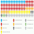
Although the incidence of small cell lung cancer (SCLC) is declining, it remains a worldwide public health problem. The Surveillance, Epidemiologic, and End Results (SEER) database reports the proportion of SCLC cases among all lung cancers in the United States decreased from 17% to 13% in the past 30 years (Fig. 38.1),1 whereas the age-adjusted rates per 100,000 decreased from 10.65 in 1990 to 6.74 in 2010.2 Among the predicted 228,190 lung cancer cases in the United States in 2013,3 an estimated 29,665 cases of SCLC were diagnosed.

Only 2% to 3% of patients with this malignancy are never smokers4,5; thus, as tobacco exposure causes SCLC in over 97% of cases, its incidence rates mirror smoking patterns. Peak cigarette consumption occurred in the 1960s, but declined following the Surgeon General’s report linking smoking to cancer and the subsequent ban on tobacco advertising on television.6 The percentage of men who smoke decreased from 50% in 1965 to 21.6% in 2011, which is a much greater proportional reduction than in women, who went from a rate of 32% to 16.5% during the same time period.6,7 Correspondingly, the incidence of SCLC in men peaked in 1984 and since has been trending steadily down, whereas in women, the incidence peaked later and only has declined slightly.8 The gender gap has narrowed such that currently about half of the patients diagnosed with SCLC are women (see Fig. 38.1).


Anatomy and Pathology

Neuroendocrine tumors of the lung encompass a spectrum of tumors, including low-grade typical carcinoid, intermediate-grade atypical carcinoid, high-grade large cell neuroendocrine carcinoma (LCNEC), and SCLC.9 Because of their shared neuroendocrine properties, these tumors have common morphologic, ultrastructural, immunohistochemical, and molecular features. Despite this, there are also important differences in clinical, epidemiologic, histologic, and molecular characteristics.

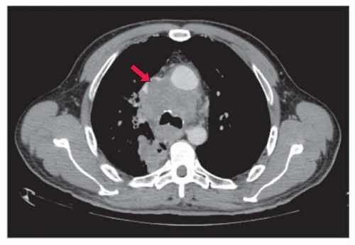
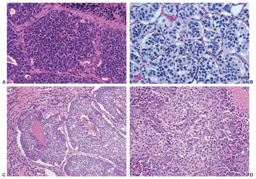
SCLC is readily diagnosed on small specimens such as bronchoscopic biopsies, fine-needle aspirates, core biopsies, and cytology. The diagnosis of SCLC is based primarily on light microscopy (Fig. 38.2A): dense sheets of small cells with scant cytoplasm, finely granular nuclear chromatin, inconspicuous or absent nucleoli, and frequent mitoses. Necrosis is common and frequently shows large areas. The tumor cells usually measure less than the diameter of three small resting lymphocytes. They are round to be fusiform in shape and have scant cytoplasm. The nuclear chromatin is finely granular and nucleoli are inconspicuous or absent.9,10,11 The mitotic rate is characteristically high, averaging 60 to 80 per 2 mm2. Crush artifact is a frequent finding in small transbronchial or mediastinal biopsy specimens and can make pathologic interpretation difficult. The tumor cells of SCLC also have a tendency to show a streaming artifact. This can also occur with non-small-cell lung cancer (NSCLC), lymphoma, and chronic inflammation. In surgically resected specimens where the tumor cells achieve better fixation, the cells of SCLC appear larger than in small biopsies.10,12 When a component of NSCLC, including adenocarcinoma, squamous cell carcinoma, large cell carcinoma, spindle cell carcinoma, and giant cell carcinoma, is present, the term combined SCLC is used with mention of the specific histology of the non-small-cell component.9,10 In resected specimens, combined SCLC may occur in up to 28% of cases.10 To diagnose combined SCLC and large cell carcinoma, the large cell carcinoma component must comprise at least 10% of the overall tumor.9,10

The following immunohistochemical stains are helpful in difficult cases. A pancytokeratin antibody such as AE1/AE3 is useful to confirm if the tumor is a carcinoma. Neuroendocrine differentiation can be demonstrated using a panel of markers such as CD56, chromogranin, and synaptophysin. However, up to 10% of SCLCs may be negative for all neuroendocrine markers.13 A high proliferation rate of 80% to 100% should be seen with Ki-67. Thyroid transcription factor-1 (TTF-1) is positive in 70% to 80% of small cell carcinomas.14,15,16 Notably, TTF-1 can be positive in extrapulmonary small cell carcinomas, so it is not useful in determining the primary site of small cell carcinomas.17


Genetics

SCLC has been characterized by frequent inactivating mutations in the critical tumor suppressor genes TP53 (75% to 90%)18 and RB1 (60% to 90%).19,20 A mouse model with conditional inactivation of these two tumor suppressors in the lung generates lung tumors histologically and biologically similar to human SCLC.21

Recent reports including exome, transcriptome, and limited whole genome sequencing have provided insights into the fuller landscape of genetic alterations in SCLC.22,23 In addition to confirming TP53 and RB1 inactivation, these studies define other alterations of interest in SCLC, with potential therapeutic implications. One consistent finding from both reports was an exceptionally high degree of genomic alteration in this tumor type, including mutations, insertions, deletions, large scale copy number alterations, and gross inter- and intrachromosomal rearrangements. MYC family member alterations, including gene amplification of MYC, MYCN, and MYCL1, as well as a recurrent gene fusion involving MYCL1, are frequent in SCLC and may represent important drivers of SCLC oncogenesis. The tumor suppressor phosphatase and tensin homolog (PTEN) appears to be inactivated in approximately 10% of SCLC, and mutations of other factors in the same signaling pathway were also identified. Other alterations implicated as potential drivers in subsets of SCLC include amplification of the tyrosine kinase FGFR1 (in 6% of cases) and of the developmental regulator and transcription factor SOX2 (in up to 27% of cases). The therapeutic implications of the large majority of
the genetic alterations documented to date in SCLC have not been defined.


Figure 38.1 Patients diagnosed with small cell lung cancer as a percentage of all lung cancer cases and the breakdown by gender.

Several reports have described the rare phenomenon of transformation to SCLC as a mechanism of acquired resistance to epidermal growth factor receptor (EGFR) tyrosine-kinase inhibitors (TKI) in patients initially treated for EGFR-mutant lung adenocarcinomas.4 Of those cases with acquired resistance to EGFR TKIs, 3% to 14% will undergo SCLC transformation. Importantly, pure SCLC lack EGFR mutations and ALK rearrangements, even in those patients with this malignancy who are never smokers.4,24 However, in the rare cases of SCLC transformation as a mechanism of acquired resistance to EGFR TKIs, there is persistence of the original EGFR mutation in the tumors confirmed on biopsy. In all cases where SCLC has been documented as a mechanism of acquired resistance to EGFR TKIs, the original tumor was a pure adenocarcinoma prior to EGFR TKI treatment, and the transformation was validated by histologic examination and confirmed by expression of neuroendocrine markers.


Figure 38.2 Histologic appearance of neuroendocrine carcinomas. (A) Small cell carcinoma. (B) Typical carcinoid. (C) Atypical carcinoid. (D) Large cell neuroendocrine carcinoma.


Screening

Screening computed tomography (CT) scans detect NSCLCs at an earlier stage compared to chest x-ray (CXR), or no screening, and can decrease lung cancer-specific mortality in heavy smokers.25 However, the natural history of SCLC is characterized by rapid tumor growth and early metastatic spread. As such, the diagnosis of SCLC in an asymptomatic patient is rare.
The National Lung Screening Trial (NLST) enrolled 53,454 Americans age 55 to 74 years with a history of at least 30 packyears of cigarette smoking, and randomly assigned them to undergo three annual screenings with either low-dose CT scans (LDCT) or standard CXR. LDCT screening identified a preponderance of adenocarcinomas, including many with bronchioloalveolar subtype. SCLCs were not detected at early stages by either LDCT or CXR in the NLST. Although the study demonstrated a reduction in lung-cancer specific mortality compared to CXR, the similar rates and patterns of detection of SCLC make it unlikely that LDCT screening will reduce mortality specific to SCLC. Therefore, screening for SCLC by any method is not recommended.25


Staging

A simple two-stage system, introduced by the Veterans’ Administration Lung Study Group (VALSG), has historically been utilized instead of the tumor, node, metastasis (TNM) system employed for most other cancer types.44 In the VALSG system, limited stage is defined as disease confined to one hemithorax
that can be “encompassed” in a “tolerable” radiation field. These patients currently are treated with a combined modality approach. All other patients are considered to have extensive-stage disease. At presentation, approximately two-thirds of patients with SCLC have extensive disease and one-third have limited-stage disease.1

In the VALSG staging system, the appropriate classification of ipsilateral pleural effusion, supraclavicular lymphadenopathy (ipsilateral or contralateral), or contralateral mediastinal lymphadenopathy as either limited or extensive stage remains controversial. Some large series have not found a survival difference between patients with an isolated ipsilateral pleural effusion and other patients with limited SCLC.45 However, analyses of two large cooperative group databases, which included over 4,000 patients, showed that the survival of individuals with an isolated effusion was similar to that of patients with extensive disease.46,47 In clinical practice, it is assumed that an effusion is malignant unless three criteria are met: the fluid is transudative, nonhemorrhagic, and cytologically negative on repeated examinations. Patients with a malignant effusion are appropriate to exclude from a combined modality treatment because hemithoracic radiotherapy to encompass the entirety of the pleura is impractical.

The presence of supraclavicular lymphadenopathy commonly is associated with extensive disease but, when encountered in patients with otherwise limited disease (5% of cases), carries a trend toward poorer survival.47 Contralateral mediastinal involvement also is usually classified as limited-stage disease. However, two studies that evaluated twice-a-day radiation regimens excluded patients with contralateral hilar disease to reduce the normal lung volume irradiated and the risk for toxicity.48,49

For patients who appear to have limited-stage SCLC, some additional tests may be appropriate to confirm this assessment. Unilateral iliac crest bone marrow aspiration and biopsy are still a routine part of staging for many oncologists and should be performed in limited-stage patients with elevations of serum lactate dehydrogenase (LDH)50,51 and evidence of myelophthisis (nucleated red blood cells, leukopenia, or thrombocytopenia) on the peripheral blood smear. If there is evidence of a pleural effusion, a thoracentesis or thoracoscopy may help confirm that the effusion is nonbloody, transudative, and cytologically negative. Effusions too small to permit image-guided sampling should not be considered in staging.52 Osseous abnormalities seen on positron-emission tomography (PET) or bone scan require confirmation with magnetic resonance imaging (MRI), CT scan, or biopsy if they represent the only disease site that makes a patient extensive stage.

For the American Joint Committee on Cancer (AJCC) seventh edition, the use of a TNM staging system for SCLC has been revisited. To establish the accuracy of outcomes based on stage, cases of completely resected SCLC were staged using the same definitions as used for NSCLC.53 The use of pathologic stage was necessary to accurately stage the patients used for this analysis. More favorable outcomes of patients have been reported in patients previously classified as very limited disease (i.e., no evidence of mediastinal metastases by CT or mediastinoscopy) treated with chemotherapy and radiation, as compared with other patients with limited-stage disease.54,55,56 The use of the TNM system is best applied for cases of early stage disease and may have less relevance for the majority of patients presenting with metastatic disease.


Clinical and Serologic Predictive and Prognostic Factors

Multivariable analyses suggest that performance status is a strong and reproducible predictive and prognostic factor.47,57,58 Poorer performance status can additionally identify individuals at higher risk for treatment-related complications. Several other clinical parameters have been proposed. Female gender has been associated with improved response and survival in patients with SCLC.47,57,58 Older age (variably defined) has not been identified as an adverse prognostic factor in patients with SCLC in most59,60,61,62 but not all36,46,47 series. Older age has been associated with decreased performance status and more comorbid illnesses and often results in compromised chemotherapy dose intensity,63,64 which may partially explain its prognostic implications. Certain metastatic sites, such as the liver,65,66,67 the brain,66,68 bone marrow,67 and bone,68 as well as the total number of metastatic sites involved,47 have been found to be of prognostic significance for patients with extensive-stage disease. Paraneoplastic Cushing syndrome has been correlated with a poor response to therapy and short survival.37 Continued use of tobacco during combined modality therapy was identified as an adverse prognostic factor in a group of 186 patients with limited disease.69 Elevation of serum LDH is found in 33% to 57% of all patients with SCLC and up to 85% of patients with extensive-stage disease and is a strong prognostic and predictive factor.46,57,65,66,68,70,71,72 Serum LDH elevation is associated with the presence of bone marrow involvement.50 Although many other serum markers have been proposed to have prognostic significance, including neuron-specific enolase,61,66 chromogranin, and precursors of gastrin-releasing peptide,73,74 none have been strong and reliable enough to warrant general use. Carcinoembryonic antigen (CEA) has been found to predict outcome in SCLC in multiple series.57,75,76


Management by Stage


General Recommendations for Initial Management

Once the pathologic diagnosis of SCLC is confirmed, a complete history and physical examination is the next step. Special attention should be paid to the cigarette smoking history. If a patient is a current smoker, he or she should be advised to quit immediately in the strongest terms and offered the most aggressive smoking cessation intervention available.69 If the patient is a never smoker, the pathologic diagnosis of SCLC should be reviewed as only 2% to 3% of never smokers develop SCLC.4,5 The National Comprehensive Cancer Network (NCCN) has compiled consensus guidelines for the initial evaluation of individuals with SCLC.52 A complete blood cell count (CBC) with platelet count, electrolytes, calcium, creatinine, blood urea nitrogen (BUN), liver function tests, and LDH are recommended. All patients should undergo a contrastenhanced CT scan of the chest, a gadolinium-enhanced MRI of the head, and whole-body PET or a bone scan. A PET scan can identify sites of metastases undetected by other modalities77,78,79 and can replace the bone scan.80

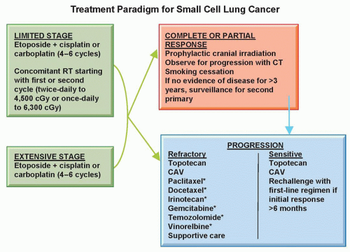
All fit patients (Karnofsky performance status greater than 60% or Eastern Cooperative Oncology Group [ECOG] performance status 0, 1, or 2) should initially receive combination chemotherapy with etoposide plus either cisplatin or carboplatin for four to six cycles (Fig. 38.4).52 Supportive data, specifics of chemotherapy regimens, duration of therapy, and alternatives for patients with contraindications or special needs are discussed in the sections that follow. Patients with limited-stage disease should receive the chemotherapy concurrently with twice-daily thoracic irradiation beginning with the first, second, or third cycle.49,81,82 Patients who achieve a response to chemotherapy should receive prophylactic whole-brain radiotherapy at the conclusion of chemotherapy or chemoradiotherapy.83 There is no routine recommendation for the treatment of patients who have a Karnofsky performance status of 50% or less or ECOG performance status 3 or 4. Because the toxicity of all treatment worsens and effectiveness lessens in patients with a low performance status, clinicians must carefully evaluate the agent(s) used and the appropriateness and goals of therapy individually. For many patients in this low performance status group, supportive care only and referral to hospice are the best options.


Figure 38.4 General treatment paradigm for small cell lung cancer. * Single agents evaluated in phase II trials with activity in SCLC.


Chemotherapy

Evolution of Chemotherapy Regimens. The sensitivity of SCLC to chemotherapy agents was recognized about 50 years ago, and the primary role of systemic treatment in SCLC was established early on. Alkylating agents, anthracyclines, vinca alkaloids, and antifolates all showed single-agent efficacy. In the 1980s, the epipodophyllotoxin, etoposide, and the platinum analogs, cisplatin and carboplatin, were introduced, and their activity ranged from 40% to 60% in previously untreated patients.84 Since then, numerous other chemotherapeutic agents have demonstrated activity in SCLC, but aside from the camptothecins, the drugs identified in the 1970s and 1980s remain the backbone of therapy. Ultimately, randomized trials of combinations demonstrated superior activity to single agents.85,86

Livingston et al.87 developed the cyclophosphamide, doxorubicin, vincristine (CAV) combination, and reported on 358 patients who received this combination followed sequentially by thoracic and brain irradiation. For patients with extensive disease, the complete response rate was 14%, the overall response rate was 57%, and the median survival was 26 weeks. For patients with limited disease, the rates were 41%, 75%, and 52 weeks, respectively. With these data, CAV became the standard chemotherapy regimen.

With the identification of etoposide as perhaps the most active agent, several modifications of the CAV regimen that included etoposide were tested. A randomized trial showed greater response duration and survival with cyclophosphamide, doxorubicin, and etoposide (CAE) compared with CAV.88 Hong et al.89 compared intensive cyclophosphamide and vincristine (CV) (with the dose of cyclophosphamide increased from 1,000 to 2,000 mg/m2) to CAV and to cyclophosphamide, etoposide, and vincristine (CEV) and reported that patients treated with CV had a shorter survival and experienced more myelosuppression than those treated on the other two arms. Five randomized trials have evaluated the addition of etoposide (CAVE) to the CAV regimen.90,91,92,93,94 In the three studies in which the doses of CAV were equivalent in each arm,90,91,92 a better response rate was evident in the arm that contained etoposide, in at least some patient subsets, with increased hematologic toxicity, although a nonstatistical improvement in survival was seen in only one study.90 Two studies intensified components of this regimen in the CAV arm compared to the CAVE arm,93 leading to equivalent myelotoxicity, response rates, and survival.

The etoposide/cisplatin (EP) regimen was tested in SCLC cases because this combination produced synergistic activity in preclinical systems. In addition, both agents could be given at full doses because of less myelosuppression with cisplatin. The first report by Sierocki et al.95 at Memorial Sloan-Kettering Cancer Center in 1979 demonstrated the activity of this combination in SCLC. Subsequent studies by Evans et al.96,97 reported response rates of 55% in patients previously treated with CAV and 86% in newly diagnosed patients. Einhorn et al.98 reported that two cycles of consolidation with EP, when added to the treatment of patients with limited disease who were responding to six cycles of CAV, produced longer survival than with CAV only. Three randomized trials have compared EP to cyclophosphamide, vincristine, and an anthracycline.99,100,101 Less myelosuppression occurred with EP, and, if given with radiation, patients experienced less esophagitis and interstitial pneumonitis. Furthermore, the largest trial showed that EP produced a better median (15 months versus 10 months) and 5-year (10% versus 3%) survival for patients with limited disease.101 Retrospective analyses and metaanalyses also support the superiority of cisplatin- or carboplatin-containing chemotherapy for SCLC.102,103,104 As a result, EP is now the standard first-line chemotherapy regimen for SCLC (Table 38.1).

Carboplatin has been substituted for cisplatin in SCLC chemotherapy regimens in an effort to decrease nonhematologic toxicities. Randomized trials comparing cisplatin and carboplatin suggest that they may have similar efficacy. The Hellenic Cooperative Oncology Group randomized 147 patients with either limited or extensive disease to receive etoposide 100 mg/m2 days 1 to 3, and cisplatin 100 mg/m2 or carboplatin 300 mg/m2.105 Concurrent radiation also was administered to responding patients starting with the third cycle. Response and survival were similar in the two arms. Nausea, vomiting, nephrotoxicity, and neurotoxicity were significantly lower in the patients who received carboplatin, as was grade 4 leukopenia. However, the sample size of this study is inadequate to confirm equivalent efficacy. A meta-analysis that evaluated individual subject data from four randomized trials with a total of 663 patients found that median overall survival (OS), median progression-free survival (PFS), and response rates were similar in the cisplatin and carboplatin arms. Although hematologic toxicities were higher in those patients that receive carboplatin, nonhematologic toxicities were increased in those that receive cisplatin.106 Based on these data, etoposide and carboplatin can
be considered an appropriate first-line regimen, particularly in patients who cannot tolerate cisplatin.


TABLE 38.1 Randomized Clinical Trials Comparing Etoposide and Cisplatin to Other Chemotherapy Regimens


Study (Ref.)


Stage


Treatment Arm


No. of Patients


Overall Response Rate (%)


Median Survival (months)


1-Year Survival (%)


2-Year Survival (%)


Fukuoka, et al.99


Limited and extensive


EP


97


78


9.9


NR


12


CAV


97


55


9.9


NR


10


CAV/EP alternating


94


76


11.8 (p = 0.056)


NR


21


Roth, et al.100


Extensive


EP


159


61


8.6


NR


NR


CAV


156


51


8.3


NR


NR


CAV/EP alternating


162


59


8.1


NR


NR


Sundstrom, et al.101


Limited and extensive


EP


218


NR


10.2 (p = 0.0004)


NR


14


Cyclophosphate, epirubicin, vincristine


218


NR


7.8


NR


6


Skarlos, et al.105


Limited and extensive


EP


71


69


12.5


NR


NR


Etoposide/carboplatin


72


78


11.8


NR


NR


Noda, et al.108


Extensive


EP


77


52


9.4


58


20


Irinotecan/cisplatin


77


65


12.8 (p = 0.002)


38


5


Hanna, et al.425


Extensive


EP


110


44


10.2


35


8


Irinotecan/cisplatin


221


48


9.3


35


8


Lara, et al.109


Extensive


EP


327


57


9.1


34


NR


Irinotecan/cisplatin


324


60


9.9


41


NR


Eckardt, et al.115


Extensive


EP


395


69


9.4


31


NR


Oral topotecan/cisplatin


389


63


9.2


31


NR


Miyamoto, et al.130


Limited and extensive


EP


45


78


12.8


53


15


Ifosfamide with EP


47


74


13.0


62


17


Loehrer, et al.131


Extensive


EP


84


67


7.3


27


5


Ifosfamide with EP


87


73


9.1 (p = 0.045)


36


13


Mavroudis, et al.135


Limited and extensive


EP


71


48


10.5


37


NR


Paclitaxel with EP


62


50


9.5


38


NR


Niell, et al.136


Extensive


EP


282


68


9.9


37


8


Paclitaxel with EP


283


75


10.6


38


11


EP, etoposide and cisplatin; CAV, cyclophosphamide, doxorubicin, and vincristine; NR, not reported.


More recently, platinum combinations with topotecan and irinotecan have emerged as potential regimens for initial therapy. Irinotecan was shown to have single-agent efficacy in Japanese studies.107 The Japan Clinical Oncology Group compared cisplatin and irinotecan to EP as initial treatment in extensive disease.108 The study was terminated after 154 of the planned 230 patients were enrolled because median (12.8 months versus 9.4 months) and 2-year (19.5% versus 5.2%) survival were significantly better in the group treated with cisplatin and irinotecan. Significant diarrhea occurred only in the irinotecan group, and myelosuppression was the most common toxicity in both groups and more frequent with EP. Two confirmatory studies were subsequently launched in the United States. In the first trial, the irinotecan/cisplatin schedule was modified in an effort to decrease toxicity, leading to equivalent response rates (48% for irinotecan/cisplatin versus 44% for EP), median time to progression (4 versus 5 months), and median OS (9 versus 10 months).425 The Southwest Oncology Group (SWOG) compared these two regimens with the same dose and schedule used in the Japanese trial and also showed, in a well-powered study, that outcomes are equivalent with the two regimens in Caucasian patients.109 High response rates have been reported in several trials using irinotecan and carboplatin with varied dosing schedules.110,111 A phase III trial comparing that regimen with etoposide/cisplatin showed improved survival, but the median survival of 7 months in the control arm, which used oral etoposide, was lower than expected.112 Many hypothesize that population-based polymorphisms in uridine 5′-diphospho (UDP)-glucuronosyltransferase (UGT1A1), the enzyme responsible for detoxifying SN-38, the active metabolite of irinotecan, may account for differences in toxicity and efficacy between Japanese and Americans.113 The regimen of topotecan plus cisplatin also has undergone phase II and III testing.114,115 Eckardt et al.115 reported the results of a randomized trial including 784 patients in which patients received oral topotecan (1.7 mg/m2 per day for 5 days) plus cisplatin, or standard etoposide and cisplatin. The response rates, median survival, and 1-year survival were identical. Severe neutropenia occurred more often with EP, but oral topotecan and cisplatin caused more anemia and thrombocytopenia.


Strategies to Improve Outcomes with Chemotherapy Regimens

Alternating Cycles of Combination Chemotherapy Regimens. The recognition of clonal heterogeneity within a tumor and the intolerability of treatment regimens that included more than four drugs due to overlapping toxicity led to trials of alternating chemotherapy combinations. The somatic mutation model developed by Goldie et al.116 predicted that the best probability of cure was achieved by the earliest possible introduction and the most rapid alternation of all active agents. If two equally effective non-cross-resistant
regimens were available, the model predicted that alternating between regimens every other cycle would be more effective than alternating after every three cycles or giving one regimen continuously for five cycles before switching to the second regimen.

Many randomized clinical trials have tested the concept of alternating multidrug combinations.100,117,118,119,120,121 The fact that the EP regimen was effective in patients who had progressed after cyclophosphamide-based chemotherapy suggested that these drug combinations were non-cross-resistant.122 With this in mind, the National Cancer Institute of Canada conducted a study in which 289 patients were randomized to CAV or CAV alternating with EP.119 Chemotherapy was given for a total of six cycles. The response rate (65% versus 47%), PFS, and median survival time (10 months versus 8 months) favored the patients who had received alternating therapy. The authors postulated that these findings could be the result of the inclusion of a more active regimen (i.e., EP) within the alternating arm, an advantage due to greater drug diversity with five effective drugs rather than three, or support of the Goldie et al. concept. A Japanese study compared CAV to EP to alternating CAV/EP in 288 patients with limited or extensive stage disease.99 Patients with limited disease received four cycles of chemotherapy followed by thoracic irradiation and were found to have improved survival with the alternating regimen compared with CAV (p = 0.058) or EP (p = 0.032). No differences in survival were noted in the patients with extensive disease. Roth et al.100 evaluated 437 patients with extensive disease in a randomized trial comparing EP for four cycles, CAV for six cycles, or CAV alternating with EP for a total of six cycles. Although a slight improvement occurred in PFS (p = 0.052) with the alternating regimen, there was no difference in response rate or OS among the treatment arms. The patients whose tumors did not respond to CAV and were crossed over to EP were twice as likely to benefit as individuals who initially received EP and were crossed over to CAV, although these differences were not significant (28% versus 14% for induction responders who relapsed and 15% versus 8% for patients with primary resistance). The modest activity seen when refractory patients were crossed over from one of these regimens to the other suggests that the CAV and EP combinations are not entirely cross-resistant, which works against a primary assumption of the Goldie et al. hypothesis. Taking all of these studies together, alternating regimens appears to have slight or no benefit over initial treatment with EP alone.

Additional studies have evaluated alternating chemotherapy introduced after achieving a response to an induction regimen.123,124,125 The National Cancer Institute of Canada designed a randomized trial in which 300 patients with limited disease received either CAV for three cycles followed by EP for three cycles or CAV alternating with EP for a total of six cycles.126 Response rates, time to treatment failure, or survival did not differ. Wolf et al.123 randomized 321 patients to treatment with ifosfamide plus etoposide given until a response plateau, followed by CAV or ifosfamide plus etoposide alternating with CAV. A total of six cycles of chemotherapy were delivered in each arm. No difference in outcome was noted.

Studies also have evaluated alternating more intensive regimens. For example, a German multicenter trial demonstrated that an alternating eight-drug regimen was superior to CAV.127 Two other European trials testing three drugs regimens alternating with four drug regimens found no survival advantage to that approach.120,128 Again, the median survival times observed in these studies were no different from those that used EP alone.99,100

Addition of a Third Chemotherapeutic Agent to Etoposide Plus Cisplatin. All efforts to add a third drug to the standard EP regimen have resulted in more toxicity with little or no improvement in survival. The three-drug regimen of etoposide, ifosfamide, and cisplatin (VIP), developed initially for refractory germ cell tumors, also has been evaluated in SCLC.129 In randomized trials comparing VIP to EP, one study, which included patients with limited and extensive disease, found no difference in survival between the two treatment groups,130 whereas another, which was larger and enrolled only patients with extensive disease, identified a significant difference in median survival (9 months versus 7 months) and 2-year survival rates (13% versus 5%).131 In both studies, myelosuppression was more severe in the ifosfamide-containing arm. Single-arm studies substituting carboplatin for cisplatin in ifosfamide, carboplatin, and etoposide (ICE) have shown impressive response rates, yet cumulative myelosuppression.132,133 A large trial comparing ICE plus a midcycle dose of vincristine to other standard therapy demonstrated an improvement in the median and 1-year survival rates.134 However, an increased rate of septicemia was noted with ifosfamide, carboplatin, and etoposide-vincristine (ICE-V) (15% versus 7% in the control arm).

Studies that added paclitaxel to EP reach the same conclusion: enhanced toxicity with similar efficacy. Two studies that compared EP to EP plus paclitaxel showed that the addition of the third drug increased toxicity, as well as treatment-related mortality, without improving survival.135,136 A German study that added paclitaxel to etoposide and carboplatin demonstrated a significantly better median survival (13 months versus 12 months) and 3-year survival (17% versus 9%) only in patients with limited disease as compared to treatment with etoposide, carboplatin, and vincristine.137

Maintenance Therapy. A large number of randomized studies have examined whether maintenance chemotherapy prolongs survival in SCLC.117,138,139,140,141,142,143,144,145,146,147,148 Three studies that randomized patients in complete remission after induction therapy to maintenance treatment or observation identified improved survival with the prolonged treatment program in some patient groups.117,141,144 The Cancer and Leukemia Group B (CALGB) randomized 258 patients to one of four chemotherapy regimens, and 57 patients in complete remission underwent a second randomization to maintenance therapy or observation. Among the 46 patients with limited disease who proceeded to the second randomization, the median survival was improved with maintenance chemotherapy (17 months versus 7 months).144 However, the initial regimens used in this study might be considered inferior to currently used treatments. In a second study, patients treated with six cycles of CAV and in complete remission were randomized to six additional cycles of the same chemotherapy or observation.141 For the patients with extensive disease, the median survival was improved by approximately 4 months with maintenance treatment. In an ECOG trial, patients were randomized to CAV alternating with another three-drug combination or CAV alone and, subsequently, those in complete remission after six to eight cycles of induction underwent a second randomization to maintenance treatment or observation.117 Patients assigned to CAV and maintenance therapy had a longer PFS and OS (p = 0.09) than patients who received only CAV with no maintenance. In contrast, for the patients who received the six-drug regimen, those who were given no maintenance survived longer than the patients who received maintenance treatment. These studies suggest that there may be patients—perhaps those with particularly chemotherapysensitive disease—who derive a benefit from maintenance. In unselected patients, however, clinical trials that have evaluated treatment programs that extend beyond six cycles of chemotherapy have not demonstrated an advantage in survival.

The Medical Research Council randomized 265 patients who had responded to six cycles of induction chemotherapy to an additional six cycles of maintenance or observation.138 Overall, there was no difference in survival between patients treated with 6 or 12 cycles of chemotherapy, although for patients in complete remission at the time of randomization, a subset analysis suggested that maintenance provides a survival benefit. Three other large studies that randomized patients responding to 5 or 6 cycles of induction to a total of 12 cycles of chemotherapy or observation found no difference in outcome.142,143,146 Another study that randomized patients with limited disease from the start of chemotherapy to a total of 6 or 12 cycles identified inferior survival in the arm treated with the longer course of therapy.140


Other studies have evaluated whether four cycles of chemotherapy are adequate.139,145,149 Spiro et al.149 designed a study that included a double randomization at diagnosis. Patients received four or eight cycles of CEV and on relapse were given additional chemotherapy or supportive care. Of the four treatment arms, patients who received four cycles of chemotherapy and only supportive care at relapse had a significantly inferior median survival of 30 weeks. Four cycles of treatment were found to be adequate, provided that chemotherapy was offered to patients appropriate for additional therapy at relapse. Two additional studies evaluated four cycles of induction with longer treatment programs. A European trial randomized patients with limited and extensive disease who responded to four cycles of EP to CAV for up to 10 additional cycles or to observation with no survival differences, although the study had limited power due to small sample size.139 ECOG enrolled 402 eligible patients with extensive-stage disease into a trial that delivered four cycles of EP followed by a randomization of patients with at least stable disease to four additional cycles of topotecan or to observation.145 Although maintenance therapy increased the time before documentation of disease progression, there was no difference in OS.

Two recent meta-analyses have been conducted evaluating maintenance chemotherapy showing small improvements in survival and an increase in toxicity.150,151 Bozcuk and colleagues150 reported an improved 1- and 2-year survival by 9% and 4%, respectively. Although Rossi et al.151 showed a significant OS benefit (hazard ratio [HR], 0.89, 95% confidence interval [CI], 0.81 to 0.92; p = 0.02) for maintenance treatment, this improvement was projected to an additional 2 weeks.151 Further, there was a high heterogeneity among the included trials.

In summary, four to six cycles of chemotherapy appear to be optimal in the management of limited and extensive SCLC. After the completion of this initial treatment, patients should be monitored closely and then offered further chemotherapy at the time of progression. Both the NCCN and the European Society of Medical Oncology Guidelines support this approach.52,152

Dose Intensification. In experimental models, numerous chemotherapy drugs display log-linear or near linear dose-response curves,153 and high-dose chemotherapy has proven effective at treating hematologic diseases. It seemed reasonable to test the hypothesis that more intensive chemotherapy could improve outcomes in SCLC. Methods used have included the use of higher chemotherapy doses without or with hematopoietic growth factor support, shortened cycle length, or extreme dose intensification with marrow or peripheral blood stem cell support.

Several investigators evaluated whether increasing the dose of drugs beyond the usual dose improves survival.154,155,156,157,158,159 Three randomized trials comparing standard versus high-dose CAV156,158 or EP157 found no difference in response rates or median survival. No hematopoietic growth factors were used in these three trials, and, as such, myelosuppression and infections were significantly more severe in the high-dose arms. A randomized trial that did utilize granulocyte-macrophage colony-stimulating factor (GM-CSF) with dose escalation found that excess toxicity actually resulted in lower drug delivery and poorer response and survival rates in the dose-intense arm.159 Only a French study with 105 limited-stage patients demonstrated improvements in PFS at 2 years (28% versus 8%) and OS (43% versus 26%) with the administration of higher drug doses.154 Few oncologists have embraced this approach.

A number of studies have evaluated whether shortening the interval between chemotherapy cycles improves survival. A multicenter study randomized 300 patients, mostly with limitedstage disease, to six cycles of ICE-V delivered every 4 weeks or every 3 weeks.160 In a second randomization, patients were given GM-CSF or placebo after each chemotherapy cycle. The delivered dose intensity was increased by 26% in the group receiving chemotherapy every 3 weeks compared to those treated every 4 weeks. The median survival (443 days versus 351 days) and the 2-year survival rate (33% versus 18%) were better in the intensified arm (p = 0.0014). GM-CSF did not reduce the incidence or the duration of febrile neutropenia, and there was no difference in survival between the patients who received GM-CSF or placebo. In a subsequent study, ICE given every 4 weeks was compared to ICE given every 2 weeks with support of GM-CSF, and autologous blood collected before the cycle was reinfused 24 hours after the chemotherapy.161 Although the median delivered dose intensity was increased by 82% without significant increased toxicity, no survival benefit was identified. Two studies compared treatment with cyclophosphamide, doxorubicin, and etoposide given either every 3 weeks, or every 2 weeks with GM-CSF support.162,163 The larger trial,163 which also included a higher percentage of limitedstage patients, showed an improvement in complete response rate and OS, but the other trial did not. No fewer than four randomized trials have shown that intensive multidrug weekly regimens are no better, yet significantly more toxic than standard regimens.164,165,166,167

Numerous reports cite the use of high-dose chemotherapy with autologous bone marrow or stem cell rescue for treating SCLC.168,169,170,171,172,173,174,175,176,177,178,179,180,181,182,183,184,185,186 These studies have included small numbers of highly selected patients.161,182,183,187,188,189 Survival has not been shown to be better than conventional treatment. As in other chemosensitive solid tumors like breast cancer, high-dose chemotherapy with stem cell support does not appear to have a role in SCLC.

Treatment After Relapse Following Initial Therapy. The strongest predictor of outcome for patients with relapsed SCLC is the duration of remission. As such, patients are distinguished as having sensitive or refractory disease. The term sensitive implies an appropriate response to initial therapy that is maintained for 3 months or more. These patients have a higher likelihood of response to any additional chemotherapy; although, at best, it is approximately half that expected in the first-line setting. Survival from the start of a second regimen averages around 6 months. Patients with refractory disease either had no response to initial therapy or progressed within 3 months after completing treatment. Their chance of response to additional therapy is less than 10%, and their median survival from the start of a second regimen is 4 months.

The only approved agent in relapsed disease is topotecan. Ardizzoni et al.190 reported a phase II trial in which topotecan, administered at a dose of 1.5 mg/m2 daily for 5 days, yielded a response rate of 38% in sensitive patients and 6% in refractory patients. Median survival from the start of second-line therapy was 7 and 5 months, respectively. A randomized trial compared topotecan, administered at that same dose and schedule, to CAV in patients who relapsed at least 2 months after initial therapy.191 The response rates for topotecan (24%) and for CAV (18%) were similar. The median survival was 6 months in both arms. Symptom improvement was better with topotecan for four of the eight symptoms queried, and as such, the U.S. Food and Drug Administration approved intravenous topotecan for sensitive relapsed SCLC. Oral topotecan has also undergone extensive testing in patients with relapsed SCLC. In a randomized phase II study, oral topotecan at a dose of 2.3 mg/m2 daily for 5 days was comparable to intravenous topotecan 1.5 mg/m2 daily for 5 days with regard to response rate (23% versus 15%), median survival (32 weeks versus 25 weeks), and symptom control.192 Subsequently, the Medical Research Council showed that oral topotecan improved survival in relapsed sensitive and refractory SCLC over best supportive care alone (26 versus 14 weeks, respectively).193 Oral topotecan led to a response rate of 7%, with a slower deterioration of quality of life and symptomatology. Another phase III study randomized 309 SCLC patients who had relapsed ≥90 days after first-line therapy to receive oral topotecan or intravenous topotecan.194 The overall response rate and median survival time were 18.3% and 33 weeks, respectively, for patients who received oral topotecan compared to 21.9% and 35 weeks, respectively, for those who received intravenous topotecan. Although oral topotecan was associated with a lower
incidence of grade 4 neutropenia, diarrhea occurred more often in this group of patients. Oral topotecan also has received regulatory approval for second-line therapy of SCLC.

Amrubicin, a third-generation synthetic 9-amino-anthracycline with diverse molecular effects including DNA intercalation, inhibition of topoisomerase II, and stabilization of topoisomerase IIa cleavable complexes, has been studied extensively and has received approval in Japan for SCLC treatment.195 A randomized phase II trial conducted there showed higher response rates and PFS compared with topotecan.196 Two North American phase II trials of amrubicin showed encouraging results.197,198 In patients with refractory SCLC, the overall response rate for single-agent amrubicin was found to be 21%, with median PFS and OS of 3.2 and 6 months, respectively. Subsequently, 76 patients with sensitive SCLC were randomized in a 2:1 fashion to receive amrubicin or topotecan. Amrubicin treatment resulted in a significantly higher response rate than topotecan (44% versus 15%, p = 0.021), which led to improved PFS (4.5 versus 3.3 months) and OS (9.2 versus 7.6 months).198 Despite phase II activity, preliminary results of the phase III study, which compared amrubicin to topotecan, were disappointing.199 Although the activity of amrubicin was apparent, there was no statistically significant difference in OS observed between the two arms. The trend favored amrubicin (HR, 0.82; 95% CI, 0.68 to 0.99; p = 0.036), especially in the subgroup of patients with primary refractory disease (HR, 0.77; 95% CI, 0.59 to 1.0; p = 0.047). Although lower hematologic adverse events were noted in the amrubicin arm, these patients had a higher incidence of infections. The role of amrubicin for the treatment of patients with relapsed SCLC remains to be defined.

Temozolomide is an oral alkylating agent that crosses the blood-brain barrier. A phase II clinical trial of temozolomide in 64 patients with relapsed sensitive or refractory SCLC has been performed.200 Temozolomide therapy was well tolerated and associated with an overall response rate of 20% in this patient population, with a 23% response rate in the sensitive group (n = 48) and a 13% response rate in the refractory cohort (n = 16). Responses also were noted in patients receiving temozolomide as third-line treatment and in those with brain metastases.200 There is an ongoing multicenter phase II study comparing temozolomide and veliparib (a poly [ADP-ribose] polymerase [PARP] inhibitor) versus temozolomide and placebo in patients with relapsed sensitive and refractory SCLC (ClinicalTrials.gov identifier: NCT01638546).

Multiple trials have been conducted using other agents in this patient population. A list of single agents and their activity in relapsed SCLC is found in Table 38.2, many of which also have been tested in combination studies. In summary, the optimal drug or combination of drugs in relapsed SCLC has not been established. Single-agent topotecan or CAV are appropriate for patients with sensitive relapse. These regimens also could be used for patients with refractory SCLC, although the response rates are lower. Agents evaluated in phase II trials with activity in SCLC can be considered. For patients with relapse 6 months or more after initial therapy, rechallenging with the same regimen as used in first-line treatment is also a consideration.52


Immunotherapy and Other Targeted Therapies

In light of the therapeutic plateau achieved with current chemotherapy, investigators have studied a wide range of novel therapies in the hopes of improving outcomes (Table 38.3). Receptor Tyrosine Kinases and Growth Factors. Small molecule kinase inhibitors are now established therapies for several diseases, but as yet, have not proven efficacious in SCLC. c-Kit protein expression has been reported in 28% to 93% of SCLC tumors.201 In vitro studies support the role of c-kit and its ligand, stem cell factor (SCF), on SCLC autocrine and paracrine growth stimulation,202 and imatinib has demonstrated growth inhibition of multiple SCLC cell lines.203 Nonetheless, three phase II studies in SCLC failed to demonstrate a single radiologic response to imatinib, even when enrollment was restricted to patients with tumors expressing c-kit protein by immunohistochemistry.201,204,205

The PI3K/AKT/mammalian target of rapamycin (mTOR) signal pathway is defective in SCLC: SCLC cells possess a constitutively active PI3K206 and harbor PI3K and PTEN mutations22,23,207; phosphorylated AKT is present in 70% of tumors in SCLC patients208; and protein expression of mTOR, S6K1, and phosphorylated 4EBP1 are elevated in SCLC cells compared to type II epithelial cells.209 Such alterations lead to growth, survival, and chemotherapy resistance in SCLC. Temsirolimus—the small molecule inhibitor of mTOR—was evaluated in an ECOG trial in which patients were randomized after initial chemotherapy to maintenance therapy with either high-dose or low-dose temsirolimus.210 Although the PFS (1.8 months versus 2.5 months) and OS were better for the high dose arm (6.5 months versus 9.0 months), these results were no better than those observed in the previous ECOG trial of topotecan maintenance therapy,145 suggesting no added benefit from temsirolimus. Similarly, a phase II trial of everolimus in relapsed SCLC did not demonstrate antitumor activity for this agent.211

Only gold members can continue reading. Log In or Register to continue

Stay updated, free articles. Join our Telegram channel

Jun 28, 2016 | Posted by in ONCOLOGY | Comments Off on Small Cell and Neuroendocrine Tumors of the Lung

Full access? Get Clinical Tree

Get Clinical Tree app for offline access