Topoisomerase Interactive Agents


Topoisomerase Interactive Agents


Khanh T. Do

Shivaani Kummar

James H. Doroshow

Yves Pommier


CLASSIFICATION, BIOCHEMICAL, AND BIOLOGIC FUNCTIONS OF TOPOISOMERASES

Nucleic acids (DNA and RNA) being long polymers, topoisomerases fulfill the need for cellular DNA to be densely packaged in the cell nucleus, transcribed, replicated, and evenly distributed between daughter cells following replication without tangles. Topoisomerases are ubiquitous and essential for all organisms as they prevent and resolve DNA and RNA entanglements and resolve DNA supercoiling during transcription and replication. This chapter first summarizes the basic elements necessary to understand the mechanism of action of topoisomerases and their inhibitors. More detailed information can be found in recent reviews1,2,3,4,5,6,7 and two recent books.8,9 The second part of the chapter summarizes the use of topoisomerase inhibitors as anticancer drugs.


Classification of Topoisomerases

Human cells contain six topoisomerase genes (Table 20.1), which have been numbered historically. The commonly used abbreviations are Top1 for topoisomerases I (Top1mt being the mitochondrial topoisomerase whose gene is encoded in the cell nucleus),10 Top2 for topoisomerases II, and Top3 for topoisomerases III. Top1 was the first eukaryotic topoisomerase discovered by Champoux and Dulbecco.11 Topoisomerases solve DNA topologic problems by cutting the DNA backbone and religating without the assistance of any additional ligase. Top1 and Top3 act by cleaving/religating a single strand of the DNA duplex, whereas Top2 enzymes cleave and religate both strands, making a four-base pair reversible staggered cut (Fig. 20.1). It is convenient to remember that odd-numbered topoisomerases (Top1 and Top3) cleave and religate one strand, whereas the even numbered topoisomerases (Top2s) cleave and religate both strands.


Biochemical Characteristics and Cleavage Complexes of the Different Topoisomerases

The DNA cutting/relegation mechanism is common to all topoisomerases and utilizes an enzyme catalytic tyrosine residue acting as a nucleophile and becoming covalently attached to the end of the broken DNA. These catalytic intermediates are referred to as cleavage complexes (see Fig. 20.1B, E). The reverse religation reaction is carried out by the attack of the ribose hydroxyl ends toward the tyrosyl-DNA bond.

Top1 (and Top1mt) attaches to the 3′-end of the break, whereas the other topoisomerases (Top2 and Top3) have opposite polarity and covalently attach to the 5′-end of the breaks (see Table 20.1 [second column] and Fig. 20.1B, E). Topoisomerases have distinct biochemical requirements. Top1 and Top1mt are the simplest, nicking/closing, and relaxing DNA as monomers in the absence of cofactor, and even at ice temperature. Top2 enzymes, on the other hand, are the most complex topoisomerases working as dimers, requiring ATP binding and hydrolysis, and a divalent metal (Mg2+) for catalysis. Top3 enzymes also require Mg2+ for catalysis but function as monomers without ATP requirement. Notably, the DNA substrates differ for Top3 enzymes. Whereas both Top1 and Top2 process double-stranded DNA, the Top3 substrates need to be single-stranded nucleic acids (DNA for Top3α and DNA or RNA for Top3β).10,12,13


Differential Topoisomerization Mechanisms: Swiveling Versus Strand Passage, DNA Versus RNA Topoisomerases

Topoisomerases use two main mechanisms to change nucleic topology. The first is by “untwisting” the DNA duplex. This mechanism is unique to Top1, which, by an enzyme-associated single-strand break, allows the broken strand to rotate around the intact strand (see Fig. 20.1B) until DNA supercoiling is dissipated. At this point, the stacking energy of adjacent DNA bases realigns the broken ends, and the 5′-hydroxyl end attacks the 3′-phosphotyrosyl end, thereby relegating the DNA. A remarkable feature of this Top1 untwisting mechanism is its extreme efficiency with a rotation speed around 6,000 rpm and relative independence from torque, thereby allowing full relaxation of DNA supercoiling.14

The second topologic mechanism is by “strand passage.” This mechanism allows the passage of a double- or a single-stranded DNA (or RNA) through the cleavage complexes. Top2α and Top2β both act by allowing the passage of an intact DNA duplex through the DNA double-strand break generated by the enzymes. After which, Top2 religates the broken duplex. Such reactions permit DNA decatenation, unknotting, and relaxation of supercoils.3 Top3 enzymes also act by strand passage but only pass one nucleic acid strand through the single-strand break generated by the enzymes. In the case of Top3α, the substrate is a single-stranded DNA segment (such as a double-Holliday junction), whereas in the case of Top3β, the substrate can be a single-stranded RNA segment, with Top3β acting as a RNA topoisomerase.13,15


TOPOISOMERASE INHIBITORS AS INTERFACIAL POISONS


Topoisomerase Inhibitors Act as Interfacial Inhibitors by Binding at the Topoisomerase-DNA Interface and Trapping Topoisomerase Cleavage Complexes

Relegation of the cleavage complexes is dependent on the structure of the ends of the broken DNA (i.e., the realignment of the broken ends). Binding the drugs at the enzyme-DNA interface misaligns the ends of the DNA and precludes relegation, resulting in the stabilization of the topoisomerase cleavage complexes (Top1cc and Top2cc). Crystal structures of drug-bound cleavage complexes have firmly established this mechanism for both Top1- and Top2-targeted drugs.16


TABLE 20.1 Classification of Human Topoisomerases and Topoisomerase Inhibitors


Type


Polarity


Mechanism


Genes


Proteins


Main Functions


Drugs


IB


3′-PY


Rotation/swiveling


TOP1


Top1


DNA supercoiling relaxation, replication, and transcription


Camptothecins, noncamptothecins


TOP1MT


Top1mt


IIA


5′-PY


Strand passage ATPase


TOP2A


Top2α


Decatenation/replication


Anthracyclines, anthracenediones, epipodophyllotoxins


TOP2B


Top2β


Transcription


IA


5′-PY


Strand passage


TOP3A


Top3α


DNA replication with BLM


None


TOP3B


Top3β


RNA topoisomerase


Top1mt, mitochondrial DNA topoisomerase; BLM, Bloom’s syndrome helicare.


It is critical to understand that the cytotoxic mechanism of topoisomerase inhibitors requires the drugs to trap the topoisomerase cleavage complexes rather than block catalytic activity. This sets apart topoisomerase inhibitors from classical enzyme inhibitors such as antifolates. Indeed, knocking out Top1 renders yeast cells totally immune to camptothecin,17,18 and reducing enzyme levels in cancer cells confers drug resistance. Conversely, in breast cancers, amplification of TOP2A, which is on the same locus as HER2, contributes to the efficacy of doxorubicin.19 Also, cellular mutations of Top1 and Top2 that renders cells insensitive to the trapping of topoisomerase cleavage complexes produce high resistance to Top1 or Top2 inhibitors. Based on this trapping of cleavage complexes mechanism, we refer to topoisomerase inhibitors as topoisomerase cleavage complex-targeted drugs.


Top1cc-Targeted Drugs (Camptothecin and Noncamptothecin Derivatives) Kill Cancer Cells by Replication Collisions

Top1cc are cytotoxic by their conversion into DNA damage by replication and transcription fork collisions. This explains why cytotoxicity is directly related to drug exposure and why arresting DNA replication protects cells from camptothecin.20,21 The collisions arise from the fact that the drugs, by slowing down the nicking/closing activity of Top1, uncouple the kinetics of Top1 with the polymerases and helicases, which lead polymerases to collide into Top1cc (Fig. 20.2A). Such collisions have two consequences. They generate double-strand breaks (replication and transcription runoff) and irreversible Top1-DNA adducts (see Fig. 20.2B). The replication double-strand breaks are repaired by homologous recombination, which explains the hypersensitivity of BRCA-deficient cancer cells to Top1cc-targeted drugs.22 The Top1-covalent complexes can be removed by two pathways, the excision pathway centered around tyrosyl-DNA-phosphodiesterase 1 (TDP1)23 and the endonuclease pathway involving 3′-flap endonucleases such as XPF-ERCC1.24 It is also possible that drug-trapped Top1cc directly generate DNA double-strand breaks when they are within 10 base pairs on opposite strands of the DNA duplex or when they occur next to a preexisting single-strand break on the opposite strand. Finally, it is not excluded that topologic defects contribute to the cytotoxicity of Top1cc-targeted drugs (the accumulation of supercoils25 and the formation of alternative structures such as R-loops) (see Fig. 20.2D).26


Figure 20.1 Mechanisms of action of topoisomerases. (A-C) Topoisomerases I (Top1 for nuclear DNA and Top1mt for mitochondrial DNA) relax supercoiled DNA (A) by reversibly cleaving one DNA strand, forming a covalent bond between the enzyme catalytic tyrosine and the 3′ end of the nicked DNA (the Top1 cleavage complex [Top1cc]) (B). This reaction allows the swiveling of the broken strand around the intact strand. Rapid religation allows the dissociation of Top1. (D-F) Topoisomerases II (Top2α and Top2β) act on two DNA duplexes (A). They act as homodimers, cleaving both strands and forming a covalent bond between their catalytic tyrosine and the 5′ end of the DNA break (Top2cc) (E). This reaction allows the passage of the intact duplex through the Top2 homodimer (red dotted arrow) (E). Top2 inhibitors trap the Top2cc and prevent the normal religation (F).


Figure 20.2 Mechanisms of action of topoisomerase inhibitors beyond the trapping of topoisomerase cleavage complexes. (A) Stalled or slow cleavage complexes lead to collisions with replication and transcription complexes. (B) Collisions of replication complexes with Top1cc on the leading strand for DNA synthesis generate DNA double-strand breaks by replication runoff. Top1cc can also form DNA double-strand breaks (DSBs) when they occur opposite to another Top1cc or preexisting nick. (C) Top2cc, which are normally held together by Top2 homodimers, can be converted to free DSBs upon Top2cc proteolysis or dimer disjunction. (D) Topologic defects resulting from functional topoisomerase deficiencies play a minor role in the anticancer activity of topoisomerase cleavage complex targeted drugs.


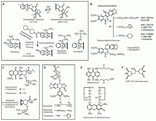
Cytotoxic Mechanisms of Top2cc-Targeted Drugs (Intercalators and Demethyl Epipodophyllotoxins)

Contrary to camptothecins, Top2 inhibitors kill cancer cells without requiring DNA replication fork collisions. Indeed, even after a 30-minute exposure, doxorubicin and other Top2cc-targeted drugs can kill over 99% of the cells, which is in vast excess of the fraction of S-phase cells in tissue culture (generally less than 50%).
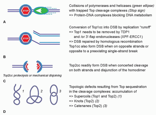
27,28 The collision mechanism in the case of Top2cc-targeted drugs (see Fig. 20.2A) appears to involve transcription and proteolysis of both Top2 and RNA polymerase II.29 Such situation would then lead to DNA double-strand breaks by disruption of the Top2 dimer interface (see Fig. 20.2C). Alternatively, the Top2 homodimer interface could be disjoined by mechanical tension (see Fig. 20.2C). Yet, it is important to bear in mind that 90% of Top2cc trapped by etoposide are not concerted and, therefore, consist in single-strand breaks,3,30,31 which is different from doxorubicin, which traps both Top2 monomers and produces a majority of DNA double-strand breaks.32 Finally, it is not excluded that topologic defects resulting from Top2 sequestration by the drug-induced cleavage complexes could contribute to the cytotoxicity of Top2cc-targeted drugs (see Fig. 20.2D). Such topologic defects would include persistent DNA knots and catenanes, potentially leading to chromosome breaks during mitosis.


TOPOISOMERASE I INHIBITORS: CAMPTOTHECINS AND BEYOND

Camptothecin is an alkaloid identified in the 1960s by Wall and Wani33 in a screen of plant extracts for antineoplastic drugs. The two water-soluble derivatives of camptothecin containing the active lactone form are topotecan and irinotecan, which are approved by the U.S. Food and Drug Administration (FDA) for the treatment of several cancers. In addition, several Top1cc-targeting drugs are in clinical development, including camptothecin derivatives and formulations (including high-molecular-weight conjugates or liposomal formulations), as well as noncamptothecin compounds that exhibit greater potency or noncross resistance to irinotecan and topotecan in preclinical cancer models.31,34,35,36


Irinotecan

Irinotecan, a prodrug containing a bulky dipiperidine side chain at C-10 (Fig. 20.3), is cleaved by a carboxylesterase-converting enzyme in the liver and other tissues to generate the active metabolite, SN-38. Irinotecan is FDA approved for the treatment of colorectal cancer in the metastatic setting as first-line treatment in combination with 5-fluorouracil/leucovorin (5-FU/LV) and as a single agent in the second-line treatment of progressive colorectal cancer after 5-FU-based therapy (see Table 20.1).37,38 Newer therapeutic uses of irinotecan include a combination with oxaliplatin and 5-FU as first-line treatment in pancreatic cancer.39 Irinotecan is additionally used in combination with cisplatin or carboplatin in extensive-stage small-cell lung cancer40,41 as well as refractory esophageal and gastroesophageal junction (GEJ) cancers, gastric cancer, cervical cancer, anaplastic gliomas and glioblastomas, and non-small-cell lung cancer (Table 20.2). Irinotecan is usually administered intravenously at a dose of 125 mg/m2 for 4 weeks with a 2-week rest period in combination with bolus 5-FU/LV, 180 mg/m2 every 2 weeks in combination with an infusion of 5-FU/LV, or 350 mg/m2 every 3 weeks as a single agent.

Diarrhea and myelosuppression are the most common toxicities associated with irinotecan administration. Two mechanisms explain irinotecan-induced diarrhea. Acute cholinergic effects resulting in abdominal cramping and diarrhea occur within 24 hours of drug administration are the result of acetylcholinesterase inhibition by the prodrug, and can be treated with the administration of atropine. Direct mucosal cytotoxicity with diarrhea is typically observed after 24 hours and can result in significant morbidity. Symptoms are managed with loperamide. Hepatic metabolism and biliary excretion accounts for >70% of the elimination of the administered dose, with renal excretion accounting for the remainder of the dose. SN-38 is glucuronidated in the liver by UGT1A1, and deficiencies in this pathway increase the risk of diarrhea and myelosuppression. Dose reductions are recommended for patients who are homozygous for the UGT1A1*28 allele, for which an FDA-approved test for detection of the UGT1A1*28 allele in patients is available.42,43 Additionally, dose reductions of irinotecan are recommended for patients with hepatic dysfunction, with bilirubin greater than 1.5 mg/mL.44


Figure 20.3 Structure of topoisomerase inhibitors. (A) Camptothecin derivatives are instable at physiologic pH with the formation of a carboxylate derivative within minutes. Irinotecan is a prodrug and needs to be converted to SN-38 to trap Top1cc. (B) Non-camptothecin derivatives in clinical trials. (C) Anthracycline derivatives. (D) Demethyl epipodophyllotoxin derivatives. (E) Other intercalating Top2 inhibitors acting by trapping Top2cc. (F) Structure of dexrazoxane, which acts as a catalytic inhibitor of Top2.


Topotecan

Topotecan contains a basic side chain at position C-9 that enhances its water solubility (see Fig. 20.3). Topotecan is approved for the treatment of ovarian cancer,45 small-cell lung cancer,46 and as a single agent and in combination with cisplatin for cervical cancer.47 Additionally, it is active in acute myeloid leukemia (AML) and myelodysplastic syndrome (see Table 20.2). Topotecan is administered intravenously as a single agent at a dose of 1.5 mg/m2 as a 30-minute infusion daily for 5 days, followed by a 2-week period of rest for the treatment of solid tumors or at a dose of 0.75 mg/m2 as a 30-minute infusion daily for 3 days in combination with cisplatin on day 1, every 3 weeks, for the treatment of cervical cancer.

Myelosuppression is the most common dose-limiting toxicity. Extensive prior radiation or previous bone marrow-suppressive chemotherapy increases the risk of topotecan-induced myelosuppression. Other toxicities include nausea, vomiting, diarrhea, fatigue, alopecia, and transient hepatic transaminitis.

Topotecan and its metabolites are primarily cleared by the kidneys, requiring dose reduction in patients with renal dysfunction. A 50% dose reduction is recommended for patients with moderate renal impairment (creatinine clearance 20 to 39 mL per minute). There are no formal guidelines for dose reductions in patients with hepatic dysfunction (defined as serum bilirubin >1.5 mg/dL to <10 mg/dL). Topotecan additionally penetrates the blood-brain barrier, achieving concentrations in cerebrospinal fluid that are approximately 30% that of plasma levels.48


Camptothecin Conjugates and Analogs

New formulations of camptothecin conjugates and analogs are currently in clinical development in an effort to improve the therapeutic index (Table 20.3). The development of camptothecin conjugates is based on the notion that the addition of a bulky conjugate would allow for a more consistent delivery system and extend the half-life of the molecule.

CRLX101, formerly IT-101, a covalent cyclodextrin-polyethylene glycol copolymer camptothecin conjugate, has plasma concentrations and area under the curve (AUC) that are approximately 100-fold higher than camptothecin, with a half-life in the range of 17 to 20 hours compared to 1.3 hours for camptothecin.49 It has demonstrated antitumor activity in preclinical studies in irinotecan-resistant tumors with complete tumor regression in human non-small-cell lung cancer, Ewing sarcoma, and
lymphoma xenograft models.50 Preliminary data from Phase 1 studies indicate that CRLX101 is well tolerated at a dose of 15 mg/m2 administered in a biweekly administration schedule.51 It is currently being studied in Phase 2 studies as a single agent and in combination with chemotherapeutic agents in lung, renal cell cancer, and gynecologic malignancies.52,53,54


TABLE 20.2 U.S. Food and Drug Administration-Approved Camptothecin Analogs


Irinotecan (Camptosar)


FDA approved for:


Metastatic colorectal cancer


First-line therapy in combination with 5-FU/LV


Diarrhea (dose reductions are recommended for patients who are homozygous for the UGT1A1*28 allele)


Second-line therapy as a single agent


Myelosuppression


Category 2Aa recommendations:


Pancreatic cancer


First-line therapy in combination with oxaliplatin, 5-FU/LV


Extensive-stage small-cell lung cancer


First-line therapy in combination with cisplatin or carboplatin


Category 2Bb recommendations:


Esophageal and GEJ cancers, gastric cancer, cervical cancer, anaplastic gliomas and glioblastomas, non-small-cell lung cancer, ovarian cancer


Topotecan (Hycamtin)


FDA approved for:


Cervical cancer


Stage IVB, recurrent, or persistent carcinoma of the cervix not amenable to curative treatment with surgery and/or radiation therapy


Myelosuppression


Ovarian cancer


Small-cell lung cancer


After failure of initial therapy


After failure of initial therapy


Class 2B recommendations:


AML, MDS


a Category 2A: Recommendations are based upon lower-level evidence, there is uniform National Comprehensive Cancer Network consensus that the intervention is appropriate.
b Category 2B: Recommendations are based upon lower-level evidence, there is National Comprehensive Cancer Network consensus that the intervention is appropriate.


MDS, myelodysplastic syndrome.


Etirinotecan pegol (NKTR-102), an irinotecan polymer conjugate, has a longer plasma circulation time with a lower maximum concentration of SN-38 compared with irinotecan. It was evaluated in a Phase 2 study in platinum-resistant refractory epithelial ovarian cancer at a dose of 145 mg/m2 administered on a schedule of every 21 days; a median progression-free survival of 5.3 months and median overall survival of 11.7 months was observed.55 Two schedules of administration, 145 mg/m2 administered every 14 days versus every 21 days, have been tested in a Phase 2 study of NKTR-102 in patients with previously treated metastatic breast cancer.56 Of the 70 patients evaluated in this study, 20 patients achieved an objective response (29%; 95% confidence interval [CI] 18.4 to 40.6). For both these studies, the most common adverse events on the 21-day administration schedule were dehydration and diarrhea. Etirinotecan pegol is currently being evaluated in several phase 2 studies in lung cancer, colorectal cancer, and high-grade gliomas,57,58,59,60 with evidence of clinical activity in refractory solid tumors. A Phase 3 trial (The BEACON Study) is underway evaluating NKTR-102 against the physicians’ choice in refractory breast cancer.61

Only gold members can continue reading. Log In or Register to continue

Stay updated, free articles. Join our Telegram channel

Jun 28, 2016 | Posted by in ONCOLOGY | Comments Off on Topoisomerase Interactive Agents

Full access? Get Clinical Tree

Get Clinical Tree app for offline access