(1)
Department of Pathology Beckley Veterans Medical Center (West Virginia), McGuire Veterans Medical Center, Virginia Commonwealth University, Richmond, VA, USA
Overview
During the embryonal stage, two types of ossification mechanism are involved. In the endochondral process, ossification starts on the framework of prefabricated chondroid tissue. This type of ossification also forms the basis for bone elongation and callus formation. The osteogenic and chondrogenic processes are so intricately connected that both osteoid and chondroid components are often seen in osteoid forming and chondroid tumors, benign or malignant.
The normal bone homeostasis features constant modeling and remodeling which is a result of concerted efforts of the three key elements (osteogenic progenitors, osteoclasts, and sinusoids) of the hematopoietic microenvironment [1–3]. Benign bone-forming tissue (also chondroid tissue) respects the sinusoid stroma as well as the adjacent benign trabeculae. Malignant osteoid (also chondroid) tissue gains bone matrix absorbing capability which is normally the domain of osteoclasts [4, 5]. This gain of function gives rise to the permeative growth pattern characteristic of malignant osteosarcoma and chondrosarcoma. In permeative growth pattern, the malignant matrix-forming tissue fills the bone marrow cavity and occasionally entraps and destroys preexisting benign trabeculae.
Malignant osteoid is defined as osteoid tissue that is rimmed by stromal cells and/or layers of osteoblasts [6] (Fig. 3.1). When chondroid component is present, the malignant nature of the osteoid can be revealed by the altered interaction pattern. Malignant osteoid is randomly distributed in chondroid lobules (Fig. 3.2). This is in contrast to the peripheral arrangement evidenced in benign tissue. This important feature can be very useful in the differentiation of chondrosarcoma and chondroblastic osteosarcoma. In the former, the reactive (benign) bone formation is in the form of attenuated semicircles which surround the malignant chondroid tissue (Fig. 3.3). In the latter, the chondroid lobules are in direct contact with sheets of spindle cells which produce lacelike osteoid. The lacelike osteoid tissue is scattered throughout the chondroid lobule.
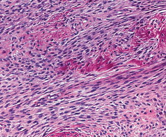
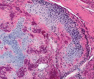
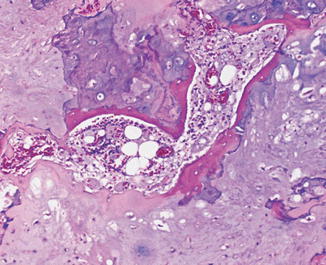
Fig 3.1
Malignant osteoid. Note the rimming by spindle stromal cells (Tumors of the Bones and Joints, Armed Force Institute of Pathology/American Registry of Pathology, 2005 with permission)
Fig 3.2
Malignant osteoid. Note randomness in the distribution of osteoid material in a chondroid lobule (Tumors of the Bones and Joints, Armed Force Institute of Pathology/American Registry of Pathology, 2005 with permission)
Fig 3.3
Reactive bone (osteoid). Attenuated semicircles surround malignant chondroid tissue (Tumors of the Bones and Joints, Armed Force Institute of Pathology/American Registry of Pathology, 2005 with permission)
Morphological Features of Low-Grade Osteosarcoma
Bone trabeculae lined by interlacing fascicles of spindle cells
Permeative growth pattern (Figs. 3.4 and 3.5)
Fig. 3.4
Low-grade osteosarcoma (Tumors of the Bones and Joints, Armed Force Institute of Pathology/American Registry of Pathology, 2005 with permission)
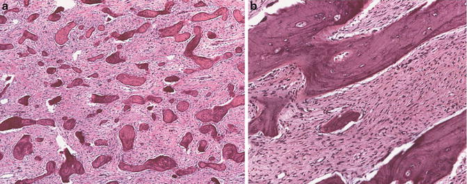
Fig. 3.5
Low-grade osteosarcoma. Permeative growth pattern by interlacing fascicles of spindle cells (Tumors of the Bones and Joints, Armed Force Institute of Pathology/American Registry of Pathology, 2005 with permission)
Discussion
Low-grade osteosarcoma is characterized by a permeative growth pattern of well-formed bony trabeculae which are rimmed by spindle cells [6].
Surface osteosarcomas are apparently derived from osteogenic stromal cells located outside the medullary cavity [6]. Strictly speaking, periosteal osteosarcoma is considered moderately differentiated. It is included in this book along with parosteal osteosarcoma. These two entities have some differences from the medullary osteosarcoma which reflects their respective stem cell location and properties. Therefore, the criteria for medullary osteosarcoma need to be modified. For instance, the tumor stroma is usually less cellular in parosteal variant, whereas periosteal osteosarcoma has no medullary involvement at all.
Related posts:

Stay updated, free articles. Join our Telegram channel

Full access? Get Clinical Tree


